
Article Figures & Data
Figures
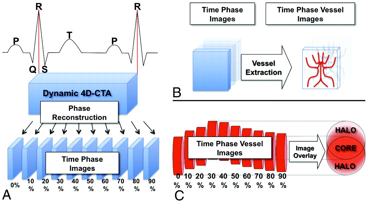
- Fig 1.
Schematic illustration of the reconstruction of 4D dynamic CTA into time-phase images and the motion-detection algorithm. A, The window center of the time-phase image is set at each 10% of the R-R interval, and the 4D-CTA image of a single heartbeat was reconstructed into 10 phase images. B, Time-phase images were digitalized for vessel extraction. The red columns on the right represent blood vessels at each phase. They are moving and pulsating according to the cardiac cycle. All of the 10 phase images are summed into a single matrix. C, The voxels in which the vessels are always present throughout the 10 phases can be identified as the CORE, and the surrounding voxels, as the HALO.
- Fig 2.
The motion map, motion index, and arterial pulse wave of the intracranial arteries. A, A posterior-cranial view of the motion map around the circle of Willis in patient 3 is shown. Red indicates the CORE, and translucent white indicates the HALO. The HALO of the bilateral P1 segment of the PCAs (white arrow) is thicker than that of the bilateral supraclinoid portion of the ICAs (double white arrows), indicating a difference in the magnitude of motion of each vessel. B, The mean ± SD of the motion index in VOI of the major cerebral vessels of the 10 subjects is shown. C, The mean ± SD of a vessel volume change in the left PCA during a single heartbeat for the 10 subjects is shown. This curve resembles a carotid artery pulse wave as well as an aortic pulse wave. D, The mean ± SD of the vessel volume change in the 10 subjects in each region of interest is shown. There is no significant difference in the amplitude among the VOI. The asterisk indicates P < .05; double asterisks, P < .001.


Tables
Patient profiles
Patient Age (yr) Sex R-R Interval Time (ms) HR (bpm) 1 53 M 1237 49 2 73 F 798 75 3 70 F 1064 56 4 65 F 982 61 5 70 F 865 69 6 73 M 729 82 7 67 F 943 64 8 69 F 769 78 9 77 M 899 67 10 68 F 644 93


