Advertisement

Main menu

  • Home
  • Content
    • Current Issue
    • Accepted Manuscripts
    • Article Preview
    • Past Issue Archive
    • Video Articles
    • AJNR Case Collection
    • Case of the Week Archive
    • Case of the Month Archive
    • Classic Case Archive
  • Special Collections
    • AJNR Awards
    • Low-Field MRI
    • Alzheimer Disease
    • ASNR Foundation Special Collection
    • Photon-Counting CT
    • View All
  • Multimedia
    • AJNR Podcasts
    • AJNR SCANtastic
    • Trainee Corner
    • MRI Safety Corner
    • Imaging Protocols
  • For Authors
    • Submit a Manuscript
    • Submit a Video Article
    • Submit an eLetter to the Editor/Response
    • Manuscript Submission Guidelines
    • Statistical Tips
    • Fast Publishing of Accepted Manuscripts
    • Graphical Abstract Preparation
    • Imaging Protocol Submission
    • Author Policies
  • About Us
    • About AJNR
    • Editorial Board
    • Editorial Board Alumni
  • More
    • Become a Reviewer/Academy of Reviewers
    • Subscribers
    • Permissions
    • Alerts
    • Feedback
    • Advertisers
    • ASNR Home

User menu

  • Alerts
  • Log in

Search

  • Advanced search
American Journal of Neuroradiology

American Journal of Neuroradiology

ASHNR American Society of Functional Neuroradiology ASHNR American Society of Pediatric Neuroradiology ASSR
  • Alerts
  • Log in

Advanced Search

  • Home
  • Content
    • Current Issue
    • Accepted Manuscripts
    • Article Preview
    • Past Issue Archive
    • Video Articles
    • AJNR Case Collection
    • Case of the Week Archive
    • Case of the Month Archive
    • Classic Case Archive
  • Special Collections
    • AJNR Awards
    • Low-Field MRI
    • Alzheimer Disease
    • ASNR Foundation Special Collection
    • Photon-Counting CT
    • View All
  • Multimedia
    • AJNR Podcasts
    • AJNR SCANtastic
    • Trainee Corner
    • MRI Safety Corner
    • Imaging Protocols
  • For Authors
    • Submit a Manuscript
    • Submit a Video Article
    • Submit an eLetter to the Editor/Response
    • Manuscript Submission Guidelines
    • Statistical Tips
    • Fast Publishing of Accepted Manuscripts
    • Graphical Abstract Preparation
    • Imaging Protocol Submission
    • Author Policies
  • About Us
    • About AJNR
    • Editorial Board
    • Editorial Board Alumni
  • More
    • Become a Reviewer/Academy of Reviewers
    • Subscribers
    • Permissions
    • Alerts
    • Feedback
    • Advertisers
    • ASNR Home
  • Follow AJNR on Twitter
  • Visit AJNR on Facebook
  • Follow AJNR on Instagram
  • Join AJNR on LinkedIn
  • RSS Feeds

AJNR Awards, New Junior Editors, and more. Read the latest AJNR updates

Research ArticlePatient Safety

Image Quality and Radiation Dose of Dual-Energy CT of the Head and Neck Compared with a Standard 120-kVp Acquisition

A.M. Tawfik, J.M. Kerl, A.A. Razek, R.W. Bauer, N.E. Nour-Eldin, T.J. Vogl and M.G. Mack
American Journal of Neuroradiology December 2011, 32 (11) 1994-1999; DOI: https://doi.org/10.3174/ajnr.A2654
A.M. Tawfik
aFrom the Department of Diagnostic and Interventional Radiology (A.M.T., J.M.K., R.B., N.E.N.-E., T.J.V., M.G.M.), Johan Wolfgang Goethe University Hospital, Frankfurt am Main, Germany
bDepartment of Diagnostic Radiology (A.M.T., A.A.R.), Mansoura University, Mansoura City, Egypt
  • Find this author on Google Scholar
  • Find this author on PubMed
  • Search for this author on this site
J.M. Kerl
aFrom the Department of Diagnostic and Interventional Radiology (A.M.T., J.M.K., R.B., N.E.N.-E., T.J.V., M.G.M.), Johan Wolfgang Goethe University Hospital, Frankfurt am Main, Germany
  • Find this author on Google Scholar
  • Find this author on PubMed
  • Search for this author on this site
A.A. Razek
bDepartment of Diagnostic Radiology (A.M.T., A.A.R.), Mansoura University, Mansoura City, Egypt
  • Find this author on Google Scholar
  • Find this author on PubMed
  • Search for this author on this site
R.W. Bauer
aFrom the Department of Diagnostic and Interventional Radiology (A.M.T., J.M.K., R.B., N.E.N.-E., T.J.V., M.G.M.), Johan Wolfgang Goethe University Hospital, Frankfurt am Main, Germany
  • Find this author on Google Scholar
  • Find this author on PubMed
  • Search for this author on this site
N.E. Nour-Eldin
aFrom the Department of Diagnostic and Interventional Radiology (A.M.T., J.M.K., R.B., N.E.N.-E., T.J.V., M.G.M.), Johan Wolfgang Goethe University Hospital, Frankfurt am Main, Germany
  • Find this author on Google Scholar
  • Find this author on PubMed
  • Search for this author on this site
T.J. Vogl
aFrom the Department of Diagnostic and Interventional Radiology (A.M.T., J.M.K., R.B., N.E.N.-E., T.J.V., M.G.M.), Johan Wolfgang Goethe University Hospital, Frankfurt am Main, Germany
  • Find this author on Google Scholar
  • Find this author on PubMed
  • Search for this author on this site
M.G. Mack
aFrom the Department of Diagnostic and Interventional Radiology (A.M.T., J.M.K., R.B., N.E.N.-E., T.J.V., M.G.M.), Johan Wolfgang Goethe University Hospital, Frankfurt am Main, Germany
  • Find this author on Google Scholar
  • Find this author on PubMed
  • Search for this author on this site
  • Article
  • Figures & Data
  • Info & Metrics
  • Responses
  • References
  • PDF
Loading

Article Figures & Data

Figures

  • Tables
  • Fig 1.
    • Download figure
    • Open in new tab
    • Download powerpoint
    Fig 1.

    Axial contrast-enhanced CT images obtained through the level of the floor of the mouth in 2 different patients. DE-derived WA image (A) and standard SE (120 kVp) image (B). Mean attenuation values (in Hounsfield units) of the sternomastoid muscle, tongue muscle, and internal jugular vein were obtained at this level by using circular regions of interest, as shown. Objective noise was obtained as the SD of the circular regions of interest drawn in air outside the patient. Noise was obtained in all anatomic levels in a similar way.
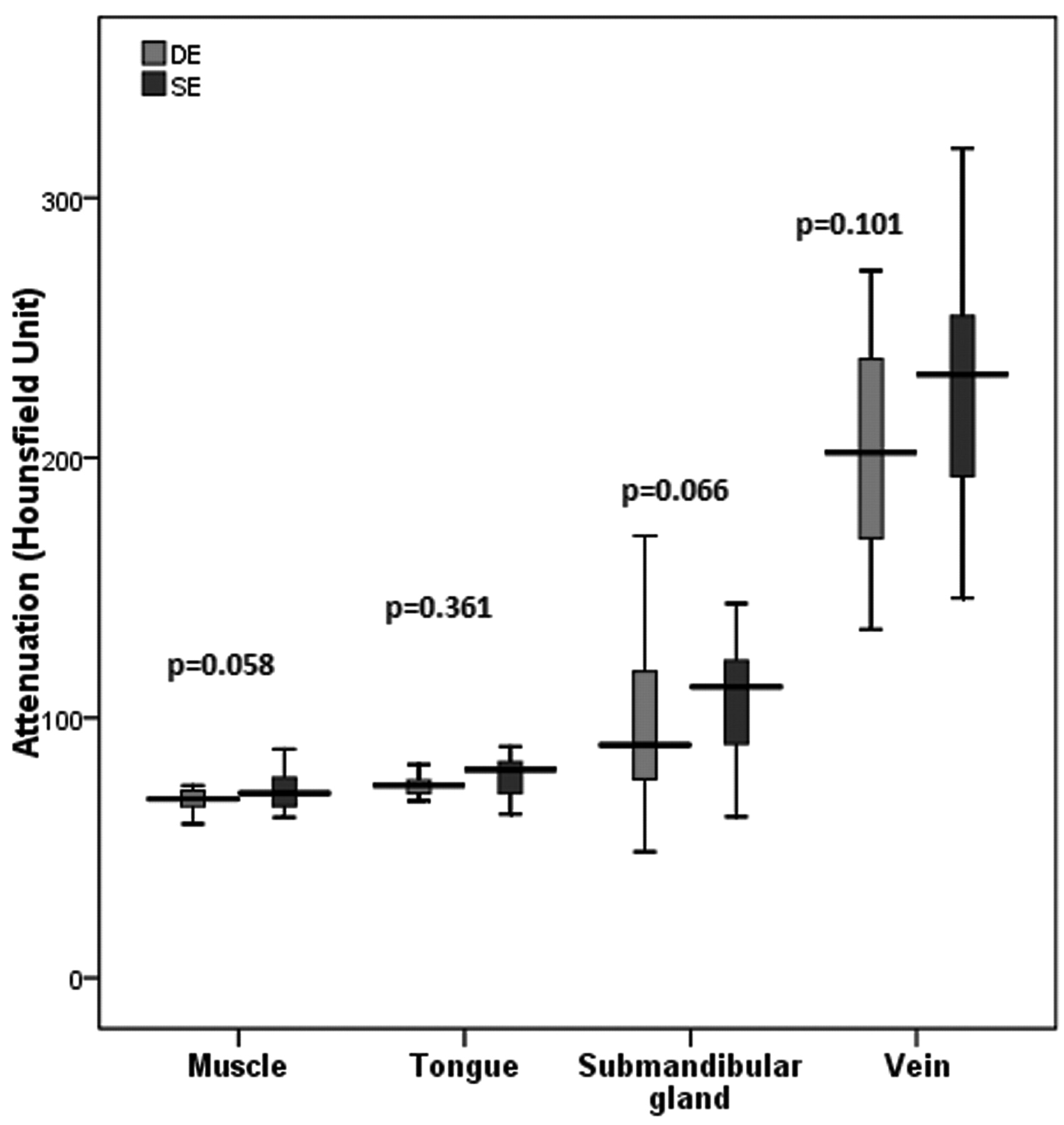
  • Fig 2.
    • Download figure
    • Open in new tab
    • Download powerpoint
    Fig 2.

    Box-and-whisker plot of attenuation measurements with DE and SECT. Boxes represent the middle 50% of cases, horizontal lines within mark median values, and whiskers represent minimal and maximal extremes. Differences between attenuation values with DECT and SECT are nonsignificant (P > .05).
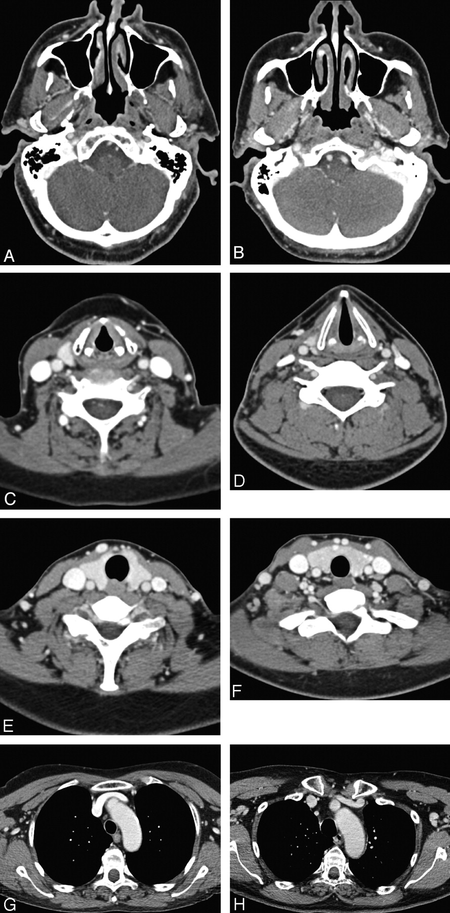
  • Fig 3.
    • Download figure
    • Open in new tab
    • Download powerpoint
    Fig 3.

    Axial contrast-enhanced CT images through the nasopharynx (A and B), arytenoids (C and D), lower thyroid (E and F), and arch-of-the-aorta (G and H) levels in different patients. On the left panel are DE-derived WA images (A, C, E, G) and on the right panel are images acquired in single energy mode (B, D, F, H). There is no perceivable difference in image noise, sharpness, or overall image quality. All images are of excellent diagnostic quality.

Tables

  • Figures
    • View popup
    Table 1:

    Objective noise with DE and standard SECTa

    Anatomic LevelNoise (HU)Difference (HU)95% CI for the DifferenceP Value
    DECTSECT
    Nasopharynx6.15 ± 1.76.25 ± 1−0.1−0.83:0.63.788
    Floor of mouth5.41 ± 15.96 ± 3.6−0.55−1.90:0.82.426
    Arytenoids4.9 ± 1.34.79 ± 0.950.1−0.39:0.76.592
    Lower thyroid8.1 ± 4.511.3 ± 4.4−3.2−8.9:2.5.270
    Arch of aorta12.2 ± 5.28.7 ± 2.33.4−2.4:9.4.243
    • ↵a Data are presented as mean ± SD. All P values are nonsignificant (>.05).

    • View popup
    Table 2:

    Subjective image-quality analysis scores for DE and SECTa

    Anatomic LevelNoiseSharpnessStreak ArtifactsOverall Image Quality
    DECTSECTPDECTSECTPDECTSECTPDECTSECTP
    Nasopharynx5 (4–5)5 (4–5).3055 (4–5)5 (4–5).552–––5 (4–5)5 (4–5).557
    Floor of mouth5 (4–5)5 (4–5).7325 (4–5)5 (4–5).154–––5 (4–5)5 (4–5).664
    Arytenoids5 (4–5)5 (4–5).7215 (4–5)5 (3–5).813–––4 (3–5)4 (4–5).896
    Lower thyroid3 (2–5)3.5 (2–5).4983 (2:5)3 (2:5).1223 (2–5)3 (2–5).1083.5 (2:5)4 (2:5).214
    Arch of aorta3 (2:5)4 (2:4).0983 (2:5)4 (2:4).0833 (2:5)4 (2:4).0613 (2:5)4 (2:4).288
    • ↵a See “Materials and Methods” section for details of scales. Data are presented as median (range). All P values are nonsignificant (>.05).

PreviousNext
Back to top

In this issue

American Journal of Neuroradiology: 32 (11)
American Journal of Neuroradiology
Vol. 32, Issue 11
1 Dec 2011
  • Table of Contents
  • Index by author
Advertisement
Print
Download PDF
Email Article

Thank you for your interest in spreading the word on American Journal of Neuroradiology.

NOTE: We only request your email address so that the person you are recommending the page to knows that you wanted them to see it, and that it is not junk mail. We do not capture any email address.

Enter multiple addresses on separate lines or separate them with commas.
Image Quality and Radiation Dose of Dual-Energy CT of the Head and Neck Compared with a Standard 120-kVp Acquisition
(Your Name) has sent you a message from American Journal of Neuroradiology
(Your Name) thought you would like to see the American Journal of Neuroradiology web site.
CAPTCHA
This question is for testing whether or not you are a human visitor and to prevent automated spam submissions.
Cite this article
A.M. Tawfik, J.M. Kerl, A.A. Razek, R.W. Bauer, N.E. Nour-Eldin, T.J. Vogl, M.G. Mack
Image Quality and Radiation Dose of Dual-Energy CT of the Head and Neck Compared with a Standard 120-kVp Acquisition
American Journal of Neuroradiology Dec 2011, 32 (11) 1994-1999; DOI: 10.3174/ajnr.A2654

Citation Manager Formats

  • BibTeX
  • Bookends
  • EasyBib
  • EndNote (tagged)
  • EndNote 8 (xml)
  • Medlars
  • Mendeley
  • Papers
  • RefWorks Tagged
  • Ref Manager
  • RIS
  • Zotero
0 Responses
Respond to this article
Share
Bookmark this article
Image Quality and Radiation Dose of Dual-Energy CT of the Head and Neck Compared with a Standard 120-kVp Acquisition
A.M. Tawfik, J.M. Kerl, A.A. Razek, R.W. Bauer, N.E. Nour-Eldin, T.J. Vogl, M.G. Mack
American Journal of Neuroradiology Dec 2011, 32 (11) 1994-1999; DOI: 10.3174/ajnr.A2654
del.icio.us logo Twitter logo Facebook logo Mendeley logo
  • Tweet Widget
  • Facebook Like
  • Google Plus One
Purchase

Jump to section

  • Article
    • Abstract
    • ABBREVIATIONS
    • Materials and Methods
    • Results
    • Discussion
    • Conclusions
    • Footnotes
    • References
  • Figures & Data
  • Info & Metrics
  • Responses
  • References
  • PDF

Related Articles

  • No related articles found.
  • PubMed
  • Google Scholar

Cited By...

  • Different Spectral Hounsfield Unit Curve and High-Energy Virtual Monochromatic Image Characteristics of Squamous Cell Carcinoma Compared with Nonossified Thyroid Cartilage
  • Dual-Energy 4-Phase CT Scan in Primary Hyperparathyroidism
  • Crossref (115)
  • Google Scholar

This article has been cited by the following articles in journals that are participating in Crossref Cited-by Linking.

  • 3D scanning applications in medical field: A literature-based review
    Abid Haleem, Mohd. Javaid
    Clinical Epidemiology and Global Health 2019 7 2
  • Assessment of an Advanced Monoenergetic Reconstruction Technique in Dual-Energy Computed Tomography of Head and Neck Cancer
    Moritz H. Albrecht, Jan-Erik Scholtz, Johannes Kraft, Ralf W. Bauer, Moritz Kaup, Patricia Dewes, Andreas M. Bucher, Iris Burck, Jens Wagenblast, Thomas Lehnert, J. Matthias Kerl, Thomas J. Vogl, Julian L. Wichmann
    European Radiology 2015 25 8
  • Virtual Monoenergetic Dual-Energy Computed Tomography
    Julian L. Wichmann, Eva-Maria Nöske, Johannes Kraft, Iris Burck, Jens Wagenblast, Anne Eckardt, Claudia Frellesen, J. Matthias Kerl, Ralf W. Bauer, Boris Bodelle, Thomas Lehnert, Thomas J. Vogl, Boris Schulz
    Investigative Radiology 2014 49 11
  • Evaluation of Cartilage Invasion by Laryngeal and Hypopharyngeal Squamous Cell Carcinoma with Dual-Energy CT
    Hirofumi Kuno, Hiroaki Onaya, Ryoko Iwata, Tatsushi Kobayashi, Satoshi Fujii, Ryuichi Hayashi, Katharina Otani, Hiroya Ojiri, Takeharu Yamanaka, Mitsuo Satake
    Radiology 2012 265 2
  • Dual-Energy CT: What the Neuroradiologist Should Know
    Alida A. Postma, Marco Das, Annika A. R. Stadler, Joachim E. Wildberger
    Current Radiology Reports 2015 3 5
  • Perfusion CT of head and neck cancer
    Ahmed Abdel Khalek Abdel Razek, Ahmed Mohamed Tawfik, Lamiaa Galal Ali Elsorogy, Nermin Yehia Soliman
    European Journal of Radiology 2014 83 3
  • Dual-Energy Computed Tomography
    Reza Forghani, Bruno De Man, Rajiv Gupta
    Neuroimaging Clinics of North America 2017 27 3
  • State-of-the-Art Imaging of Salivary Gland Tumors
    Ahmed Abdel Khalek Abdel Razek, Suresh K. Mukherji
    Neuroimaging Clinics of North America 2018 28 2
  • Dual-Energy CT in Head and Neck Imaging
    Elise D. Roele, Veronique C. M. L. Timmer, Lauretta A. A. Vaassen, Anna M. J. L. van Kroonenburgh, A. A. Postma
    Current Radiology Reports 2017 5 5
  • Dual-Energy CT Applications in Head and Neck Imaging
    Thomas J. Vogl, Boris Schulz, Ralf W. Bauer, Timo Stöver, Robert Sader, Ahmed M. Tawfik
    American Journal of Roentgenology 2012 199 5_supplement

More in this TOC Section

  • Safety of Intrathecal Gadobutrol in Various Doses
  • Impact of Kidney Function on CNS Gadolinium Deposition in Patients Receiving Repeated Doses of Gadobutrol
  • Contrast-Induced Acute Kidney Injury in Radiologic Management of Acute Ischemic Stroke in the Emergency Setting
Show more PATIENT SAFETY

Similar Articles

Advertisement

Indexed Content

  • Current Issue
  • Accepted Manuscripts
  • Article Preview
  • Past Issues
  • Editorials
  • Editor's Choice
  • Fellows' Journal Club
  • Letters to the Editor
  • Video Articles

Cases

  • Case Collection
  • Archive - Case of the Week
  • Archive - Case of the Month
  • Archive - Classic Case

More from AJNR

  • Trainee Corner
  • Imaging Protocols
  • MRI Safety Corner
  • Book Reviews

Multimedia

  • AJNR Podcasts
  • AJNR Scantastics

Resources

  • Turnaround Time
  • Submit a Manuscript
  • Submit a Video Article
  • Submit an eLetter to the Editor/Response
  • Manuscript Submission Guidelines
  • Statistical Tips
  • Fast Publishing of Accepted Manuscripts
  • Graphical Abstract Preparation
  • Imaging Protocol Submission
  • Evidence-Based Medicine Level Guide
  • Publishing Checklists
  • Author Policies
  • Become a Reviewer/Academy of Reviewers
  • News and Updates

About Us

  • About AJNR
  • Editorial Board
  • Editorial Board Alumni
  • Alerts
  • Permissions
  • Not an AJNR Subscriber? Join Now
  • Advertise with Us
  • Librarian Resources
  • Feedback
  • Terms and Conditions
  • AJNR Editorial Board Alumni

American Society of Neuroradiology

  • Not an ASNR Member? Join Now

© 2025 by the American Society of Neuroradiology All rights, including for text and data mining, AI training, and similar technologies, are reserved.
Print ISSN: 0195-6108 Online ISSN: 1936-959X

Powered by HighWire