Advertisement

Main menu

  • Home
  • Content
    • Current Issue
    • Accepted Manuscripts
    • Article Preview
    • Past Issue Archive
    • Video Articles
    • AJNR Case Collection
    • Case of the Week Archive
    • Case of the Month Archive
    • Classic Case Archive
  • Special Collections
    • AJNR Awards
    • Low-Field MRI
    • Alzheimer Disease
    • ASNR Foundation Special Collection
    • Photon-Counting CT
    • View All
  • Multimedia
    • AJNR Podcasts
    • AJNR SCANtastic
    • Trainee Corner
    • MRI Safety Corner
    • Imaging Protocols
  • For Authors
    • Submit a Manuscript
    • Submit a Video Article
    • Submit an eLetter to the Editor/Response
    • Manuscript Submission Guidelines
    • Statistical Tips
    • Fast Publishing of Accepted Manuscripts
    • Graphical Abstract Preparation
    • Imaging Protocol Submission
    • Author Policies
  • About Us
    • About AJNR
    • Editorial Board
    • Editorial Board Alumni
  • More
    • Become a Reviewer/Academy of Reviewers
    • Subscribers
    • Permissions
    • Alerts
    • Feedback
    • Advertisers
    • ASNR Home

User menu

  • Alerts
  • Log in

Search

  • Advanced search
American Journal of Neuroradiology

American Journal of Neuroradiology

ASHNR American Society of Functional Neuroradiology ASHNR American Society of Pediatric Neuroradiology ASSR
  • Alerts
  • Log in

Advanced Search

  • Home
  • Content
    • Current Issue
    • Accepted Manuscripts
    • Article Preview
    • Past Issue Archive
    • Video Articles
    • AJNR Case Collection
    • Case of the Week Archive
    • Case of the Month Archive
    • Classic Case Archive
  • Special Collections
    • AJNR Awards
    • Low-Field MRI
    • Alzheimer Disease
    • ASNR Foundation Special Collection
    • Photon-Counting CT
    • View All
  • Multimedia
    • AJNR Podcasts
    • AJNR SCANtastic
    • Trainee Corner
    • MRI Safety Corner
    • Imaging Protocols
  • For Authors
    • Submit a Manuscript
    • Submit a Video Article
    • Submit an eLetter to the Editor/Response
    • Manuscript Submission Guidelines
    • Statistical Tips
    • Fast Publishing of Accepted Manuscripts
    • Graphical Abstract Preparation
    • Imaging Protocol Submission
    • Author Policies
  • About Us
    • About AJNR
    • Editorial Board
    • Editorial Board Alumni
  • More
    • Become a Reviewer/Academy of Reviewers
    • Subscribers
    • Permissions
    • Alerts
    • Feedback
    • Advertisers
    • ASNR Home
  • Follow AJNR on Twitter
  • Visit AJNR on Facebook
  • Follow AJNR on Instagram
  • Join AJNR on LinkedIn
  • RSS Feeds

AJNR Awards, New Junior Editors, and more. Read the latest AJNR updates

Review ArticleReview Articles
Open Access

Pseudotumor Cerebri: Brief Review of Clinical Syndrome and Imaging Findings

A.J. Degnan and L.M. Levy
American Journal of Neuroradiology December 2011, 32 (11) 1986-1993; DOI: https://doi.org/10.3174/ajnr.A2404
A.J. Degnan
aFrom the Department of Radiology, George Washington University Hospital, Washington, DC.
  • Find this author on Google Scholar
  • Find this author on PubMed
  • Search for this author on this site
L.M. Levy
aFrom the Department of Radiology, George Washington University Hospital, Washington, DC.
  • Find this author on Google Scholar
  • Find this author on PubMed
  • Search for this author on this site
  • Article
  • Figures & Data
  • Info & Metrics
  • Responses
  • References
  • PDF
Loading

Article Figures & Data

Figures

  • Tables
  • Fig 1.
    • Download figure
    • Open in new tab
    • Download powerpoint
    Fig 1.

    This 31-year-old woman presenting with headache is found to have an empty sella on sagittal T1-weighted MR imaging.
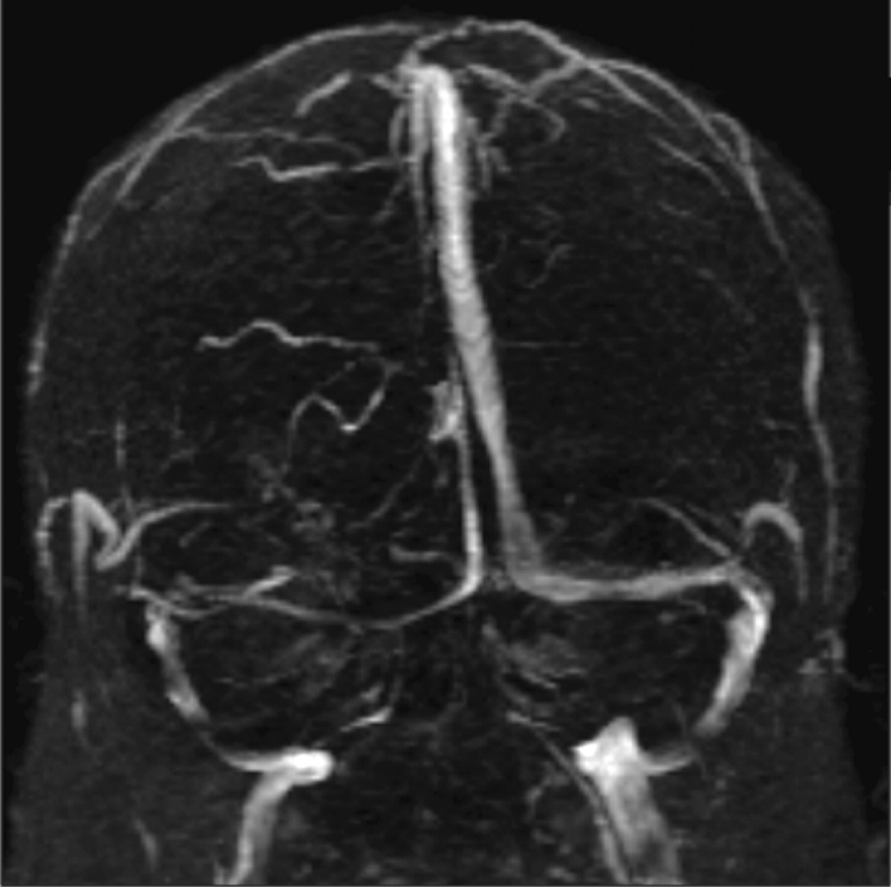
  • Fig 2.
    • Download figure
    • Open in new tab
    • Download powerpoint
    Fig 2.

    A narrowed right transverse sinus is noted in this 32-year-old woman, seen on MR venography, in addition to ONS enlargement and a partially empty sella on axial MR imaging.

  • Fig 3.
    • Download figure
    • Open in new tab
    • Download powerpoint
    Fig 3.

    Protrusion of the right optic nerve head and horizontal tortuosity of the optic nerve are seen in this 21-year-old woman on axial T2-weighted MR imaging. Clinically, the patient presented with headaches, vision changes, and papilledema noted on examination.
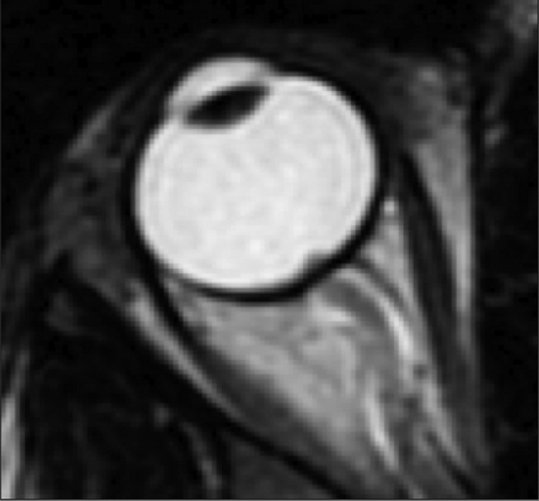
  • Fig 4.
    • Download figure
    • Open in new tab
    • Download powerpoint
    Fig 4.

    A, The ONS is widened with expanded CSF hyperintensity surrounding the optic nerve, seen on axial T2-weighted MR imaging in conjunction with posterior flattening of the globes. ONS widening is thought to coincide with papilledema, which is seen in this 27-year-old woman who presented with headaches. B, Coronal T2-weighted MR imaging in a 55-year-old woman with headache demonstrates increased peri-ONS space marked by hyperintense signal intensity surrounding the optic nerve.

Tables

  • Figures
    • View popup
    Table 1:

    Confounding conditions that may present as similar to IIHa

    Medical disorders
    • Addison disease

    • Hypoparathyroidism

    • Chronic obstructive pulmonary disease

    • Right heart failure with pulmonary hypertension

    • Obstructive sleep apnea

    • Pickwickian syndrome

    • Polycystic ovary syndrome

    • Systemic lupus erythematosus

    • Uremia

    • Severe iron deficiency anemia

    Medications
    • Tetracycline and related compounds (minocycline, doxycycline)

    • Vitamin A (at doses >25 000 IU daily) and related compounds (isotretinoin [Accutane], vitamin supplements, excessive intake of liver, all-trans retinoic acid)

    • Anabolic steroids

    • Corticosteroid withdrawal following prolonged administration

    • Growth hormone administration in deficient patients

    • Nalidixic acid

    • Lithium

    • Oral contraceptive use

    • Levonorgestrel implant system

    • Amiodarone

    • Cyclosporine

    • Cytarabine

    Obstruction to venous drainage
    • Cerebral venous sinus thrombosis

    • Jugular vein thrombosis

    • Superior vena cava syndrome

    • Jugular vein ligation following bilateral radical neck dissection

    • Increased right heart pressure

    • Glomus tumor

    • Compression by tumor process (eg, meningioma)

    Infections
    • HIV infection, borreliosis

    • Postvaricella infection in children

    • ↵a Adapted from Friedman and Jacobson,7 Szitkar,8 Wall,22 and Alperin et al.79

    • View popup
    Table 2:

    Criteria for diagnosing IIHa

    1) If symptoms are present, they may only reflect those of generalized intracranial hypertension or papilledema
    2) If signs are present, they may reflect only those of generalized intracranial hypertension or papilledema
    3) Documented elevated ICP measured in the lateral decubitus position (findings of assessment of ICP by lumbar puncture are considered abnormal if above 20 cm H2O in normal-weight individuals and 25 mm H2O in obese individuals20); MRI abnormal if above 20 cm H2O in normal-weight individuals and 25 mm H2O in obese individuals20)
    4) Normal CSF composition
    5) No evidence of hydrocephalus, mass, structural, or vascular lesion on MRI or contrast-enhanced CT for typical patients and on MRI and MR venography for all others
    6) No other cause of intracranial hypertension identified
    • ↵a Adapted from Friedman and Jacobson.7

    • View popup
    Table 3:

    Putative theories to explain elevated ICP in PTCa

    Proximal EtiologyResult
    Increased interstitial fluid (ISF) volumeIncreased cerebral volume
    Increased blood volume
    Increased tissue volume
    Increased CSF production rateIncreased CSF volume
    Increased CSF outflow resistance
    Loss of cerebral autoregulationIncreased cerebral arterial pressure
    Increased cerebral venous pressureIncreased venous blood volume and increased ISF
    Reduced CSF outflow and increased CSF volume
    • ↵a (Adapted from Walker.10)

    • View popup
    Table 4:

    Imaging Findings in PTC

    ReferencesSensitivitySpecificity
    Empty sellaAgid et al, 20066626.7%94.6%
    Yuh et al, 2000642.5%
    Partially empty sella/decreased pituitary heightAgid et al, 20066653.3%75%
    Yuh et al, 20006480%92%
    Brodsky and Vaphiades, 19986270%
    Flattened posterior globe/scleraAgid et al, 20066643.3%100%
    Brodsky and Vaphiades, 19986280%
    Jinkins et al, 19967166.7%
    Enlarged ONS (perioptic subarachnoid space)Agid et al, 20066666.7%82.1%
    Brodsky and Vaphiades, 19986245%
    Increased tortuosity of optic nerveAgid et al, 20066640%91.1%
    Brodsky and Vaphiades, 19986240%
    Enhancement of optic nerveAgid et al, 2006666.7%98.2%
    Brodsky and Vaphiades, 19986250%
    Intraocular protrusion of optic nerve headAgid et al, 2006663.3%100%
    Brodsky and Vaphiades, 19986230%
    Slitlike ventriclesAgid et al, 2006663.3%100%
PreviousNext
Back to top

In this issue

American Journal of Neuroradiology: 32 (11)
American Journal of Neuroradiology
Vol. 32, Issue 11
1 Dec 2011
  • Table of Contents
  • Index by author
Advertisement
Print
Download PDF
Email Article

Thank you for your interest in spreading the word on American Journal of Neuroradiology.

NOTE: We only request your email address so that the person you are recommending the page to knows that you wanted them to see it, and that it is not junk mail. We do not capture any email address.

Enter multiple addresses on separate lines or separate them with commas.
Pseudotumor Cerebri: Brief Review of Clinical Syndrome and Imaging Findings
(Your Name) has sent you a message from American Journal of Neuroradiology
(Your Name) thought you would like to see the American Journal of Neuroradiology web site.
CAPTCHA
This question is for testing whether or not you are a human visitor and to prevent automated spam submissions.
Cite this article
A.J. Degnan, L.M. Levy
Pseudotumor Cerebri: Brief Review of Clinical Syndrome and Imaging Findings
American Journal of Neuroradiology Dec 2011, 32 (11) 1986-1993; DOI: 10.3174/ajnr.A2404

Citation Manager Formats

  • BibTeX
  • Bookends
  • EasyBib
  • EndNote (tagged)
  • EndNote 8 (xml)
  • Medlars
  • Mendeley
  • Papers
  • RefWorks Tagged
  • Ref Manager
  • RIS
  • Zotero
0 Responses
Respond to this article
Share
Bookmark this article
Pseudotumor Cerebri: Brief Review of Clinical Syndrome and Imaging Findings
A.J. Degnan, L.M. Levy
American Journal of Neuroradiology Dec 2011, 32 (11) 1986-1993; DOI: 10.3174/ajnr.A2404
del.icio.us logo Twitter logo Facebook logo Mendeley logo
  • Tweet Widget
  • Facebook Like
  • Google Plus One
Purchase

Jump to section

  • Article
    • Abstract
    • ABBREVIATIONS
    • Etiology
    • Clinical Syndrome
    • Imaging Findings
    • Recommended Imaging Protocol
    • Summary
    • References
  • Figures & Data
  • Info & Metrics
  • Responses
  • References
  • PDF

Related Articles

  • No related articles found.
  • PubMed
  • Google Scholar

Cited By...

  • Idiopathic intracranial hypertension in two twin sisters
  • Effectiveness of radiology modalities in diagnosing and characterizing brain disorders
  • The Monro-Kellie Doctrine: A Review and Call for Revision
  • Optic ataxia in a patient with HaNDL syndrome
  • Empty Sella Is a Sign of Symptomatic Lateral Sinus Stenosis and Not Intracranial Hypertension
  • Mastoid osteoma with stenosis of transverse and sigmoid sinuses as a cause of pseudotumor cerebri
  • The Occipital Emissary Vein: A Possible Marker for Pseudotumor Cerebri
  • Contrast-Enhanced 3D-FLAIR Imaging of the Optic Nerve and Optic Nerve Head: Novel Neuroimaging Findings of Idiopathic Intracranial Hypertension
  • Idiopathic intracranial hypertension: consensus guidelines on management
  • A new index for the assessment of transverse sinus stenosis for diagnosing idiopathic intracranial hypertension
  • Quantifying the Cerebral Hemodynamics of Dural Arteriovenous Fistula in Transverse Sigmoid Sinus Complicated by Sinus Stenosis: A Retrospective Cohort Study
  • Obesity in Children and Adolescents: Health Effects and Imaging Implications
  • Structural Brain Changes following Long-Term 6{degrees} Head-Down Tilt Bed Rest as an Analog for Spaceflight
  • Meta-Analysis of CSF Diversion Procedures and Dural Venous Sinus Stenting in the Setting of Medically Refractory Idiopathic Intracranial Hypertension
  • Volumetric Assessment of Optic Nerve Sheath and Hypophysis in Idiopathic Intracranial Hypertension
  • MR Imaging of Papilledema and Visual Pathways: Effects of Increased Intracranial Pressure and Pathophysiologic Mechanisms
  • Magnetic resonance imaging of CNS in 15 043 children with GH deficiency in KIGS (Pfizer International Growth Database)
  • MRI Evidence of Impaired CSF Homeostasis in Obesity-Associated Idiopathic Intracranial Hypertension
  • Update on the pathophysiology and management of idiopathic intracranial hypertension
  • Crossref (201)
  • Google Scholar

This article has been cited by the following articles in journals that are participating in Crossref Cited-by Linking.

  • Idiopathic intracranial hypertension: consensus guidelines on management
    Susan P Mollan, Brendan Davies, Nick C Silver, Simon Shaw, Conor L Mallucci, Benjamin R Wakerley, Anita Krishnan, Swarupsinh V Chavda, Satheesh Ramalingam, Julie Edwards, Krystal Hemmings, Michelle Williamson, Michael A Burdon, Ghaniah Hassan-Smith, Kathleen Digre, Grant T Liu, Rigmor Højland Jensen, Alexandra J Sinclair
    Journal of Neurology, Neurosurgery & Psychiatry 2018 89 10
  • Meta-Analysis of CSF Diversion Procedures and Dural Venous Sinus Stenting in the Setting of Medically Refractory Idiopathic Intracranial Hypertension
    S.R. Satti, L. Leishangthem, M.I. Chaudry
    American Journal of Neuroradiology 2015 36 10
  • European Headache Federation guideline on idiopathic intracranial hypertension
    Jan Hoffmann, Susan P Mollan, Koen Paemeleire, Christian Lampl, Rigmor H Jensen, Alexandra J Sinclair
    The Journal of Headache and Pain 2018 19 1
  • Meningoceles in Idiopathic Intracranial Hypertension
    Omer Y. Bialer, Mario Perez Rueda, Beau B. Bruce, Nancy J. Newman, Valérie Biousse, Amit M. Saindane
    American Journal of Roentgenology 2014 202 3
  • Microgravity-Induced Fluid Shift and Ophthalmic Changes
    Emily Nelson, Lealem Mulugeta, Jerry Myers
    Life 2014 4 4
  • Headache arising from idiopathic changes in CSF pressure
    Anne Ducros, Valérie Biousse
    The Lancet Neurology 2015 14 6
  • MR Imaging of Papilledema and Visual Pathways: Effects of Increased Intracranial Pressure and Pathophysiologic Mechanisms
    N. Passi, A.J. Degnan, L.M. Levy
    American Journal of Neuroradiology 2013 34 5
  • Morphometric and volumetric MRI changes in idiopathic intracranial hypertension
    Jan Hoffmann, Hans-Jürgen Huppertz, Christoph Schmidt, Hagen Kunte, Lutz Harms, Randolf Klingebiel, Edzard Wiener
    Cephalalgia 2013 33 13
  • Idiopathic Intracranial Hypertension and Obesity
    Laura E. Andrews, Grant T. Liu, Melissa W. Ko
    Hormone Research in Paediatrics 2014 81 4
  • MRI Evidence of Impaired CSF Homeostasis in Obesity-Associated Idiopathic Intracranial Hypertension
    N. Alperin, S. Ranganathan, A.M. Bagci, D.J. Adams, B. Ertl-Wagner, E. Saraf-Lavi, E.M. Sklar, B.L. Lam
    American Journal of Neuroradiology 2013 34 1

More in this TOC Section

  • An Atlas of Neonatal Neurovascular Imaging Anatomy as Depicted with Microvascular Imaging: The Intracranial Arteries
  • An Atlas of Neonatal Neurovascular Imaging Anatomy as Depicted with Microvascular Imaging: The Intracranial Veins
  • Clinical Translation of Hyperpolarized 13C Metabolic Probes for Glioma Imaging
Show more Review articles

Similar Articles

Advertisement

Indexed Content

  • Current Issue
  • Accepted Manuscripts
  • Article Preview
  • Past Issues
  • Editorials
  • Editor's Choice
  • Fellows' Journal Club
  • Letters to the Editor
  • Video Articles

Cases

  • Case Collection
  • Archive - Case of the Week
  • Archive - Case of the Month
  • Archive - Classic Case

More from AJNR

  • Trainee Corner
  • Imaging Protocols
  • MRI Safety Corner
  • Book Reviews

Multimedia

  • AJNR Podcasts
  • AJNR Scantastics

Resources

  • Turnaround Time
  • Submit a Manuscript
  • Submit a Video Article
  • Submit an eLetter to the Editor/Response
  • Manuscript Submission Guidelines
  • Statistical Tips
  • Fast Publishing of Accepted Manuscripts
  • Graphical Abstract Preparation
  • Imaging Protocol Submission
  • Evidence-Based Medicine Level Guide
  • Publishing Checklists
  • Author Policies
  • Become a Reviewer/Academy of Reviewers
  • News and Updates

About Us

  • About AJNR
  • Editorial Board
  • Editorial Board Alumni
  • Alerts
  • Permissions
  • Not an AJNR Subscriber? Join Now
  • Advertise with Us
  • Librarian Resources
  • Feedback
  • Terms and Conditions
  • AJNR Editorial Board Alumni

American Society of Neuroradiology

  • Not an ASNR Member? Join Now

© 2025 by the American Society of Neuroradiology All rights, including for text and data mining, AI training, and similar technologies, are reserved.
Print ISSN: 0195-6108 Online ISSN: 1936-959X

Powered by HighWire