
Article Figures & Data
Figures
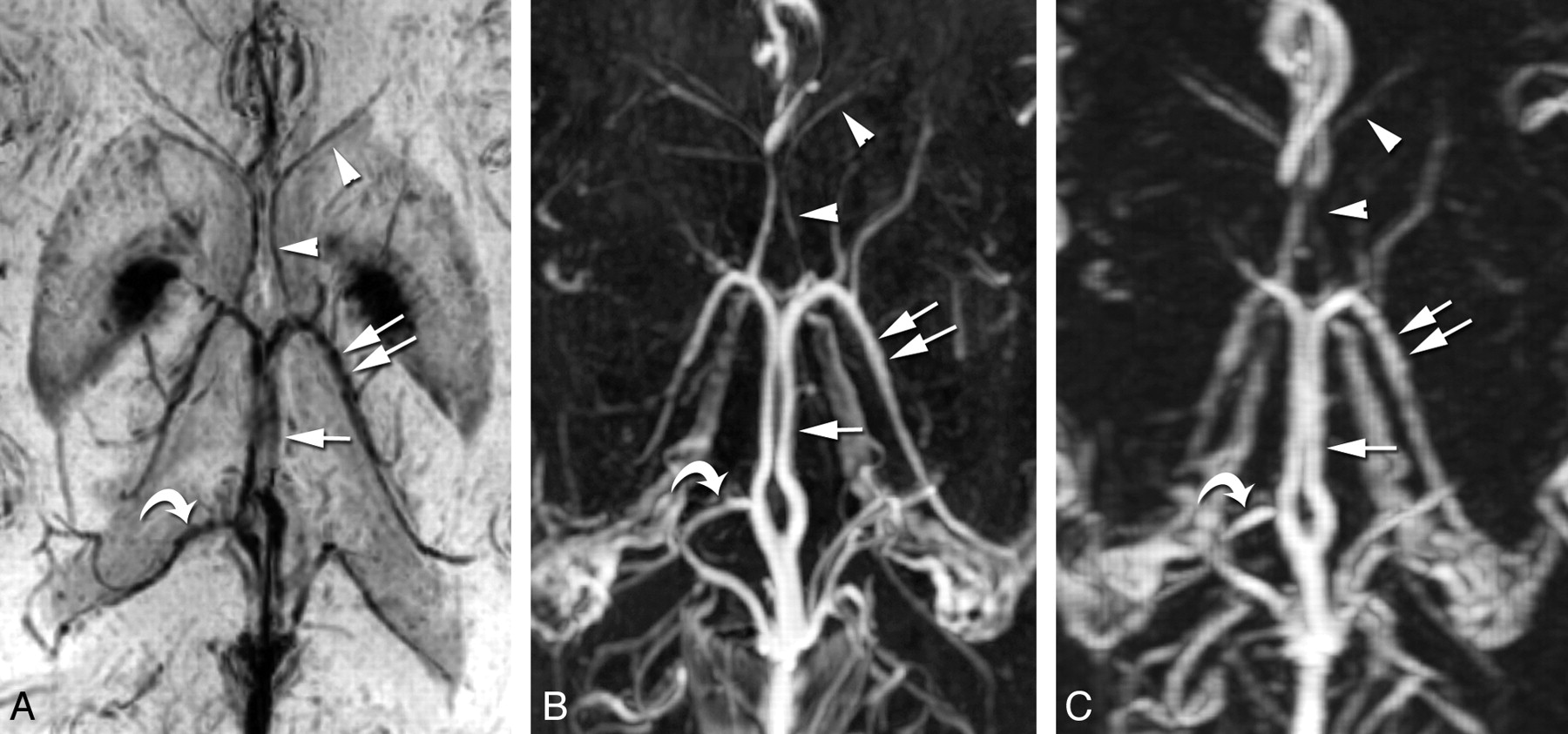
- Fig 1.
The ICV (arrow) and TSV (double arrows) are clearly and completely displayed with the PSI-MRV (A), 3D CE-FSPGR (B), and 3D CE-MRV (C) in this patient. The septal veins (arrowheads) are delineated best on the PSI-MRV, followed by the 3D CE-FSPGR and then the 3D CE-MRV. The medial atrial vein (curved arrows) is easily identified on PSI-MRV, compared with 3D CE-FSPGR and 3D CE-MRV, because of the easy localization of the lateral ventricles and less arterial pollution around the vein of Galen.
- Fig 2.
Anterior caudate nucleus veins (arrow) are shown qualitatively best on 3D CE-FSPGR (B), followed by PSI-MRV (A), and then 3D CE-MRV (C). The superior choroidal vein (arrowhead) is clearly shown on 3D CE-FSPGR (B), poorly shown on 3D CE-MRV (C), and invisible on PSI-MRV (A) in this patient.


Tables
Parameter PSI 3D CE-MRV 3D CE-FSPGR Orientation Axial Sagittal Axial TR (ms) 40 3.1 7.8 TE (ms) 26 0.9 3.8 Flip angle 20° 25° 25° Slab thickness (mm) 80 160 160–180 Section thickness (mm) 2 1 0.7 FOV (mm) 240 260 240 Pixel matrix 320 × 288 288 × 256 320 × 256 NEX 0.75 1 2 Acquisition time (min/sec) 4.57 1.50 5.47–7.43 - Table 2:
A comparison of the visualization of ICV and tributaries on the 3 different MRV techniques
Veins MRV PSI-MRV/3D CE-MRV PSI-MRV/3D CE-FSPGR 3D CE-MRV/3D CE-FSPGR PSI-MRV 3D CE-MRV 3D CE-FSPGR Range Mean Range Mean Range Mean ICV 3–3 3.00 ± 0.00 3–3 3.00 ± 0.00 3–3 3.00 ± 0.00 None None None TSV 3–3 3.00 ± 0.00 3–3 3.00 ± 0.00 3–3 3.00 ± 0.00 None None None SV 2–3 2.90 ± 0.30 1–3 2.44 ± 0.52 2–3 2.73 ± 0.50 PSIa PSIa FSPGRa Anterior caudate nucleus veins 1–3 2.34 ± 0.81 0–3 1.44 ± 0.74 1–3 2.46 ± 0.57 PSIa FSPGRb FSPGRa Superior choroidal vein 0–1 0.86 ± 0.35 0–2 1.65 ± 0.66 0–2 1.85 ± 0.51 3D CE-MRVa FSPGRa FSPGRa Medial atrial vein 0–3 1.93 ± 0.94 0–3 1.50 ± 0.91 0–3 2.09 ± 1.04 PSIa FSPGRc FSPGRc


