Advertisement

Main menu

  • Home
  • Content
    • Current Issue
    • Accepted Manuscripts
    • Article Preview
    • Past Issue Archive
    • Video Articles
    • AJNR Case Collection
    • Case of the Week Archive
    • Case of the Month Archive
    • Classic Case Archive
  • Special Collections
    • AJNR Awards
    • Low-Field MRI
    • Alzheimer Disease
    • ASNR Foundation Special Collection
    • Photon-Counting CT
    • View All
  • Multimedia
    • AJNR Podcasts
    • AJNR SCANtastic
    • Trainee Corner
    • MRI Safety Corner
    • Imaging Protocols
  • For Authors
    • Submit a Manuscript
    • Submit a Video Article
    • Submit an eLetter to the Editor/Response
    • Manuscript Submission Guidelines
    • Statistical Tips
    • Fast Publishing of Accepted Manuscripts
    • Graphical Abstract Preparation
    • Imaging Protocol Submission
    • Author Policies
  • About Us
    • About AJNR
    • Editorial Board
    • Editorial Board Alumni
  • More
    • Become a Reviewer/Academy of Reviewers
    • Subscribers
    • Permissions
    • Alerts
    • Feedback
    • Advertisers
    • ASNR Home

User menu

  • Alerts
  • Log in

Search

  • Advanced search
American Journal of Neuroradiology

American Journal of Neuroradiology

ASHNR American Society of Functional Neuroradiology ASHNR American Society of Pediatric Neuroradiology ASSR
  • Alerts
  • Log in

Advanced Search

  • Home
  • Content
    • Current Issue
    • Accepted Manuscripts
    • Article Preview
    • Past Issue Archive
    • Video Articles
    • AJNR Case Collection
    • Case of the Week Archive
    • Case of the Month Archive
    • Classic Case Archive
  • Special Collections
    • AJNR Awards
    • Low-Field MRI
    • Alzheimer Disease
    • ASNR Foundation Special Collection
    • Photon-Counting CT
    • View All
  • Multimedia
    • AJNR Podcasts
    • AJNR SCANtastic
    • Trainee Corner
    • MRI Safety Corner
    • Imaging Protocols
  • For Authors
    • Submit a Manuscript
    • Submit a Video Article
    • Submit an eLetter to the Editor/Response
    • Manuscript Submission Guidelines
    • Statistical Tips
    • Fast Publishing of Accepted Manuscripts
    • Graphical Abstract Preparation
    • Imaging Protocol Submission
    • Author Policies
  • About Us
    • About AJNR
    • Editorial Board
    • Editorial Board Alumni
  • More
    • Become a Reviewer/Academy of Reviewers
    • Subscribers
    • Permissions
    • Alerts
    • Feedback
    • Advertisers
    • ASNR Home
  • Follow AJNR on Twitter
  • Visit AJNR on Facebook
  • Follow AJNR on Instagram
  • Join AJNR on LinkedIn
  • RSS Feeds

AJNR Awards, New Junior Editors, and more. Read the latest AJNR updates

Research ArticleTechnical Note
Open Access

Visualization of the Internal Cerebral Veins on MR Phase-Sensitive Imaging: Comparison with 3D Gadolinium-Enhanced MR Venography and Fast-Spoiled Gradient Recalled Imaging

J. Sun, J. Wang, L. Jie, H. Wang and X. Gong
American Journal of Neuroradiology November 2011, 32 (10) E191-E193; DOI: https://doi.org/10.3174/ajnr.A2308
J. Sun
  • Find this author on Google Scholar
  • Find this author on PubMed
  • Search for this author on this site
J. Wang
  • Find this author on Google Scholar
  • Find this author on PubMed
  • Search for this author on this site
L. Jie
  • Find this author on Google Scholar
  • Find this author on PubMed
  • Search for this author on this site
H. Wang
  • Find this author on Google Scholar
  • Find this author on PubMed
  • Search for this author on this site
X. Gong
  • Find this author on Google Scholar
  • Find this author on PubMed
  • Search for this author on this site
  • Article
  • Figures & Data
  • Info & Metrics
  • Responses
  • References
  • PDF
Loading

Abstract

SUMMARY: The purpose of this article is to compare the visualization quality of ICV and their tributaries on PSI-MRV, 3D CE-MRV, and 3D CE-FSPGR. PSI-MRV has been shown to delineate the ICV, TSV, SV, anterior caudate nucleus veins, and medial atrial veins effectively. Due to its advantages of no contrast material injection, acceptable examination times, and no arterial contamination, PSI-MRV should be considered in the evaluation of deep cerebral veins.

Abbreviations

3D CE-FSPGR
3D contrast-enhanced fast-spoiled gradient recalled imaging
3D CE-MRV
3D contrast-enhanced MR venography
ICV
internal cerebral veins
MinIP
minimum intensity projection
MIP
maximum intensity projection
MRV
MR venography
PSI
phase-sensitive imaging
PSI-MRV
phase-sensitive imaging-based MR venography
SV
septal veins
SWI
susceptibility-weighted imaging
TSV
thalamostriate veins

Deep cerebral veins have received increasing attention due to their importance in preoperational planning.1,2 Various MRV technologies have been developed for imaging the deep cerebral veins, including the ICV, noninvasively. However, displaying the ICV and their tributaries is still a challenge for noncontrast or contrast MRV, though the image quality has improved with the advent of higher field systems.3,4 SWI uses the paramagnetic deoxygenated hemoglobin in veins, which causes a shift in resonance frequency between the venous vessel and surrounding tissue, as an intrinsic contrast agent. In SWI-based venography, high spatial resolution increases the venous blood volume fraction within a voxel and leads to a better visualization of small venous structures.5,6 SWI is called PSI, as implemented on the Signa Excite HD scanner (GE Healthcare, Milwaukee, Wisconsin).7 The purpose of this study was to evaluate the applicability of PSI-MRV in detecting the ICV and their tributaries through a comparison with 3D CE-MRV and 3D CE-FSPGR.

Technique

Patients

After obtaining the approval of the hospital ethics committee and written informed consent of patients, 65 consecutive patients with supratentorial meningioma were examined with 3D CE-MRV for presurgical evaluation of intracranial dural sinuses. PSI-MRV and 3D CE-FSPGR were performed before and after 3D CE-MRV.

Twelve patients were excluded because the meningioma caused severe distortion of the third or lateral ventricular area. Nine patients were excluded because motion artifacts occurred in at least 1 of the 3 MRV examinations. Four patients who underwent repeat surgery were excluded because their images could not be evaluated due to susceptibility artifacts caused by hemorrhage around the ventricular area. A total of 40 patients (21 women and 19 men; age range, 22-77 years; mean age, 53.9 years) were enrolled.

Imaging Examinations

All MR imaging studies were performed on a 3T system (Signa Excite HD, GE Healthcare) equipped with an 8-channel phased array head coil. The MR imaging scanning field covered the bilateral hemispheres, including the ICV and their main tributaries. After conventional T1-weighted and T2-weighted imaging, PSI-MRV, 3D CE-MRV, and 3D CE-FSPGR were performed in order. 3D CE-MRV consisted of 2 scans with an acquisition time of 59 seconds each. A dose of 0.05-mmoL/kg gadolinium contrast material was administered intravenously with an injector at a rate of 3 mL/s immediately after the first scan. A repeat scan with the same parameters was obtained after a 20-second delay. 3D CE-FSPGR was performed after the 3D CE-MRV sequence. The imaging parameters are listed in Table 1.

View this table:
  • View inline
  • View popup
Table 1:

Imaging parameters in PSI, 3D CE-MRV, and 3D CE-FSPGR sequences

Image Reconstruction

The MR images were postprocessed with an Advantage Workstation version 4.2 (GE Healthcare). For PSI-MRV, images were reformatted by using the MinIP technique with 15- to 30-mm thickness in the transverse plane. Reconstructed images were saved once the ICV and tributaries were optimally displayed. A subtraction was performed between the pre- and postcontrast 3D CE-MRV scannings to get images without background pollution. The subtracted images were subsequently reconstructed in the transverse plane by using an MIP algorithm. The MIP images used the same thickness and orientation as the prior PSI-MRV images. 3D CE-FSPGR images were also reformatted with the MIP algorithm in the transverse plane like the 3D CE-MRV images. The same technician performed all image reconstructions.

Image Evaluation

The reconstructed images of PSI-MRV, 3D CE-MRV, and 3D CE-FSPGR were evaluated by a consensus of 2 radiologists with previous experience in PSI and CE-MRV. Source images were reviewed when the radiologists needed to trace or define a vein. Evaluation from another experienced radiologist was obtained when the 2 reviewers failed to agree. The veins evaluated in this study included the ICV, TSV, SV, anterior caudate nucleus vein, superior choroidal vein, and medial atrial vein. Visualization of the veins was qualitatively graded in 4 levels: intense and continuous = 3, faint and continuous = 2, partially visible = 1, invisible = 0. The Wilcoxon signed rank test was used to analyze differences in the visualization quality of the ICV and its tributaries among the 3 MRV techniques.

Results

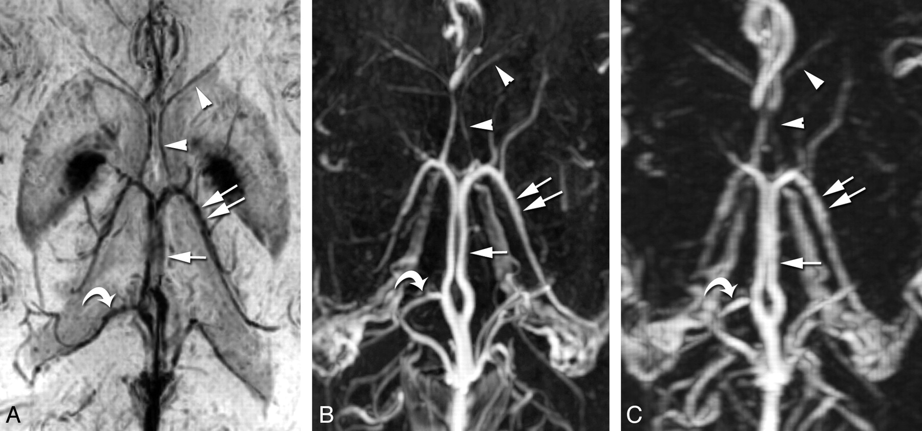
The ICV and TSV were clearly and completely displayed at low signal intensity with PSI-MRV in all 80 lateral hemispheres of the 40 patients. They were equally well shown on the 3D CE-MRV and 3D CE-FSPGR images. The SV were delineated best on PSI-MRV, with an average score of 2.9, followed by 3D CE-FSPGR (2.73) and then 3D CE-MRV (2.44) (Table 2 and Fig 1 A-C). There were significant differences among the 3 MRV modalities (P < .001 for each pair of MRV images). Anterior caudate nucleus veins were shown qualitatively better on PSI-MRV (2.34) than on 3D CE-MRV (1.44) with significant differences (P < .001), but inferior to the veins on 3D CE-FSPGR (2.46) (P < .01). The superior choroidal vein was poorly delineated in all patients on PSI-MRV, with an average score of 0.86, which was inferior to scores of veins demonstrated on the 3D CE-MRV or 3D CE-FSPGR (P < .001 for each pair) (Table 2 and Fig 2 A−C). For visualization of the medial atrial vein, PSI-MRV was better than 3D CE-MRV (P < .001) but worse than 3D CE-FSPGR, with a statistical difference (P < .05).

View this table:
  • View inline
  • View popup
Table 2:

A comparison of the visualization of ICV and tributaries on the 3 different MRV techniques

Fig 1.
  • Download figure
  • Open in new tab
  • Download powerpoint
Fig 1.

The ICV (arrow) and TSV (double arrows) are clearly and completely displayed with the PSI-MRV (A), 3D CE-FSPGR (B), and 3D CE-MRV (C) in this patient. The septal veins (arrowheads) are delineated best on the PSI-MRV, followed by the 3D CE-FSPGR and then the 3D CE-MRV. The medial atrial vein (curved arrows) is easily identified on PSI-MRV, compared with 3D CE-FSPGR and 3D CE-MRV, because of the easy localization of the lateral ventricles and less arterial pollution around the vein of Galen.

Fig 2.
  • Download figure
  • Open in new tab
  • Download powerpoint
Fig 2.

Anterior caudate nucleus veins (arrow) are shown qualitatively best on 3D CE-FSPGR (B), followed by PSI-MRV (A), and then 3D CE-MRV (C). The superior choroidal vein (arrowhead) is clearly shown on 3D CE-FSPGR (B), poorly shown on 3D CE-MRV (C), and invisible on PSI-MRV (A) in this patient.

Discussion

On the basis of similar principles of SWI, PSI has been introduced to evaluate the variants of ICV noninvasively.8 With this study, all 3 MRV techniques provided equally good demonstration of the ICV and TSV. The SV were best shown with PSI-MRV among the 3 MRV procedures. The anterior caudate nucleus veins, which comprised several small veins draining inward and backward from the head of the caudate nucleus to the TSV, SV, or ICV, were observed on PSI-MRV with a better image quality than on 3D CE-MRV. The medial atrial vein runs along the medial wall of the atrium and joins the ICV at its terminal portion. With 3D CE-MRV and 3D CE-FSPGR, this vein was commonly confused with other vessels around the vein of Galen. Thus, the reviewers had to carefully identify the vein with the aid of the original image. However, it was quite easy to identify this vein on PSI-MRV because PSI-MRV showed the lateral ventricles clearly and reduced pollution from small arteries around the vein of Galen. The PSI-MRV display of the superior choroidal vein was poor compared with the 3D CE-MRV and 3D CE-FSPGR sequences, possibly because CSF signals obscure the vessel passing the ventricle in PSI MinIP images.

Compared with 3D CE-MRV and 3D CE-FSPGR, using PSI to evaluate deep cerebral veins has several advantages. First, no injection of contrast material is required on PSI-MRV. Second, the PSI-MRV image is free from arterial contamination, which is a big issue on 3D CE-MRV and 3D CE-FSPGR. Finally, the examination can be repeated immediately if the MRV fails for various reasons, because PSI scanning is free of any contamination from brain parenchymal or meningeal contrast enhancement. The examination duration for PSI-MRV is relatively longer than for 3D CE-MRV, but it is almost commensurate with 3D CE-FSPGR. Most patients can tolerate the examination duration.

The basal vein of Rosenthal, another important component of the deep cerebral veins, was not included in this study. A 2-mm-thickness image paralleling the ICV was insufficient to show this vein because it forms an angle to the ICV. An additional acquisition is needed for evaluating the basal vein of Rosenthal. The original image of 3D CE-MRV in this study was designed in the sagittal plane, which enables reliable visualization of the relationship between a meningioma and the superior sagittal sinus. The in-plane spatial resolution of the reconstructed transverse images may underestimate the small tributaries of ICV.

Acknowledgment

We thank Mrs. Trudie Loo for her written polishing of this article.

Footnotes

  • This study was supported by the Science and Technology Department of Zhejiang Province, China (No. 2009R10009).

Indicates open access to non-subscribers at www.ajnr.org

References

  1. 1.↵
    1. Giordano M,
    2. Wrede KH,
    3. Stieglitz LH,
    4. et al
    . Identification of venous variants in the pineal region with 3D preoperative computed tomography and magnetic resonance imaging navigation: a statistical study of venous anatomy in living patients. J Neurosurg 2007;106:1006–11
    CrossRefPubMed
  2. 2.↵
    1. Kiliç T,
    2. Ozduman K,
    3. Cavdar S,
    4. et al
    . The galenic venous system: surgical anatomy and its angiographic and magnetic resonance venographic correlations. Eur J Radiol 2005;56:212–19
    CrossRefPubMed
  3. 3.↵
    1. Kirchhof K,
    2. Welzel T,
    3. Jansen Q,
    4. et al
    . More reliable noninvasive visualization of the cerebral veins and dural sinuses: comparison of three MR angiographic techniques. Radiology 2002;224:804–10
    CrossRefPubMed
  4. 4.↵
    1. Lettau M,
    2. Sartor K,
    3. Heiland S,
    4. et al
    . 3T high-spatial-resolution contrast-enhanced MR angiography of the intracranial venous system with parallel imaging. AJNR Am J Neuroradiol 2009;30:185–87
    Abstract/FREE Full Text
  5. 5.↵
    1. Reichenbach JR,
    2. Venkatesan R,
    3. Schillinger DJ,
    4. et al
    . Small vessels in the human brain: MR venography with deoxyhemoglobin as an intrinsic contrast agent. Radiology 1997;204:272–77
    PubMed
  6. 6.↵
    1. Reichenbach JR,
    2. Jonetz-Mentzel L,
    3. Fitzek C,
    4. et al
    . High-resolution blood oxygen-level dependent MR venography (HRBV): a new technique. Neuroradiology 2001;43:364–69
    CrossRefPubMed
  7. 7.↵
    1. Kurosaki M,
    2. Tabuchi S,
    3. Akatsuka K,
    4. et al
    . Application of phase sensitive imaging (PSI) for hemorrhage diagnosis in pituitary adenomas. Neurol Res 2010;32:614–19
    CrossRefPubMed
  8. 8.↵
    1. Fujii S,
    2. Kanasaki Y,
    3. Matsusue E,
    4. et al
    . Demonstration of cerebral venous variations in the region of the third ventricle on phase-sensitive imaging. AJNR Am J Neuroradiol 2010;31:55–59
    Abstract/FREE Full Text
  • Received May 23, 2010.
  • Accepted after revision August 14, 2010.
  • © 2011 by American Journal of Neuroradiology
View Abstract
PreviousNext
Back to top

In this issue

American Journal of Neuroradiology: 32 (10)
American Journal of Neuroradiology
Vol. 32, Issue 10
1 Nov 2011
  • Table of Contents
  • Index by author
Advertisement
Print
Download PDF
Email Article

Thank you for your interest in spreading the word on American Journal of Neuroradiology.

NOTE: We only request your email address so that the person you are recommending the page to knows that you wanted them to see it, and that it is not junk mail. We do not capture any email address.

Enter multiple addresses on separate lines or separate them with commas.
Visualization of the Internal Cerebral Veins on MR Phase-Sensitive Imaging: Comparison with 3D Gadolinium-Enhanced MR Venography and Fast-Spoiled Gradient Recalled Imaging
(Your Name) has sent you a message from American Journal of Neuroradiology
(Your Name) thought you would like to see the American Journal of Neuroradiology web site.
CAPTCHA
This question is for testing whether or not you are a human visitor and to prevent automated spam submissions.
Cite this article
J. Sun, J. Wang, L. Jie, H. Wang, X. Gong
Visualization of the Internal Cerebral Veins on MR Phase-Sensitive Imaging: Comparison with 3D Gadolinium-Enhanced MR Venography and Fast-Spoiled Gradient Recalled Imaging
American Journal of Neuroradiology Nov 2011, 32 (10) E191-E193; DOI: 10.3174/ajnr.A2308

Citation Manager Formats

  • BibTeX
  • Bookends
  • EasyBib
  • EndNote (tagged)
  • EndNote 8 (xml)
  • Medlars
  • Mendeley
  • Papers
  • RefWorks Tagged
  • Ref Manager
  • RIS
  • Zotero
0 Responses
Respond to this article
Share
Bookmark this article
Visualization of the Internal Cerebral Veins on MR Phase-Sensitive Imaging: Comparison with 3D Gadolinium-Enhanced MR Venography and Fast-Spoiled Gradient Recalled Imaging
J. Sun, J. Wang, L. Jie, H. Wang, X. Gong
American Journal of Neuroradiology Nov 2011, 32 (10) E191-E193; DOI: 10.3174/ajnr.A2308
del.icio.us logo Twitter logo Facebook logo Mendeley logo
  • Tweet Widget
  • Facebook Like
  • Google Plus One
Purchase

Jump to section

  • Article
    • Abstract
    • Abbreviations
    • Technique
    • Results
    • Discussion
    • Acknowledgment
    • Footnotes
    • References
  • Figures & Data
  • Info & Metrics
  • Responses
  • References
  • PDF

Related Articles

  • No related articles found.
  • PubMed
  • Google Scholar

Cited By...

  • No citing articles found.
  • Crossref (6)
  • Google Scholar

This article has been cited by the following articles in journals that are participating in Crossref Cited-by Linking.

  • Minimally invasive endovascular stent-electrode array for high-fidelity, chronic recordings of cortical neural activity
    Thomas J Oxley, Nicholas L Opie, Sam E John, Gil S Rind, Stephen M Ronayne, Tracey L Wheeler, Jack W Judy, Alan J McDonald, Anthony Dornom, Timothy J H Lovell, Christopher Steward, David J Garrett, Bradford A Moffat, Elaine H Lui, Nawaf Yassi, Bruce C V Campbell, Yan T Wong, Kate E Fox, Ewan S Nurse, Iwan E Bennett, Sébastien H Bauquier, Kishan A Liyanage, Nicole R van der Nagel, Piero Perucca, Arman Ahnood, Katherine P Gill, Bernard Yan, Leonid Churilov, Christopher R French, Patricia M Desmond, Malcolm K Horne, Lynette Kiers, Steven Prawer, Stephen M Davis, Anthony N Burkitt, Peter J Mitchell, David B Grayden, Clive N May, Terence J O'Brien
    Nature Biotechnology 2016 34 3
  • The Ovine Cerebral Venous System: Comparative Anatomy, Visualization, and Implications for Translational Research
    Anke Hoffmann, Michael H. Stoffel, Björn Nitzsche, Donald Lobsien, Johannes Seeger, Holm Schneider, Johannes Boltze, Piotr Walczak
    PLoS ONE 2014 9 4
  • Time-resolved MR angiography of the intracranial venous system: an alternative MR venography technique
    Hasan Yiğit, Aynur Turan, Elif Ergün, Pınar Koşar, Uğur Koşar
    European Radiology 2012 22 5
  • Susceptibility-Weighted Imaging of the Anatomic Variation of Thalamostriate Vein and Its Tributaries
    Xiao-fen Zhang, Jian-ce Li, Xin-dong Wen, Chuan-gen Ren, Ming Cai, Cheng-chun Chen, Heye Zhang
    PLOS ONE 2015 10 10
  • Susceptibility-weighted imaging of the venous networks around the brain stem
    Ming Cai, Xiao-Fen Zhang, Hui-Huang Qiao, Zhong-Xiao Lin, Chuan-Gen Ren, Jian-Ce Li, Cheng-Chun Chen, Nu Zhang
    Neuroradiology 2015 57 2
  • Anatomy imaging and hemodynamics research on the cerebral vein and venous sinus among individuals without cranial sinus and jugular vein diseases
    Lu Liu, Yan Wu, Kaiyuan Zhang, Ran Meng, Jiangang Duan, Chen Zhou, Xunming Ji
    Frontiers in Neuroscience 2022 16

More in this TOC Section

  • 3D Fast Spin-Echo T1 Black-Blood Imaging for the Diagnosis of Cervical Artery Dissection
  • The Kety-Schmidt Technique for Quantitative Perfusion and Oxygen Metabolism Measurements in the MR Imaging Environment
  • A Swine Model to Analyze Arterial Structural Changes Induced by Mechanical Thrombectomy
Show more Technical Note

Similar Articles

Advertisement

Indexed Content

  • Current Issue
  • Accepted Manuscripts
  • Article Preview
  • Past Issues
  • Editorials
  • Editor's Choice
  • Fellows' Journal Club
  • Letters to the Editor
  • Video Articles

Cases

  • Case Collection
  • Archive - Case of the Week
  • Archive - Case of the Month
  • Archive - Classic Case

More from AJNR

  • Trainee Corner
  • Imaging Protocols
  • MRI Safety Corner
  • Book Reviews

Multimedia

  • AJNR Podcasts
  • AJNR Scantastics

Resources

  • Turnaround Time
  • Submit a Manuscript
  • Submit a Video Article
  • Submit an eLetter to the Editor/Response
  • Manuscript Submission Guidelines
  • Statistical Tips
  • Fast Publishing of Accepted Manuscripts
  • Graphical Abstract Preparation
  • Imaging Protocol Submission
  • Evidence-Based Medicine Level Guide
  • Publishing Checklists
  • Author Policies
  • Become a Reviewer/Academy of Reviewers
  • News and Updates

About Us

  • About AJNR
  • Editorial Board
  • Editorial Board Alumni
  • Alerts
  • Permissions
  • Not an AJNR Subscriber? Join Now
  • Advertise with Us
  • Librarian Resources
  • Feedback
  • Terms and Conditions
  • AJNR Editorial Board Alumni

American Society of Neuroradiology

  • Not an ASNR Member? Join Now

© 2025 by the American Society of Neuroradiology All rights, including for text and data mining, AI training, and similar technologies, are reserved.
Print ISSN: 0195-6108 Online ISSN: 1936-959X

Powered by HighWire