
Article Figures & Data
Figures
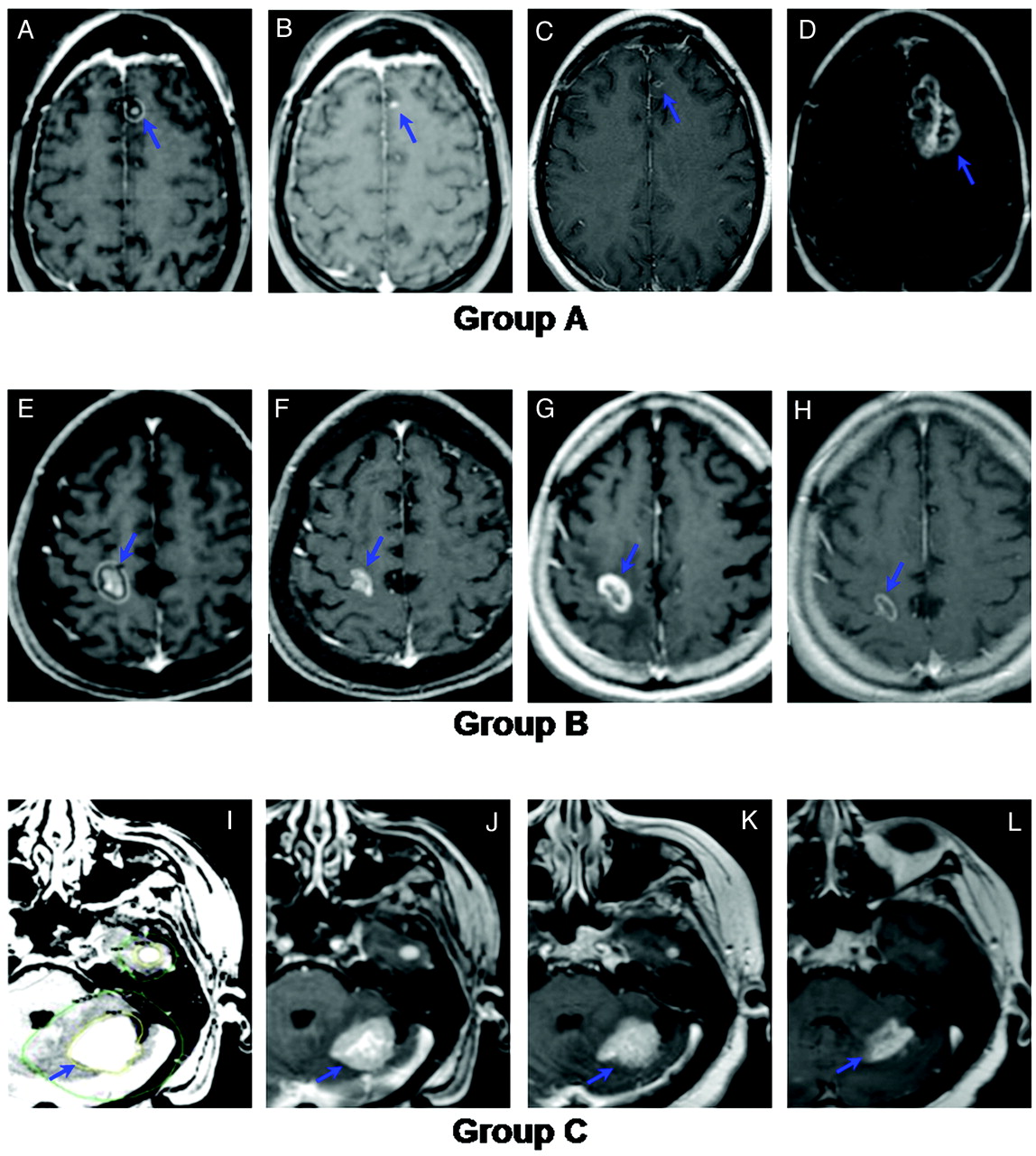
- Fig 1.
Examples illustrating the study definitions of groups A, B, and C. A−D, Representative images from a group A lesion (increased in follow-up to a volume >120% of initial size). A, Gamma knife treatment plan for a 36-year-old woman nonsmoker with non-small cell lung carcinoma. The lesion was treated with 18 Gy to the 50% isodose line. B, Initial MR image. C, Six-month follow-up MR image. D, Twelve-month follow-up MR image. E−H, Representative images from a group B lesion (size fluctuated throughout follow-up but never increased beyond 120% of the initial size). E, Gamma Knife treatment plan for a 75-year-old man with colorectal adenocarcinoma. The lesion was treated with 20 Gy to the 50% isodose line. F, Initial MR image. G, Three-month follow-up MR image. H, Six-month follow-up MR image. I–L, Representative images from a group C lesion (remained stable or decreased in size throughout follow-up). I, Gamma Knife treatment plan for a 76-year-old woman with 2 metastatic breast cancer lesions. Both lesions were treated with 20 Gy to the 50% isodose line. J, Initial MR image. K, Six-month follow-up MR image. L, Twelve-month follow-up MR image.
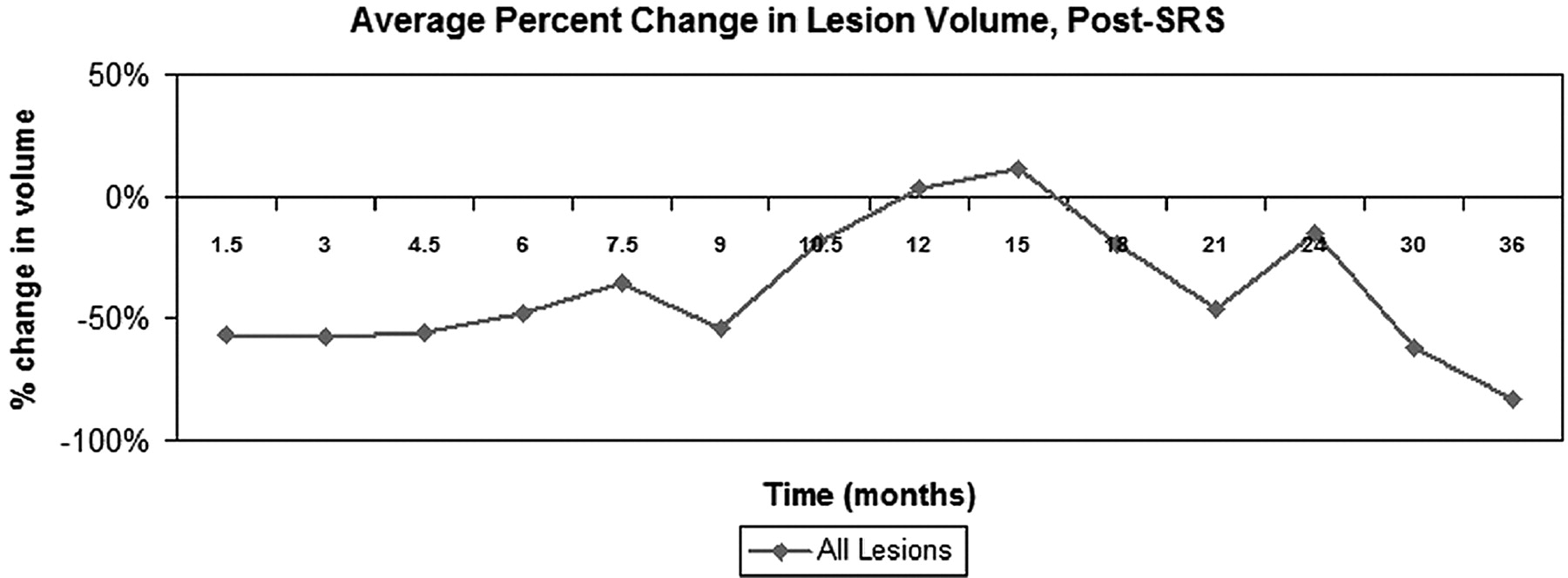
- Fig 2.
Average change in lesional size with time, relative to initial treatment volume, all lesions. Lesions decreased or remained stable in size for the first 9 months post-SRS. Subsequently, they increased in size until approximately 18 months post-SRS, at which point they began to decrease in size once again.
- Fig 3.
Average change in lesional size with time, relative to the initial treatment volume, separated by histopathology (radiosensitive: lung, breast, colon, other; radioresistant: melanoma, renal). Radiosensitive tumors were more likely to increase in size during the first 12–18 months post-SRS than radioresistant tumors. However, this difference did not extend, consistently, beyond 18 months.
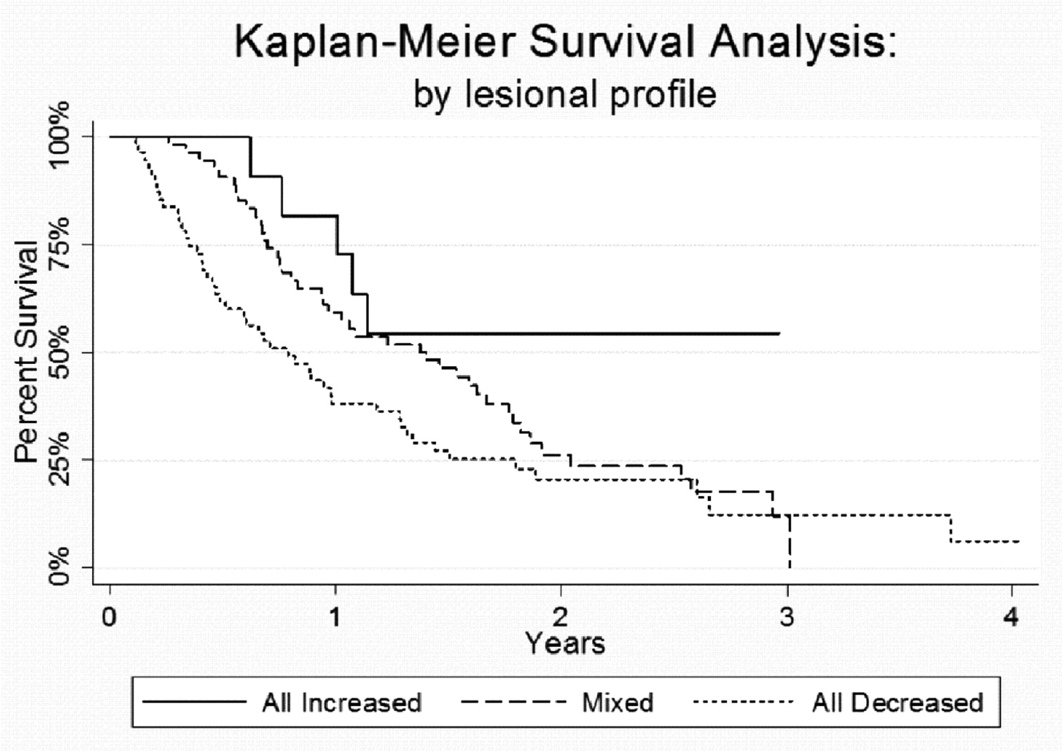
- Fig 4.
Kaplan-Meier survival curve, by lesional profile, demonstrates that patients with lesions that all increased in size following SRS had significantly improved survival (P = .035).
- Fig 5.
Example of radiologic and histopathologic changes in enlarging lesions. A, Gamma Knife treatment plan for a 56-year-old man with metastatic melanoma. The lesion was treated with 22 Gy to the 50% isodose line. B, Twelve-month follow-up T1 postcontrast MR imaging. C, Twelve-month FLAIR image. D and E, Twelve-month DWI/apparent diffusion coefficient images, respectively. F, Twelve-month FDG-PET image. G, Histopathology from stereotactic image-guided biopsies of the lesion. Specimens from the central, T1 hypointense portion of the lesion demonstrate coagulative necrosis. Specimens from the peripheral, T1 hypointense portion of the lesion demonstrate reactive gliosis and demyelination. Specimens from the T1 contrast-enhancing portion of the lesion demonstrate vascular hyalinization. This constellation of histopathologic findings suggests a diagnosis of radiation-induced changes.


Tables
Lesion Cohorta (n = 516) Patient Cohorta (n = 120) Median Survival (mo) Age Median 59.7 yr Range 31–89 yr Sex Male 53 (44%) Female 67 (56%) Race Caucasian 106 (88%) Other 14 (12%) Primary pathology NSCLCa 175 (34%) 47 (39%) 11.8 SCLCa 20 (4%) 6 (5%) 6.8 Breast 116 (22%) 18 (15%) 12.3 Melanoma 123 (24%) 24 (20%) 13.7 Renal 51 (10%) 11 (9%) 8.5 Colon 13 (2.5%) 6 (5%) 13.0 Other 18 (3.5%) 8 (7%) 10.0 Radiosensitiveb 342 (66%) 85 (71%) 12.3 Radioresistantc 174 (34%) 35 (29%) 12.9 - Table 2:
Average percentage change in lesion volume with time, relative to initial treatment volume
Time since SRS (No.)a Average % Change in Lesion Volume 6 Weeks (442) −56.5% 3 Months (299) −57.3% 4.5 Months (222) −55.7% 6 Months (211) −47.8% 7.5 Months (113) −35.2% 9 Months (107) −54.3% 10.5 Months (88) −18.7% 12 Months (67) +3.6% 15 Months (36) +11.6% 18 Months (52) −20.1% 21 Months (35) −46.2% 24 Months (19) −15.3% 30 Months (18) −62.0% 36 Months (11) −82.9% -
Note:—Bold type indicates that lesions were larger than their initial treatment volumes at these timepoints.
-
↵a Number of lesions.
-
- Table 3:
Timing of lesional increases in size (percentage of total group A or B lesions beginning to increase in size at a given timepoint)
Time since SRS Group A (n = 81 lesions)a Group B (n = 83 lesions)a 6 weeks 30.9% 0% 3 months 16.0% 20.5% 4.5 months 7.4% 29% 6 months 12.3% 33% 7.5 months 4.9% 8.5% 9 months 6.2% 2.5% 10.5 months 3.7% 1% 12 months 6.2% 2.5% 13.5 months 2.5% 1% 15 months 1.2% 0% 16.5 months 2.5% 1% 18 months 3.7% 0% 19.5 months 0% 0% 21 months 2.5% 1% 22.5 months 0% 0% 24 months 0% 0% -
↵a Group A denotes lesions that increased in size to >120% of the pretreatment volume (ie, each timepoint is compared with the initial treatment volume). Group B denotes lesions whose volumes fluctuated in size during follow-up but never increased to a volume exceeding 120% of the pretreatment volume (ie, each timepoint is compared with both the initial treatment and prior time point volumes).
-


