Advertisement

Main menu

  • Home
  • Content
    • Current Issue
    • Accepted Manuscripts
    • Article Preview
    • Past Issue Archive
    • Video Articles
    • AJNR Case Collection
    • Case of the Week Archive
    • Case of the Month Archive
    • Classic Case Archive
  • Special Collections
    • AJNR Awards
    • Low-Field MRI
    • Alzheimer Disease
    • ASNR Foundation Special Collection
    • Photon-Counting CT
    • View All
  • Multimedia
    • AJNR Podcasts
    • AJNR SCANtastic
    • Trainee Corner
    • MRI Safety Corner
    • Imaging Protocols
  • For Authors
    • Submit a Manuscript
    • Submit a Video Article
    • Submit an eLetter to the Editor/Response
    • Manuscript Submission Guidelines
    • Statistical Tips
    • Fast Publishing of Accepted Manuscripts
    • Graphical Abstract Preparation
    • Imaging Protocol Submission
    • Author Policies
  • About Us
    • About AJNR
    • Editorial Board
    • Editorial Board Alumni
  • More
    • Become a Reviewer/Academy of Reviewers
    • Subscribers
    • Permissions
    • Alerts
    • Feedback
    • Advertisers
    • ASNR Home

User menu

  • Alerts
  • Log in

Search

  • Advanced search
American Journal of Neuroradiology

American Journal of Neuroradiology

ASHNR American Society of Functional Neuroradiology ASHNR American Society of Pediatric Neuroradiology ASSR
  • Alerts
  • Log in

Advanced Search

  • Home
  • Content
    • Current Issue
    • Accepted Manuscripts
    • Article Preview
    • Past Issue Archive
    • Video Articles
    • AJNR Case Collection
    • Case of the Week Archive
    • Case of the Month Archive
    • Classic Case Archive
  • Special Collections
    • AJNR Awards
    • Low-Field MRI
    • Alzheimer Disease
    • ASNR Foundation Special Collection
    • Photon-Counting CT
    • View All
  • Multimedia
    • AJNR Podcasts
    • AJNR SCANtastic
    • Trainee Corner
    • MRI Safety Corner
    • Imaging Protocols
  • For Authors
    • Submit a Manuscript
    • Submit a Video Article
    • Submit an eLetter to the Editor/Response
    • Manuscript Submission Guidelines
    • Statistical Tips
    • Fast Publishing of Accepted Manuscripts
    • Graphical Abstract Preparation
    • Imaging Protocol Submission
    • Author Policies
  • About Us
    • About AJNR
    • Editorial Board
    • Editorial Board Alumni
  • More
    • Become a Reviewer/Academy of Reviewers
    • Subscribers
    • Permissions
    • Alerts
    • Feedback
    • Advertisers
    • ASNR Home
  • Follow AJNR on Twitter
  • Visit AJNR on Facebook
  • Follow AJNR on Instagram
  • Join AJNR on LinkedIn
  • RSS Feeds

AJNR Awards, New Junior Editors, and more. Read the latest AJNR updates

Research ArticleNeurointervention

Improved Microsurgical Creation of Venous Pouch Arterial Bifurcation Aneurysms in Rabbits

C. Sherif, S. Marbacher, S. Erhardt and J. Fandino
American Journal of Neuroradiology January 2011, 32 (1) 165-169; DOI: https://doi.org/10.3174/ajnr.A2235
C. Sherif
  • Find this author on Google Scholar
  • Find this author on PubMed
  • Search for this author on this site
S. Marbacher
  • Find this author on Google Scholar
  • Find this author on PubMed
  • Search for this author on this site
S. Erhardt
  • Find this author on Google Scholar
  • Find this author on PubMed
  • Search for this author on this site
J. Fandino
  • Find this author on Google Scholar
  • Find this author on PubMed
  • Search for this author on this site
  • Article
  • Figures & Data
  • Info & Metrics
  • Responses
  • References
  • PDF
Loading

Article Figures & Data

Figures

  • Tables
  • Fig 1.
    • Download figure
    • Open in new tab
    • Download powerpoint
    Fig 1.

    Microsurgical steps during venous pouch arterial bifurcation aneurysm creation. A, Suturing sequence at the posterior aspect of the anastomosis: 1) right to left CCA, 2) venous pouch to left CCA, 3) venous pouch to right CCA. B, Suturing sequence at the anterior aspect: 4) right to left CCA, 5) venous pouch to left CCA, 6) venous pouch to right CCA.
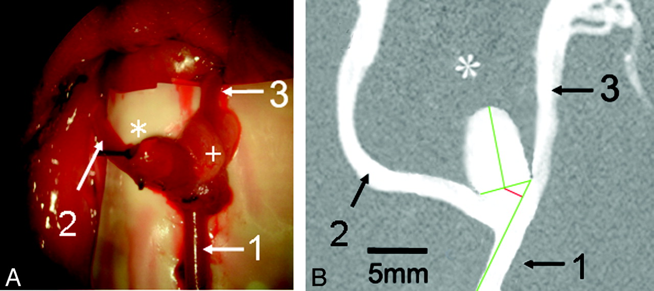
  • Fig 2.
    • Download figure
    • Open in new tab
    • Download powerpoint
    Fig 2.

    Morphology of the venous pouch arterial bifurcation aneurysm. A, View under the operation microscope after microsurgery. B, Calibrated DSA 4 weeks after operation. A, 1) Proximal left CCA, 2) right CCA, 3) distal left CCA. Asterisk indicates the dome of the venous pouch aneurysm; the plus sign, the fat pad for sealing the suture line. B, DSA shows the aneurysm and parent-vessel patency after 4 weeks. Green lines indicate the aneurysm neck width and aneurysm length and the contour of the in-flow parent-artery (left CCA). Red line shows the distance of the neck plane center from the parent-artery contour.

Tables

  • Figures
  • Angiographic evaluation

    Aneurysm No.PatencyParent Artery ø (mm)Length (mm)Neck Width (mm)Aspect RatioOhshima Shift (mm)Ohshima Type
    1No
    2Yes2.408.404.701.791.60B
    3Yes1.907.703.702.081.70B
    4Yes2.607.503.901.921.80B
    5Yes2.707.504.101.831.60B
    6Yes2.508.004.201.902.10B
    7Yes1.908.604.302.001.80B
    8No
    9Yes2.608.004.002.002.10B
    10Yes2.408.104.101.981.50B
    11Yes1.907.603.901.951.30B
    12Yes1.607.403.801.951.40B
    13Yes1.907.404.101.802.10B
    14Yes2.407.104.101.731.80B
    15Yes2.307.904.201.881.50B
    16Yes2.508.004.801.672.00B
PreviousNext
Back to top

In this issue

American Journal of Neuroradiology: 32 (1)
American Journal of Neuroradiology
Vol. 32, Issue 1
1 Jan 2011
  • Table of Contents
  • Index by author
Advertisement
Print
Download PDF
Email Article

Thank you for your interest in spreading the word on American Journal of Neuroradiology.

NOTE: We only request your email address so that the person you are recommending the page to knows that you wanted them to see it, and that it is not junk mail. We do not capture any email address.

Enter multiple addresses on separate lines or separate them with commas.
Improved Microsurgical Creation of Venous Pouch Arterial Bifurcation Aneurysms in Rabbits
(Your Name) has sent you a message from American Journal of Neuroradiology
(Your Name) thought you would like to see the American Journal of Neuroradiology web site.
CAPTCHA
This question is for testing whether or not you are a human visitor and to prevent automated spam submissions.
Cite this article
C. Sherif, S. Marbacher, S. Erhardt, J. Fandino
Improved Microsurgical Creation of Venous Pouch Arterial Bifurcation Aneurysms in Rabbits
American Journal of Neuroradiology Jan 2011, 32 (1) 165-169; DOI: 10.3174/ajnr.A2235

Citation Manager Formats

  • BibTeX
  • Bookends
  • EasyBib
  • EndNote (tagged)
  • EndNote 8 (xml)
  • Medlars
  • Mendeley
  • Papers
  • RefWorks Tagged
  • Ref Manager
  • RIS
  • Zotero
0 Responses
Respond to this article
Share
Bookmark this article
Improved Microsurgical Creation of Venous Pouch Arterial Bifurcation Aneurysms in Rabbits
C. Sherif, S. Marbacher, S. Erhardt, J. Fandino
American Journal of Neuroradiology Jan 2011, 32 (1) 165-169; DOI: 10.3174/ajnr.A2235
del.icio.us logo Twitter logo Facebook logo Mendeley logo
  • Tweet Widget
  • Facebook Like
  • Google Plus One
Purchase

Jump to section

  • Article
    • Abstract
    • Abbreviations
    • Materials and Methods
    • Results
    • Discussion
    • Conclusions
    • Acknowledgments
    • Footnotes
    • References
  • Figures & Data
  • Info & Metrics
  • Responses
  • References
  • PDF

Related Articles

  • No related articles found.
  • PubMed
  • Google Scholar

Cited By...

  • Testing Stenting and Flow Diversion Using a Surgical Elastase-Induced Complex Fusiform Aneurysm Model
  • Creation of sidewall aneurysm in rabbits: aneurysm patency and growth follow-up
  • 3D Computerized Occlusion Rating of Embolized Experimental Aneurysms Using Noninvasive 1.5T MR Imaging
  • Complex Bilobular, Bisaccular, and Broad-Neck Microsurgical Aneurysm Formation in the Rabbit Bifurcation Model for the Study of Upcoming Endovascular Techniques
  • Quantitative Angiographic and Histopathologic Evaluation of Experimental Aneurysms
  • Crossref
  • Google Scholar

This article has not yet been cited by articles in journals that are participating in Crossref Cited-by Linking.

More in this TOC Section

  • Connectivity for remote neurovascular procedures
  • Cavernous dural AVF treated by transfacial route
  • A Retrospective Study in Tentorial DAVFs
Show more NEUROINTERVENTION

Similar Articles

Advertisement

Indexed Content

  • Current Issue
  • Accepted Manuscripts
  • Article Preview
  • Past Issues
  • Editorials
  • Editor's Choice
  • Fellows' Journal Club
  • Letters to the Editor
  • Video Articles

Cases

  • Case Collection
  • Archive - Case of the Week
  • Archive - Case of the Month
  • Archive - Classic Case

More from AJNR

  • Trainee Corner
  • Imaging Protocols
  • MRI Safety Corner
  • Book Reviews

Multimedia

  • AJNR Podcasts
  • AJNR Scantastics

Resources

  • Turnaround Time
  • Submit a Manuscript
  • Submit a Video Article
  • Submit an eLetter to the Editor/Response
  • Manuscript Submission Guidelines
  • Statistical Tips
  • Fast Publishing of Accepted Manuscripts
  • Graphical Abstract Preparation
  • Imaging Protocol Submission
  • Evidence-Based Medicine Level Guide
  • Publishing Checklists
  • Author Policies
  • Become a Reviewer/Academy of Reviewers
  • News and Updates

About Us

  • About AJNR
  • Editorial Board
  • Editorial Board Alumni
  • Alerts
  • Permissions
  • Not an AJNR Subscriber? Join Now
  • Advertise with Us
  • Librarian Resources
  • Feedback
  • Terms and Conditions
  • AJNR Editorial Board Alumni

American Society of Neuroradiology

  • Not an ASNR Member? Join Now

© 2025 by the American Society of Neuroradiology All rights, including for text and data mining, AI training, and similar technologies, are reserved.
Print ISSN: 0195-6108 Online ISSN: 1936-959X

Powered by HighWire