
Article Figures & Data
Figures
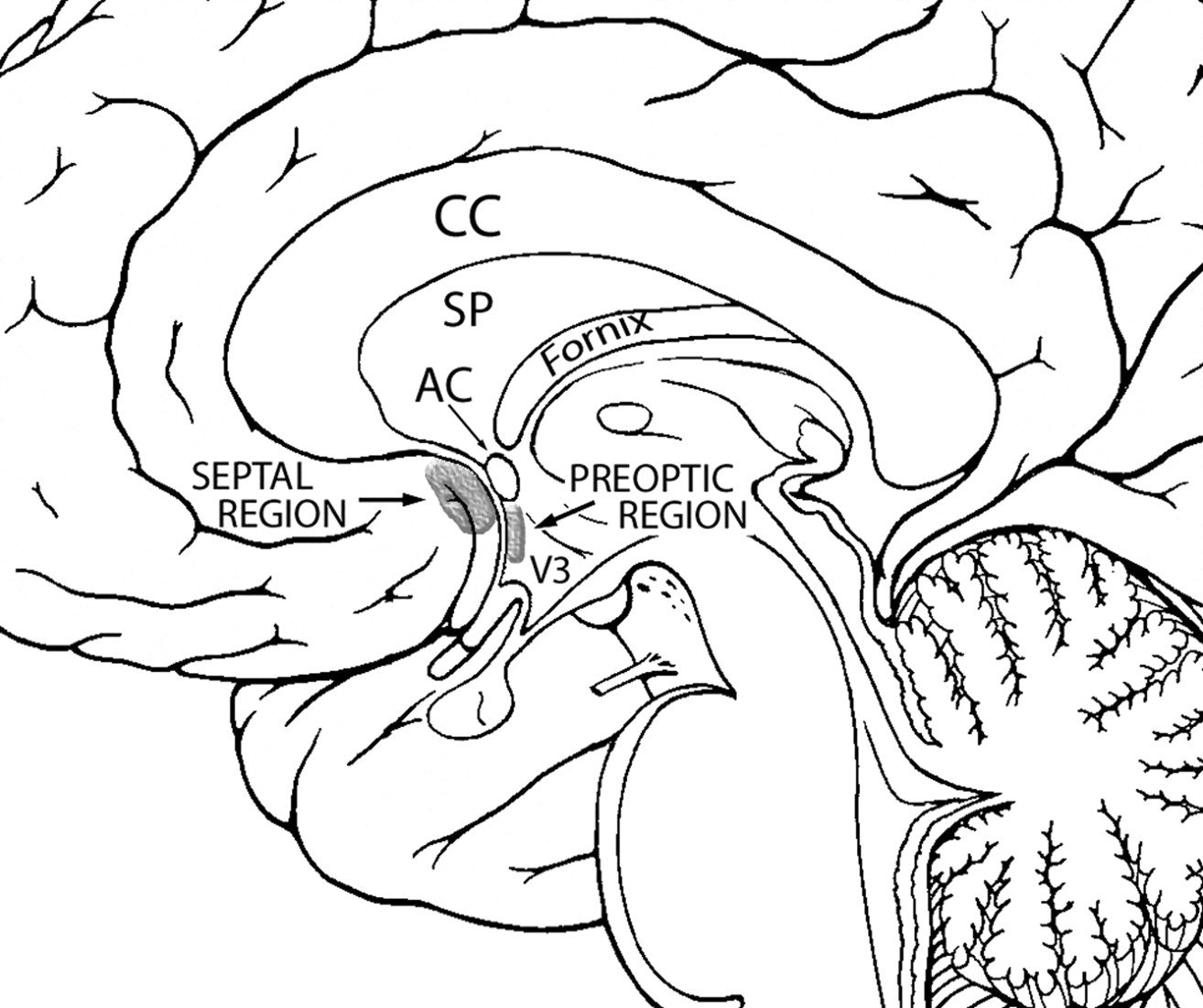
- Fig 1.
Diagram of the midsagittal view of the brain displaying the locations of the septal and preoptic regions. Modified from Martin J.22
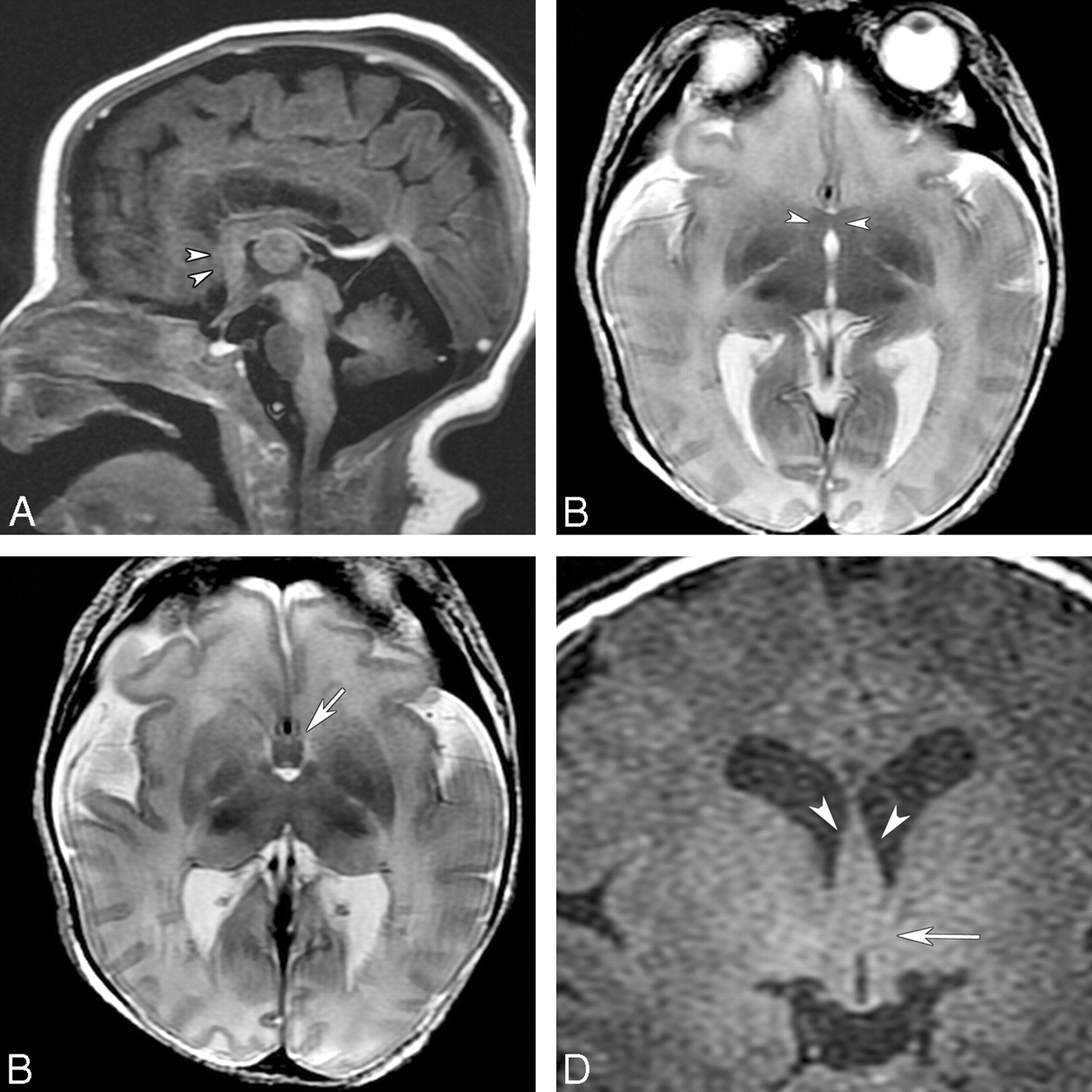
- Fig 2.
MR imaging of a 10-year-old boy with learning disabilities, a SMMCI, CNPAS, precocious puberty, and other endocrinopathies. A, T1-weighted midsagittal image shows hypoplasia of the rostrum of the CC and a rectangular area of abnormality in the subcallosal region, anterior to the hypothalamic region (arrowheads). The dysplastic-appearing fornix is anterior to this region (arrows). B, Axial T2-weighted image shows well-developed anterior and posterior interhemispheric fissures and an azygous ACA flow void in the anterior interhemispheric fissure. There is an area of midline fusion just anterior to the AC, which appears as a dark bowlike band (arrowheads). Further anterior to this region are the dysplastic fornix (arrows) and cortical gray matter that is continuous in the middle. C, Coronal SPGR image anterior to the AC shows an area of fusion of the septal region (arrow). D, Coronal SPGR image at the level to the AC shows dysplastic thickened fornices (arrow) traveling below the SP and inferiorly an area of midline fusion in the preoptic region and basal structures (curved arrow).
- Fig 3.
MR imaging of an 8-day-old term neonate with atrial and ventricular septal defects and SMMCI. A, Sagittal T1-weighted image shows a thin CC (black arrows), a thickened fornix, and dysplastic subcallosal areas (arrowheads). A small ectopic pituitary gland is noted near the chiasm (white arrow). B, Axial T2-weighted image shows an area of fusion in the septal and preoptic regions (arrowheads). C, An azygous ACA is present in the anterior IHF. Axial T2-weighted image slightly superior shows the presence of the SP (white arrow) and a thin genu of the CC. D, Coronal T2-weighted image at the level of the AC shows fusion of the midline region (white arrow) below the fornices (black arrow).
- Fig 4.
MR imaging of a 6-day-old term neonate initially diagnosed as having choanal atresia, vertebral anomalies, and coactation of aorta. A, Sagittal FSPGR image shows an area of fusion in the subcallosal region (arrowheads). B, Axial T2-weighted image shows the abnormal fusion in the same region (arrowheads). An azygous ACA is present in the anterior IHF. C, Axial T2-weighted image slightly superior shows thickened fornices (arrow) and partial fusion of the thalami. D, Coronal FSPGR image shows thickened fornices (arrowheads) and an area of fusion in the preoptic area (arrow).


Tables
Neuroimaging features comparing lobar versus septopreoptic HPE
Lobar Septopreoptic Cortical nonseparation Basal frontal Septal and preoptic regions CC Rostrum and genu absent; anterior body variably present; splenium present Rostrum absent or hypoplastic; genu hypoplastic; body and splenium present Anterior interhemispheric fissure and falx Hypoplastic anteriorly Fairly deep IHF Ventricles Rudimentary frontal horns; third ventricle formed Normal or small frontal horns; third ventricle formed Dorsal cyst Absent Absent SP Absent Present or dysplastic, rarely absent Hypothalamus Often fused to some degree (83%) Anterior hypothalamus often fused Cerebral vasculature Azygous anterior cerebral artery (more anteriorly displaced) Azygous anterior cerebral artery (more posteriorly placed)


