
Article Figures & Data
Figures
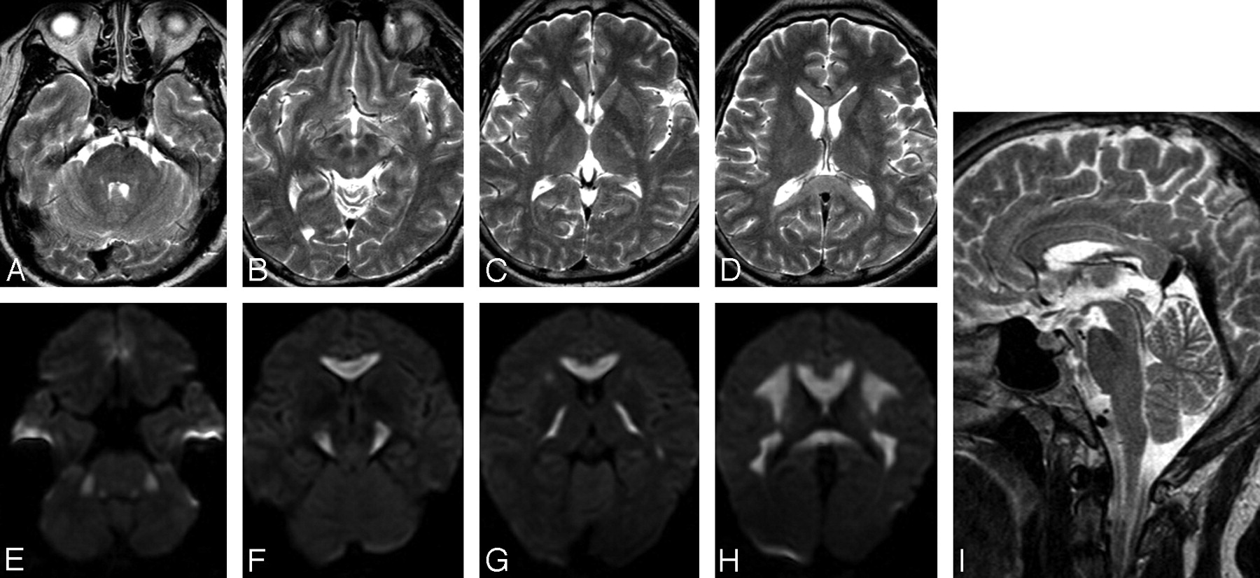
- Fig 1.
MR images obtained on day 5. A–D, On axial T2WI, the lesions appear hyperintense in the middle cerebellar peduncles, cerebral peduncles, internal capsules, and hemispheric white matter. E–H, On corresponding DWI, the lesions appear hyperintense. I, On sagittal T2WI, hyperintense lesions are seen throughout the corpus callosum.
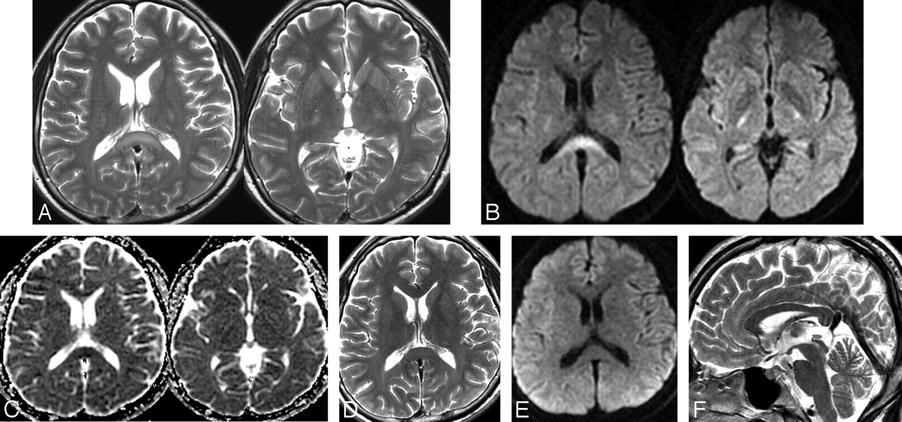
- Fig 2.
Follow-up MR images obtained on days 17 (A–C) and 30 (D–F). A, On axial T2WI, hyperintense lesions are seen in the splenium and internal capsules. B and C, On axial DWI and an ADC map, the lesions appear hyperintense in DWI and hypointense on the ADC map in the splenium and internal capsule. D–F, On follow-up MR images obtained 3 months after onset, note the disappearance of all signal-intensity abnormalities on axial T2WI, DWI, and sagittal T2WI.


In this issue


Jump to section
Related Articles
Cited By...
- Marchiafava-Bignami disease presenting as reversible coma
- Clinical Features of Cytotoxic Lesions of the Corpus Callosum Associated with Aneurysmal Subarachnoid Hemorrhage
- Acute Marchiafava-Bignami disease: clinical and serial MRI correlation
- Diagnosis and management of Marchiafava-Bignami disease: a review of CT/MRI confirmed cases
- Unusual MRI findings in a case of Marchiafava Bignami disease