
Article Figures & Data
Figures
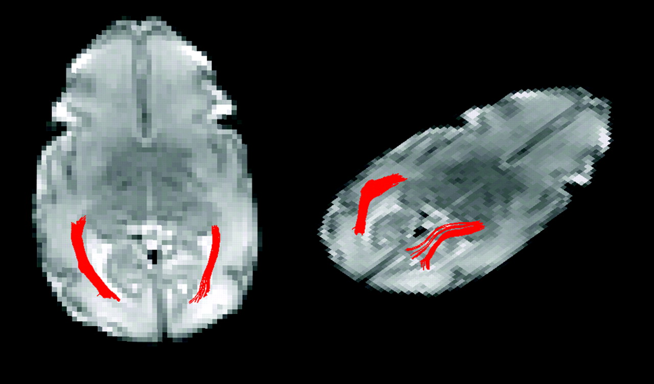
- Fig 1.
Two views of the left and right optic radiations from a single subject born at 28 4/7 weeks gestation and imaged at 33 3/7 weeks gestation.
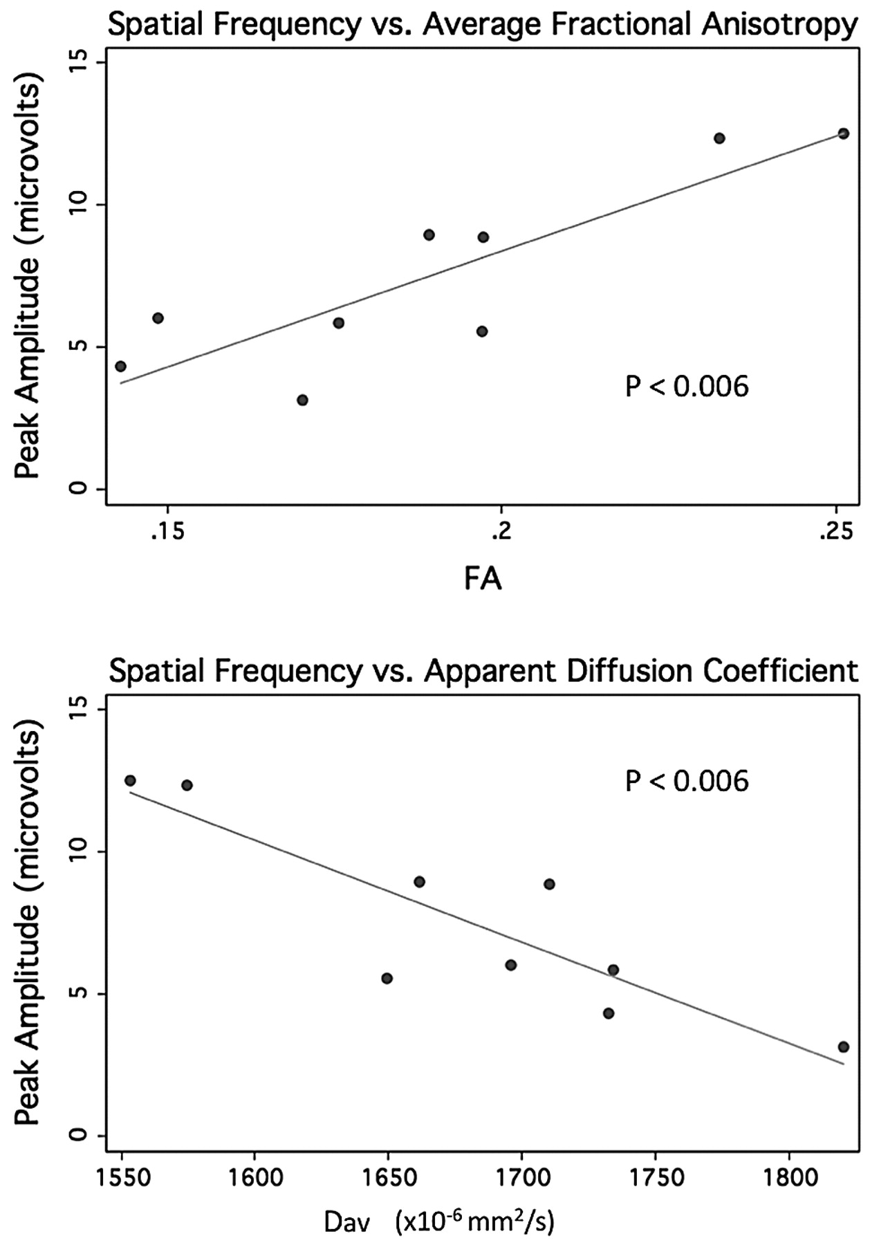
- Fig 2.
Peak amplitude from spatial frequency sweep trials versus average FA (top) and versus apparent diffusion coefficient derived from the early MR imaging in 9 preterm neonates.
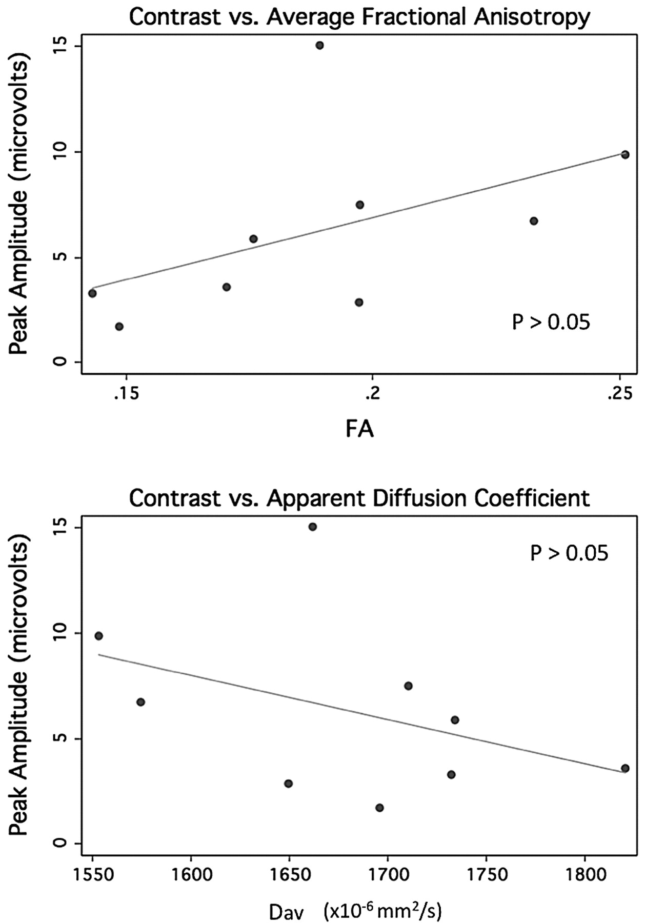
- Fig 3.
Peak amplitude from contrast sweep trials versus average FA (top) and versus apparent diffusion coefficient derived from the early MR imaging in 9 preterm neonates.
- Fig 4.
Peak amplitude from vernier offset sweep trials versus average FA (top) and versus apparent diffusion coefficient derived from the early MR imaging in 9 preterm neonates.


Tables
General characteristics of 9 preterm newborns evaluated using MR imaging and sVEP
General Characteristics Postmenstrual Age at MRI (weeks) Conventional MRI Results Corrected Age at sVEP (mo) Outcome 29 Weeks 31 Normal 8.1 Normal exam Male 40 1/7 BSID III 85 Singleton (28 mo) 913 g 26 3/7 Weeks 34 Normal 11.9 Normal exam Female 39 6/7 BSID III 115 Singleton (29 mo) 900 g 28 2/7 Weeks 29 1/7 IVH with PVHI 9 Mild diplegia Male 32 6/7 BSID III 100 Singleton (30 mo) 1190 g 31 1/7 Weeks 33 3/7 Normal 18.3 Normal exam Twin BSID III 90 875 g (37 mo) 32 3/7 Weeks 33 4/7 Moderate WMI 10.5 Normal exam Twin BSID III 125 1770 g (31 mo) 32 3/7 Weeks 34 1/7 Moderate WMI 10.5 Normal exam Twin 39 4/7 BSID III 135 1840 g (31 mo) 28 4/7 Weeks 33 3/7 Normal 13.4 Normal exam Singleton 36 3/7 BSID III 130 1115 g (30 mo) 28 6/7 Weeks 34 5/7 Normal 9 Normal exam Twin BSID III 135 1040 g (14 mo) 28 6/7 Weeks 34 2/7 Normal 9 Normal exam Twin 36 2/7 BSID III 120 1150 g (14 mo)


