
Article Figures & Data
Figures
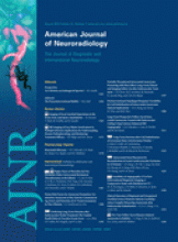
- Fig 1.
A, Conventional anatomy demonstrating paired thalamic and midbrain perforating arteries. B, AOP arising as a single unpaired trunk from P1 supplying the bilateral paramedian thalami and rostral midbrain. Reprinted with permission from Amirsys Inc.
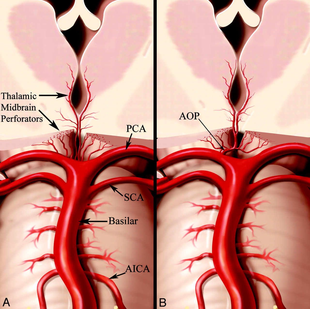
- Fig 2.
Collective extent of all 4 infarct patterns at the level of the thalamus (A) and midbrain (B) from all 37 cases superimposed.
- Fig 3.
Case 2 (A and B) and case 16 (C and D). Axial FLAIR MR images at the level the thalamus (A and C) and midbrain (B and D) demonstrate bilateral paramedian thalamic and midbrain involvement (pattern 1). Notice the hyperintense signal intensity along the pial surface of the midbrain interpeduncular fossa representing the V sign (B and D).
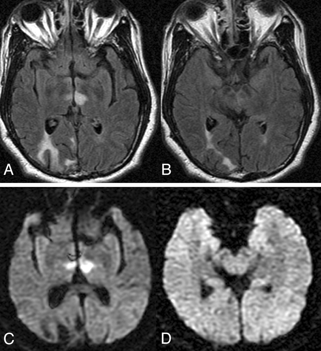
- Fig 4.
Case 22. Axial FLAIR (A and B) and DWI (C and D) images at the level of the thalamus (A and C) and midbrain (B and D) demonstrate infarction of the bilateral paramedian thalami without midbrain involvement (pattern 2).
- Fig 5.
Axial FLAIR MR images through the midbrain from cases 31 (A), 5 (B), and 9 (C) show a V-shaped hyperintense signal intensity along the pial surface of the midbrain at the interpeduncular fossa (the V sign).
- Fig 6.
DSA of the left vertebral injection, lateral (A) and anteroposterior (B) views, and a coronal CTA image (C) from case 23 demonstrate a large unpaired thalamic perforating artery (arrows) arising from the proximal P1 segment supplying the bilateral thalami (ie, an AOP).


In this issue


Jump to section
Related Articles
Cited By...
- Bilateral paramedian thalamic infarction in the setting of uncontrolled atrial fibrillation with rapid ventricular response
- Thalamic warning syndrome and the artery of Percheron
- Role of neurorehabilitation in the recovery of bilateral thalamic stroke related to the artery of Percheron anatomical variant
- Teaching NeuroImages: Comatose patient with bilateral thalamic infarct due to internal carotid artery occlusion
- CT perfusion: stroke, seizure or both?
- Artery of Percheron infarct: hiding in plain sight
- Artery of Percheron occlusion with first-pass recanalisation of the first segment of posterior cerebral artery
- Stroke masquerading as cardiac arrest: the artery of Percheron
- Clinical Reasoning: A 71-year-old man receiving treatment for cryptococcal meningitis, developing new-onset lethargy
- Artery of Percheron: an unusual stroke presentation
- Acute Basilar Artery Occlusion: Differences in Characteristics and Outcomes after Endovascular Therapy between Patients with and without Underlying Severe Atherosclerotic Stenosis
- Artery of percheron occlusion causing bilateral thalamic ischaemic infarcts
- Teaching NeuroImages: Acute Parinaud syndrome
- Fluctuating drowsiness following cardiac catheterisation: artery of Percheron ischaemic stroke causing bilateral thalamic infarcts
- Posterior circulation cerebrovascular syndromes: diagnosis and management
- Contemporary therapeutic strategies for occlusion of the artery of Percheron: a review of the literature
- Decreased consciousness: bilateral thalamic infarction and its relation to the artery of Percheron
- Unilateral midbrain infarct presenting as dorsal midbrain syndrome
- Current differential diagnoses and treatment options of vascular occlusions presenting as bilateral thalamic infarcts: a review of the literature
- Teaching NeuroImages: Comatose patient with bilateral thalamic infarct due to internal carotid artery occlusion
- Occlusion of the artery of Percheron: an unusual cause of bilateral stroke
- Bilateral infarction of paramedian thalami: a report of two cases of artery of Percheron occlusion and review of the literature