
Article Figures & Data
Figures
- Fig 1.
MRA technique sensitivity for any aneurysm remnant, larger (class 3 or 4) remnants, and the detection of growth since previous DSA; 95% confidence intervals are shown.
- Fig 2.
MRA technique specificity for any aneurysm remnant, larger (class 3 or 4) remnants, and the detection of growth since previous DSA; 95% confidence intervals are shown.
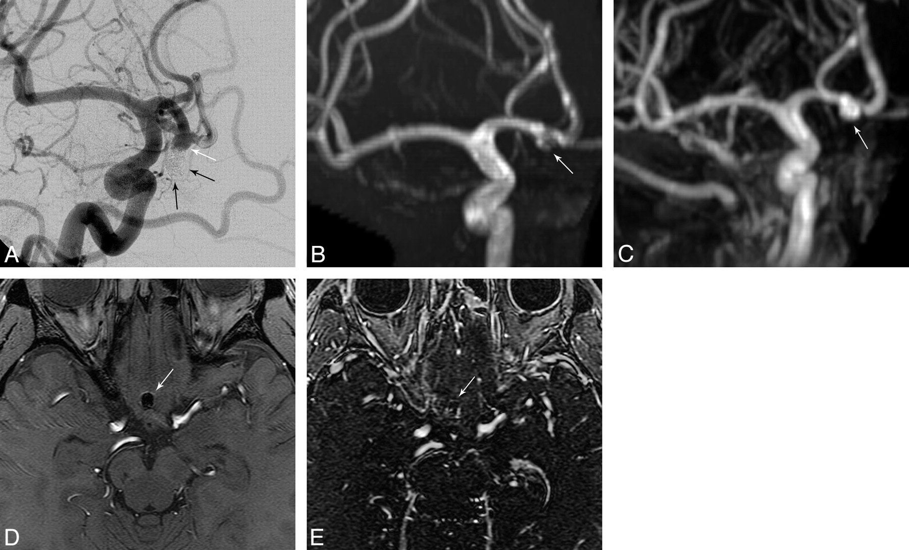
- Fig 3.
DSA (A), 3T TOF-MRA MIP (B), 3T CE-MRA MIP (C), 3T TOF-MRA source image (D), and 3T CE-MRA source image (E). A class 4 coiled anterior communicating artery remnant/recurrence (white arrows in A, B, and C) is better depicted as a large remnant with CE-MRA (C) than with TOF-MRA (B). Digitally subtracted coils are seen in A (black arrows). However, by using our clinical scanning protocols, localization of the coil mass is improved with TOF-MRA (D, white arrow) relative to CE-MRA (E). This may be related to better visualization of the surrounding brain tissue with TOF than with CE-MRA because of a shorter TR and higher flip angle of the CE-MRA and a longer TE with TOF-MRA.


Tables
1.5T TOF 1.5T CE 3T TOF 3T CE TE 6.9 Minimum 3.4 Minimum TR 36 6.6 38 8.6 Flip angle 25° 45° 25° 40° Sequence TOF SPGR Fast TOF SPGR TOF SPGR Fast TOF SPGR Matrix/NEX 256/224/1 256/224/1 512/256/1 416/224/1 Nominal section thickness (mm) 1.4 (3 slabs) 1.2 1.4 (3 slabs) 1.2 Zero-fill interpolation Yes Yes Yes Yes LOC/overlap 32/6 48 32/6 48 FOV (cm) 18 22 18 22 Receiver bandwidth (± kHz) 16 32 16 32 MRA Technique P Value Comparing Sensitivities P Value Comparing Specificities Aneurysm class: 1 vs 2 ,3, 4 (for any remnant) 1.5 TOF vs 1.5 CE .3173 .1797 1.5 TOF vs 3 TOF .3173 1 1.5 TOF vs 3 CE 1 .2568 1.5 CE vs 3 TOF .3173 .1797 1.5 CE vs 3 CE .4142 1 3 TOF vs 3 CE 1 1 Aneurysm class: 1, 2 vs 3, 4 (for larger remnants) 1.5 TOF vs 1.5 CE .0455 1 1.5 TOF vs 3 TOF 1 1 1.5 TOF vs 3 CE .0455 .5637 1.5 CE vs 3 TOF .1025 1 1.5 CE vs 3 CE 1 .5637 3 TOF vs 3 CE 1 1 Aneurysm occlusion: better or same vs worse 1.5 TOF vs 1.5 CE 1 .6547 1.5 TOF vs 3 TOF .3173 .3173 1.5 TOF vs 3 CE .1573 .5637 1.5 CE vs 3 TOF .3173 .5637 1.5 CE vs 3 CE .1573 1 3 TOF vs 3 CE .3173 .5637
In this issue


Jump to section
Related Articles
Cited By...
- MRA versus DSA for the follow-up imaging of intracranial aneurysms treated using endovascular techniques: a meta-analysis
- Long-Term Results and Follow-Up Examinations after Endovascular Embolization for Unruptured Cerebral Aneurysms
- Pointwise Encoding Time Reduction with Radial Acquisition with Subtraction-Based MRA during the Follow-Up of Stent-Assisted Coil Embolization of Anterior Circulation Aneurysms
- Visualization of Aneurysmal Neck and Dome after Coiling with 3D Multifusion Imaging of Silent MRA and FSE-MR Cisternography
- Clinical Evaluation of Highly Accelerated Compressed Sensing Time-of-Flight MR Angiography for Intracranial Arterial Stenosis
- Multiparametric MRI of intracranial aneurysms treated with the Woven EndoBridge (WEB): a case of Faradays cage?
- Voxel based analysis of recurrence dynamics in intracranial aneurysms after coiling
- Autologous adipose-derived mesenchymal stem cells improve healing of coiled experimental saccular aneurysms: an angiographic and histopathological study
- Follow-up of intracranial aneurysms treated by a WEB flow disrupter: a comparative study of DSA and contrast-enhanced MR angiography
- Imaging Follow-Up of Intracranial Aneurysms Treated by Endovascular Means: Why, When, and How?
- Is Visual Evaluation of Aneurysm Coiling a Reliable Study End Point?: Systematic Review and Meta-Analysis
- Is long-term follow-up of adequately coil-occluded ruptured cerebral aneurysms always necessary? A single-center study of recurrences after endovascular treatment
- MRA Versus DSA for Follow-Up of Coiled Intracranial Aneurysms: A Meta-Analysis
- Standard of practice: embolization of ruptured and unruptured intracranial aneurysms
- Review of 2 Decades of Aneurysm-Recurrence Literature, Part 2: Managing Recurrence after Endovascular Coiling
- Follow-Up of Coiled Intracranial Aneurysms: Comparison of 3D Time-of-Flight MR Angiography at 3T and 1.5T in a Large Prospective Series
- Temporal Evolution of Susceptibility Artifacts from Coiled Aneurysms on MR Angiography: An In Vivo Canine Study
- Use of CT Angiography in Comparison with Other Imaging Techniques for the Determination of Embolus and Remnant Size in Experimental Aneurysms Embolized with Hydrogel Filaments
- Observer Agreement Regarding the Necessity of Retreatment of Previously Coiled Recurrent Cerebral Aneurysms