Advertisement

Main menu

  • Home
  • Content
    • Current Issue
    • Accepted Manuscripts
    • Article Preview
    • Past Issue Archive
    • Video Articles
    • AJNR Case Collection
    • Case of the Week Archive
    • Case of the Month Archive
    • Classic Case Archive
  • Special Collections
    • AJNR Awards
    • Low-Field MRI
    • Alzheimer Disease
    • ASNR Foundation Special Collection
    • Photon-Counting CT
    • View All
  • Multimedia
    • AJNR Podcasts
    • AJNR SCANtastic
    • Trainee Corner
    • MRI Safety Corner
    • Imaging Protocols
  • For Authors
    • Submit a Manuscript
    • Submit a Video Article
    • Submit an eLetter to the Editor/Response
    • Manuscript Submission Guidelines
    • Statistical Tips
    • Fast Publishing of Accepted Manuscripts
    • Graphical Abstract Preparation
    • Imaging Protocol Submission
    • Author Policies
  • About Us
    • About AJNR
    • Editorial Board
    • Editorial Board Alumni
  • More
    • Become a Reviewer/Academy of Reviewers
    • Subscribers
    • Permissions
    • Alerts
    • Feedback
    • Advertisers
    • ASNR Home

User menu

  • Alerts
  • Log in

Search

  • Advanced search
American Journal of Neuroradiology

American Journal of Neuroradiology

ASHNR American Society of Functional Neuroradiology ASHNR American Society of Pediatric Neuroradiology ASSR
  • Alerts
  • Log in

Advanced Search

  • Home
  • Content
    • Current Issue
    • Accepted Manuscripts
    • Article Preview
    • Past Issue Archive
    • Video Articles
    • AJNR Case Collection
    • Case of the Week Archive
    • Case of the Month Archive
    • Classic Case Archive
  • Special Collections
    • AJNR Awards
    • Low-Field MRI
    • Alzheimer Disease
    • ASNR Foundation Special Collection
    • Photon-Counting CT
    • View All
  • Multimedia
    • AJNR Podcasts
    • AJNR SCANtastic
    • Trainee Corner
    • MRI Safety Corner
    • Imaging Protocols
  • For Authors
    • Submit a Manuscript
    • Submit a Video Article
    • Submit an eLetter to the Editor/Response
    • Manuscript Submission Guidelines
    • Statistical Tips
    • Fast Publishing of Accepted Manuscripts
    • Graphical Abstract Preparation
    • Imaging Protocol Submission
    • Author Policies
  • About Us
    • About AJNR
    • Editorial Board
    • Editorial Board Alumni
  • More
    • Become a Reviewer/Academy of Reviewers
    • Subscribers
    • Permissions
    • Alerts
    • Feedback
    • Advertisers
    • ASNR Home
  • Follow AJNR on Twitter
  • Visit AJNR on Facebook
  • Follow AJNR on Instagram
  • Join AJNR on LinkedIn
  • RSS Feeds

AJNR Awards, New Junior Editors, and more. Read the latest AJNR updates

Research ArticleBrain
Open Access

Initial Experience in Using Continuous Arterial Spin-Labeled MR Imaging for Early Detection of Alzheimer Disease

C.A. Raji, C. Lee, O.L. Lopez, J. Tsay, J.F. Boardman, E.D. Schwartz, W.S. Bartynski, H.M. Hefzy, H.M. Gach, W. Dai and J.T. Becker
American Journal of Neuroradiology May 2010, 31 (5) 847-855; DOI: https://doi.org/10.3174/ajnr.A1955
C.A. Raji
  • Find this author on Google Scholar
  • Find this author on PubMed
  • Search for this author on this site
C. Lee
  • Find this author on Google Scholar
  • Find this author on PubMed
  • Search for this author on this site
O.L. Lopez
  • Find this author on Google Scholar
  • Find this author on PubMed
  • Search for this author on this site
J. Tsay
  • Find this author on Google Scholar
  • Find this author on PubMed
  • Search for this author on this site
J.F. Boardman
  • Find this author on Google Scholar
  • Find this author on PubMed
  • Search for this author on this site
E.D. Schwartz
  • Find this author on Google Scholar
  • Find this author on PubMed
  • Search for this author on this site
W.S. Bartynski
  • Find this author on Google Scholar
  • Find this author on PubMed
  • Search for this author on this site
H.M. Hefzy
  • Find this author on Google Scholar
  • Find this author on PubMed
  • Search for this author on this site
H.M. Gach
  • Find this author on Google Scholar
  • Find this author on PubMed
  • Search for this author on this site
W. Dai
  • Find this author on Google Scholar
  • Find this author on PubMed
  • Search for this author on this site
J.T. Becker
  • Find this author on Google Scholar
  • Find this author on PubMed
  • Search for this author on this site
  • Article
  • Figures & Data
  • Info & Metrics
  • Responses
  • References
  • PDF
Loading

Article Figures & Data

Figures

  • Tables
  • Fig 1.
    • Download figure
    • Open in new tab
    • Download powerpoint
    Fig 1.

    Midaxial CASL image shows the regions assessed by the reader: 1, anterior cingulate gyrus; 2, middle frontal lobe; 3. caudate nucleus; 4, putamen and globus pallidus; 5, thalamus; 6, superior and medial temporal lobes; 7, parietal lobes; 8, posterior cingulate gyrus; 9, occipital lobes. Many of these regions, such as the cingulate gyrus, temporal lobes, and thalamus, are common targets of AD pathology. Reproduction of Fig 1 with permission from Lee et al.47

  • Fig 2.
    • Download figure
    • Open in new tab
    • Download powerpoint
    Fig 2.

    Examples of control (A) and AD (B) CASL images. Please refer to text for details.
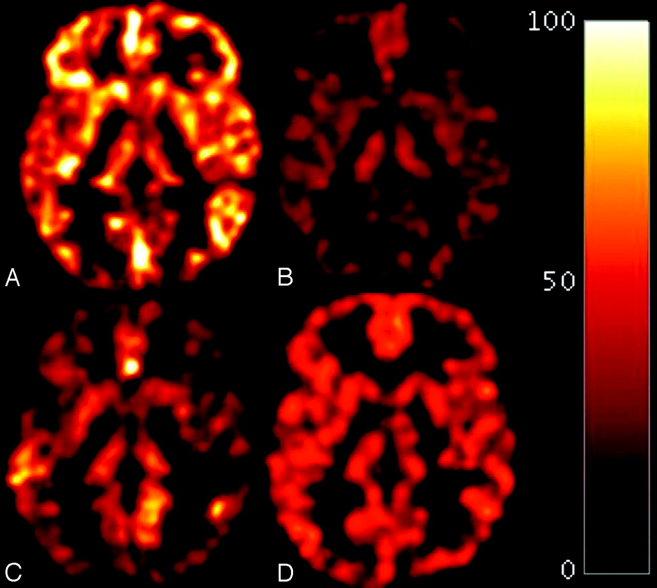
  • Fig 3.
    • Download figure
    • Open in new tab
    • Download powerpoint
    Fig 3.

    Axial CASL MR images obtained at the level of the thalamus. A, True-negative. B, True-positive. C, False-negative. D, false-positive. The images are obtained from the OsiriX Imaging Software with a standard Hot Iron scale of 0- to 100-mL blood/g tissue/min, the same scale used by the readers to view the scans shown in Fig 2.

Tables

  • Figures
    • View popup
    Table 1:

    Subject characteristics

    Normal CognitionProbable ADχ2/T Test ValueaPValue, Cohen D or ϕ
    Number1913
    Age (SD and range)82.31 ± 3.8783.1 ± 3.35−.63.53, .23
    (76.17–93.34)(77.22–88.32)
    Male/female8/118/51.17.47, .28
    Caucasian/AA18/113/0.71.59, .41
    Education (beyond 12th grade/12th grade)11/88/5.04.56, .84
    Modified MMSE94.73 ± 4.4488.9 ± 6.313.08<.01, 1.11
    (85–99)(76–97)
    Hypertension (±)11/810/31.23.23, .27
    Heart disease17/210/3.92.32, .34
    Type II diabetes mellitus17/210/2.25.63, .09
    MRI infarcts (±)12/79/4.13.53, .72
    Small vessel ischemic disease11/88/5.04.57, .84
    • a df = 30.

    • View popup
    Table 2:

    Summary of individual and average reader metrics of diagnostic utility

    Sensitivity (%)Specificity (%)Accuracy (area under ROC curve,%)
    CASL
        Reader 1697472
        Reader 2775365
        Reader 3924770
        Reader 41004271
        Average (%)855470
    SPGR
        Reader 1775365
        Reader 2547966
        Reader 3810054
        Reader 4854766
        Average (%)567063
    • View popup
    Table 3:

    Most commonly identified regions in correct abnormal CASL scan classifications by reader

    Reader 1 (%)Reader 2 (%)Reader 3 (%)Reader 4 (%)
    Frontal lobes (100)Frontal lobes (100)Parietal cortex (100)Frontal lobes (100)
    Caudate (100)Caudate (100)Frontal lobes (92)Caudate (100)
    Lentiform (100)Thalamus (90)Thalamus (83)Lentiform (100)
    Thalamus (89)Lentiform (80)Medial temporal lobe (75)Thalamus (85)
    Medial temporal lobe (78)Medial temporal lobe (80)Caudate (67)Parietal cortex (85)
    Precuneus (78)Posterior cingulate (60)Lentiform (67)Precuneus (77)
    Anterior cingulate (67)Parietal cortex (50)Precuneus (33)Posterior cingulate (77)
    Posterior cingulate (67)Anterior cingulate (40)Anterior cingulate (25)Anterior cingulate (69)
    Parietal cortex (67)Precuneus (20)Posterior cingulate (25)Medial temporal lobe (38)
    • View popup
    Table 4:

    Sensitivity, specificity, and accuracy values across all 4 readers based upon visual identification of lower rCBF in the frontal lobes with CASL

    CASLSensitivity (%)Specificity (%)Accuracy (area under ROC curve,%)
    Reader 1697974
    Reader 2926378
    Reader 3926880
    Reader 41006382
    Average (%)886879
    • View popup
    Table 5:

    Correlation between reader confidence and correct image classification

    ModalityReader 1Reader 2Reader 3Reader 4
    CASLr = 0.46, P = .008r = 0.44, P = .01r= 0.34, P = .06r= 0.42, P = .02
    SPGRr = 0.16, P = .39r = 0.32, P = .07r= −0.14, P= .45r= −0.16, P= .36
PreviousNext
Back to top

In this issue

American Journal of Neuroradiology: 31 (5)
American Journal of Neuroradiology
Vol. 31, Issue 5
1 May 2010
  • Table of Contents
  • Index by author
Advertisement
Print
Download PDF
Email Article

Thank you for your interest in spreading the word on American Journal of Neuroradiology.

NOTE: We only request your email address so that the person you are recommending the page to knows that you wanted them to see it, and that it is not junk mail. We do not capture any email address.

Enter multiple addresses on separate lines or separate them with commas.
Initial Experience in Using Continuous Arterial Spin-Labeled MR Imaging for Early Detection of Alzheimer Disease
(Your Name) has sent you a message from American Journal of Neuroradiology
(Your Name) thought you would like to see the American Journal of Neuroradiology web site.
CAPTCHA
This question is for testing whether or not you are a human visitor and to prevent automated spam submissions.
Cite this article
C.A. Raji, C. Lee, O.L. Lopez, J. Tsay, J.F. Boardman, E.D. Schwartz, W.S. Bartynski, H.M. Hefzy, H.M. Gach, W. Dai, J.T. Becker
Initial Experience in Using Continuous Arterial Spin-Labeled MR Imaging for Early Detection of Alzheimer Disease
American Journal of Neuroradiology May 2010, 31 (5) 847-855; DOI: 10.3174/ajnr.A1955

Citation Manager Formats

  • BibTeX
  • Bookends
  • EasyBib
  • EndNote (tagged)
  • EndNote 8 (xml)
  • Medlars
  • Mendeley
  • Papers
  • RefWorks Tagged
  • Ref Manager
  • RIS
  • Zotero
0 Responses
Respond to this article
Share
Bookmark this article
Initial Experience in Using Continuous Arterial Spin-Labeled MR Imaging for Early Detection of Alzheimer Disease
C.A. Raji, C. Lee, O.L. Lopez, J. Tsay, J.F. Boardman, E.D. Schwartz, W.S. Bartynski, H.M. Hefzy, H.M. Gach, W. Dai, J.T. Becker
American Journal of Neuroradiology May 2010, 31 (5) 847-855; DOI: 10.3174/ajnr.A1955
del.icio.us logo Twitter logo Facebook logo Mendeley logo
  • Tweet Widget
  • Facebook Like
  • Google Plus One
Purchase

Jump to section

  • Article
    • Abstract
    • Abbreviations
    • Materials and Methods
    • Results
    • Discussion
    • Conclusions
    • Acknowledgments
    • Appendix
    • Footnotes
    • References
  • Figures & Data
  • Info & Metrics
  • Responses
  • References
  • PDF

Related Articles

  • No related articles found.
  • PubMed
  • Google Scholar

Cited By...

  • Head-to-Head Visual Comparison between Brain Perfusion SPECT and Arterial Spin-Labeling MRI with Different Postlabeling Delays in Alzheimer Disease
  • Crossref (26)
  • Google Scholar

This article has been cited by the following articles in journals that are participating in Crossref Cited-by Linking.

  • A neuroradiologist’s guide to arterial spin labeling MRI in clinical practice
    M. Grade, J. A. Hernandez Tamames, F. B. Pizzini, E. Achten, X. Golay, M. Smits
    Neuroradiology 2015 57 12
  • Association of brain amyloid-β with cerebral perfusion and structure in Alzheimer’s disease and mild cognitive impairment
    Niklas Mattsson, Duygu Tosun, Philip S. Insel, Alix Simonson, Clifford R Jack, Laurel A. Beckett, Michael Donohue, William Jagust, Norbert Schuff, Michael W. Weiner
    Brain 2014 137 5
  • Cerebral blood flow measured by arterial spin labeling MRI at resting state in normal aging and Alzheimer’s disease
    Nan Zhang, Marc L. Gordon, Terry E. Goldberg
    Neuroscience & Biobehavioral Reviews 2017 72
  • Direct comparison of fluorodeoxyglucose positron emission tomography and arterial spin labeling magnetic resonance imaging in Alzheimer&#039;s disease
    Erik S. Musiek, Yufen Chen, Marc Korczykowski, Babak Saboury, Patricia M. Martinez, Janet S. Reddin, Abass Alavi, Daniel Y. Kimberg, David A. Wolk, Per Julin, Andrew B. Newberg, Steven E. Arnold, John A. Detre
    Alzheimer's & Dementia 2012 8 1
  • Arterial spin labeling MRI
    David A. Wolk, John A. Detre
    Current Opinion in Neurology 2012 25 4
  • Reliability of two‐dimensional and three‐dimensional pseudo‐continuous arterial spin labeling perfusion MRI in elderly populations: Comparison with 15o‐water positron emission tomography
    Emily Kilroy, Liana Apostolova, Collin Liu, Lirong Yan, John Ringman, Danny J.J. Wang
    Journal of Magnetic Resonance Imaging 2014 39 4
  • Cerebral Hypoperfusion and Hypometabolism Detected by Arterial Spin Labeling MRI and FDG‐PET in Early‐Onset Alzheimer&#039;s Disease
    Sebastien Verclytte, Renaud Lopes, Pierre Lenfant, Adeline Rollin, Franck Semah, Xavier Leclerc, Florence Pasquier, Christine Delmaire
    Journal of Neuroimaging 2016 26 2
  • Contribution of Neuroimaging to the Diagnosis of Alzheimer’s Disease and Vascular Dementia
    Gustavo Román, Belén Pascual
    Archives of Medical Research 2012 43 8
  • Physical activity, body mass index, and brain atrophy in Alzheimer&#039;s disease
    Christina P. Boyle, Cyrus A. Raji, Kirk I. Erickson, Oscar L. Lopez, James T. Becker, H. Michael Gach, W.T. Longstreth, Leonid Teverovskiy, Lewis H. Kuller, Owen T. Carmichael, Paul M. Thompson
    Neurobiology of Aging 2015 36
  • Evolution of the diagnostic criteria for degenerative and cognitive disorders
    Oscar L. Lopez, Eric McDade, Mario Riverol, James T. Becker
    Current Opinion in Neurology 2011 24 6

More in this TOC Section

  • Evaluating the Effects of White Matter Multiple Sclerosis Lesions on the Volume Estimation of 6 Brain Tissue Segmentation Methods
  • Quiet PROPELLER MRI Techniques Match the Quality of Conventional PROPELLER Brain Imaging Techniques
  • Predictors of Reperfusion in Patients with Acute Ischemic Stroke
Show more BRAIN

Similar Articles

Advertisement

Indexed Content

  • Current Issue
  • Accepted Manuscripts
  • Article Preview
  • Past Issues
  • Editorials
  • Editor's Choice
  • Fellows' Journal Club
  • Letters to the Editor
  • Video Articles

Cases

  • Case Collection
  • Archive - Case of the Week
  • Archive - Case of the Month
  • Archive - Classic Case

More from AJNR

  • Trainee Corner
  • Imaging Protocols
  • MRI Safety Corner
  • Book Reviews

Multimedia

  • AJNR Podcasts
  • AJNR Scantastics

Resources

  • Turnaround Time
  • Submit a Manuscript
  • Submit a Video Article
  • Submit an eLetter to the Editor/Response
  • Manuscript Submission Guidelines
  • Statistical Tips
  • Fast Publishing of Accepted Manuscripts
  • Graphical Abstract Preparation
  • Imaging Protocol Submission
  • Evidence-Based Medicine Level Guide
  • Publishing Checklists
  • Author Policies
  • Become a Reviewer/Academy of Reviewers
  • News and Updates

About Us

  • About AJNR
  • Editorial Board
  • Editorial Board Alumni
  • Alerts
  • Permissions
  • Not an AJNR Subscriber? Join Now
  • Advertise with Us
  • Librarian Resources
  • Feedback
  • Terms and Conditions
  • AJNR Editorial Board Alumni

American Society of Neuroradiology

  • Not an ASNR Member? Join Now

© 2025 by the American Society of Neuroradiology All rights, including for text and data mining, AI training, and similar technologies, are reserved.
Print ISSN: 0195-6108 Online ISSN: 1936-959X

Powered by HighWire