Advertisement

Main menu

  • Home
  • Content
    • Current Issue
    • Accepted Manuscripts
    • Article Preview
    • Past Issue Archive
    • Video Articles
    • AJNR Case Collection
    • Case of the Week Archive
    • Case of the Month Archive
    • Classic Case Archive
  • Special Collections
    • AJNR Awards
    • Low-Field MRI
    • Alzheimer Disease
    • ASNR Foundation Special Collection
    • Photon-Counting CT
    • View All
  • Multimedia
    • AJNR Podcasts
    • AJNR SCANtastic
    • Trainee Corner
    • MRI Safety Corner
    • Imaging Protocols
  • For Authors
    • Submit a Manuscript
    • Submit a Video Article
    • Submit an eLetter to the Editor/Response
    • Manuscript Submission Guidelines
    • Statistical Tips
    • Fast Publishing of Accepted Manuscripts
    • Graphical Abstract Preparation
    • Imaging Protocol Submission
    • Author Policies
  • About Us
    • About AJNR
    • Editorial Board
    • Editorial Board Alumni
  • More
    • Become a Reviewer/Academy of Reviewers
    • Subscribers
    • Permissions
    • Alerts
    • Feedback
    • Advertisers
    • ASNR Home

User menu

  • Alerts
  • Log in

Search

  • Advanced search
American Journal of Neuroradiology

American Journal of Neuroradiology

ASHNR American Society of Functional Neuroradiology ASHNR American Society of Pediatric Neuroradiology ASSR
  • Alerts
  • Log in

Advanced Search

  • Home
  • Content
    • Current Issue
    • Accepted Manuscripts
    • Article Preview
    • Past Issue Archive
    • Video Articles
    • AJNR Case Collection
    • Case of the Week Archive
    • Case of the Month Archive
    • Classic Case Archive
  • Special Collections
    • AJNR Awards
    • Low-Field MRI
    • Alzheimer Disease
    • ASNR Foundation Special Collection
    • Photon-Counting CT
    • View All
  • Multimedia
    • AJNR Podcasts
    • AJNR SCANtastic
    • Trainee Corner
    • MRI Safety Corner
    • Imaging Protocols
  • For Authors
    • Submit a Manuscript
    • Submit a Video Article
    • Submit an eLetter to the Editor/Response
    • Manuscript Submission Guidelines
    • Statistical Tips
    • Fast Publishing of Accepted Manuscripts
    • Graphical Abstract Preparation
    • Imaging Protocol Submission
    • Author Policies
  • About Us
    • About AJNR
    • Editorial Board
    • Editorial Board Alumni
  • More
    • Become a Reviewer/Academy of Reviewers
    • Subscribers
    • Permissions
    • Alerts
    • Feedback
    • Advertisers
    • ASNR Home
  • Follow AJNR on Twitter
  • Visit AJNR on Facebook
  • Follow AJNR on Instagram
  • Join AJNR on LinkedIn
  • RSS Feeds

AJNR Awards, New Junior Editors, and more. Read the latest AJNR updates

Research ArticleHead and Neck

Osteoradionecrosis of the Hyoid Bone: Imaging Findings

J.S. Yoo, D.I. Rosenthal, K. Mitchell and L.E. Ginsberg
American Journal of Neuroradiology April 2010, 31 (4) 761-766; DOI: https://doi.org/10.3174/ajnr.A1892
J.S. Yoo
  • Find this author on Google Scholar
  • Find this author on PubMed
  • Search for this author on this site
D.I. Rosenthal
  • Find this author on Google Scholar
  • Find this author on PubMed
  • Search for this author on this site
K. Mitchell
  • Find this author on Google Scholar
  • Find this author on PubMed
  • Search for this author on this site
L.E. Ginsberg
  • Find this author on Google Scholar
  • Find this author on PubMed
  • Search for this author on this site
  • Article
  • Figures & Data
  • Info & Metrics
  • Responses
  • References
  • PDF
Loading

Article Figures & Data

Figures

  • Tables
  • Fig 1.
    • Download figure
    • Open in new tab
    • Download powerpoint
    Fig 1.

    Patient 4. A 64-year-old man radiated for squamous cell carcinoma of the left lateral oropharyngeal wall. A, CT examination with contrast obtained before radiation therapy shows the lower aspect of the lesion in the vallecula, lying adjacent to the hyoid bone (arrow). There is a nodal metastasis in the left neck (arrowhead), also adjacent to the hyoid. B and C, Axial contrast-enhanced CT images obtained 5 months following treatment with 70 Gy show a defect in the lateral pharyngeal wall (arrow, B) and an exposed piece of hyoid (arrow in C). At this time, the hyoid was otherwise intact. D, CT bone window on follow-up imaging shows a missing resorbed hyoid.

  • Fig 2.
    • Download figure
    • Open in new tab
    • Download powerpoint
    Fig 2.

    Patient 10. A 67-year-old man after radiation therapy at an outside hospital for base-of-tongue cancer. A and B, Axial contrast-enhanced CT scans show an air-filled soft-tissue defect in the left posterior tongue (arrow, A). Lower down, this communicates with the hyoid (B). There is an unrelated nodal recurrence in the right neck (arrowhead, A). B, Soft-tissue CT image more inferiorly shows air (arrowheads) and fluid (arrows) surrounding the hyoid. C, CT bone window shows intraosseous air (arrowheads) and a hyoid fracture (arrow).

  • Fig 3.
    • Download figure
    • Open in new tab
    • Download powerpoint
    Fig 3.

    Patient 9. A 70-year-old man previously radiated for base-of-tongue carcinoma and subsequently re-irradiated for neck recurrence. A, Contrast-enhanced CT examination 6 months after the completion of the second course of radiation demonstrates an air-filled necrotic cavity in the tongue base without surrounding enhancement (arrow). B, CT image more inferiorly shows that the cavity contains fluid and internal air bubbles (arrow, B) and is in close contact with the hyoid. The hyoid was reported as being at risk for ORN. C, CT bone window obtained 2 months after B reveals that the hyoid is now fragmented (arrow).
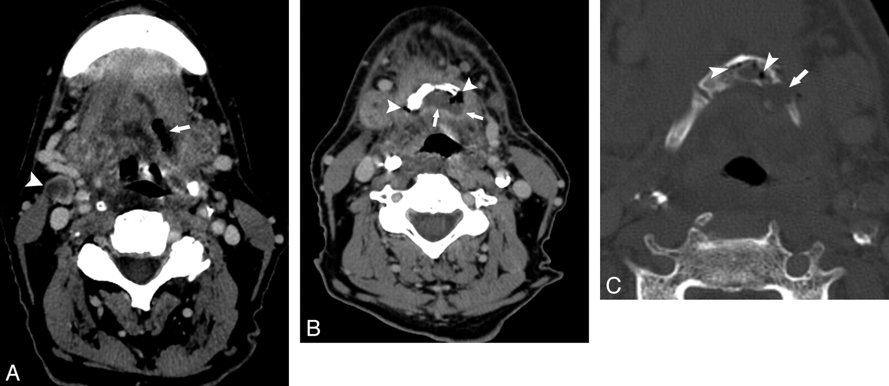
  • Fig 4.
    • Download figure
    • Open in new tab
    • Download powerpoint
    Fig 4.

    Patient 3. A 66-year-old man status post 60 Gy for treatment of metastatic squamous cell carcinoma to the left neck, primary unknown, who subsequently developed a primary malignancy in the right oropharyngeal wall, for which an additional dose of 50.2 Gy was administered. A, CT bone window before XRT shows an intact hyoid. B and C, Contrast-enhanced CT, soft-tissue and bone windows, respectively, 5 months following completion of the second course of XRT. There is an enhancing process surrounding the right side of the hyoid (arrows, B) and an exposed fragment of hyoid surrounded by air (arrowhead, B and C). Hyoid fractures can be seen (arrow in C). D, Contrast-enhanced CT image 5 months after B and C shows dramatically increased enhancement extending to the left of midline (arrows). Because of these findings and relentless aspiration, a total laryngectomy was performed; at histologic examination, there was only necrosis and inflammation.

  • Fig 5.
    • Download figure
    • Open in new tab
    • Download powerpoint
    Fig 5.

    Patient 1. A 67-year-old man, 34 months following an unknown dose of XRT at an outside facility for treatment of squamous cell carcinoma of the tongue base. A, Axial bone window CT scan shows typical fragmentation and intraosseous air characteristic of hyoid ORN. B, PET/CT, obtained concurrent to A, demonstrates intense focal FDG uptake (SUV=7.5), which was regarded as suspicious for tumor. The patient was offered hyoidectomy but refused, and his symptoms of dysphagia and odynophagia gradually improved with hyperbaric O2 during the follow-up period of 14 months, after which the patient was lost to follow-up.

Tables

  • Figures
  • Summary of patients with hyoid ORN

    No.Age/SexDose (Gy)Primary Adjacent to HyoidPost-XRT ORN DX (mo)Preceding Soft-Tissue UlcerationAdjacent EnhancementPost-ORN Follow-Up (mo)
    167/MOSH-N/A–34NoYes12
    246/M71Yes5NoNo6
    366/Ma60, 50.2No2NoYes23
    464/M70.0Yes5YesNo30
    545/M66.8Yes5.5YesNo1
    664/M70.6Yes13NoNo16
    754/M70.1Yes8NoNo7
    849/M71.6Yes26NoNo16
    970/M60, 20–6NoNo14
    1067/MOSH-N/A–1NoNo21
    1147/MOSH-N/A–32YesYes7
    1267/M41, 63Yes10YesYes2
    1361/M50, 60–17YesYes4
    • a Patient was originally radiated for metastasis to the neck (60 Gy), primary unknown, and subsequently re-irradiated (50.2 Gy) for a pharyngeal wall squamous cell carcinoma.

PreviousNext
Back to top

In this issue

American Journal of Neuroradiology: 31 (4)
American Journal of Neuroradiology
Vol. 31, Issue 4
1 Apr 2010
  • Table of Contents
  • Index by author
Advertisement
Print
Download PDF
Email Article

Thank you for your interest in spreading the word on American Journal of Neuroradiology.

NOTE: We only request your email address so that the person you are recommending the page to knows that you wanted them to see it, and that it is not junk mail. We do not capture any email address.

Enter multiple addresses on separate lines or separate them with commas.
Osteoradionecrosis of the Hyoid Bone: Imaging Findings
(Your Name) has sent you a message from American Journal of Neuroradiology
(Your Name) thought you would like to see the American Journal of Neuroradiology web site.
CAPTCHA
This question is for testing whether or not you are a human visitor and to prevent automated spam submissions.
Cite this article
J.S. Yoo, D.I. Rosenthal, K. Mitchell, L.E. Ginsberg
Osteoradionecrosis of the Hyoid Bone: Imaging Findings
American Journal of Neuroradiology Apr 2010, 31 (4) 761-766; DOI: 10.3174/ajnr.A1892

Citation Manager Formats

  • BibTeX
  • Bookends
  • EasyBib
  • EndNote (tagged)
  • EndNote 8 (xml)
  • Medlars
  • Mendeley
  • Papers
  • RefWorks Tagged
  • Ref Manager
  • RIS
  • Zotero
0 Responses
Respond to this article
Share
Bookmark this article
Osteoradionecrosis of the Hyoid Bone: Imaging Findings
J.S. Yoo, D.I. Rosenthal, K. Mitchell, L.E. Ginsberg
American Journal of Neuroradiology Apr 2010, 31 (4) 761-766; DOI: 10.3174/ajnr.A1892
del.icio.us logo Twitter logo Facebook logo Mendeley logo
  • Tweet Widget
  • Facebook Like
  • Google Plus One
Purchase

Jump to section

  • Article
    • Abstract
    • Abbreviations
    • Materials and Methods
    • Results
    • Discussion
    • Conclusions
    • References
  • Figures & Data
  • Info & Metrics
  • Responses
  • References
  • PDF

Related Articles

  • No related articles found.
  • PubMed
  • Google Scholar

Cited By...

  • Osteoradionecrosis after Radiation Therapy for Head and Neck Cancer: Differentiation from Recurrent Disease with CT and PET/CT Imaging
  • Crossref (22)
  • Google Scholar

This article has been cited by the following articles in journals that are participating in Crossref Cited-by Linking.

  • Osteoradionecrosis after Radiation Therapy for Head and Neck Cancer: Differentiation from Recurrent Disease with CT and PET/CT Imaging
    L. Alhilali, A.R. Reynolds, S. Fakhran
    American Journal of Neuroradiology 2014 35 7
  • Osteoradionecrosis of the mandible: through a radiologist's eyes
    S.S. Deshpande, M.H. Thakur, K. Dholam, A. Mahajan, S. Arya, S. Juvekar
    Clinical Radiology 2015 70 2
  • Adverse Radiation Therapy Effects in the Treatment of Head and Neck Tumors
    Pedro H. P. Rocha, Raphael M. Reali, Marcos Decnop, Soraia A. Souza, Lorine A. B. Teixeira, Ademar Lucas Júnior, Maíra O. Sarpi, Murilo B. Cintra, Marco C. Pinho, Marcio R. T. Garcia
    RadioGraphics 2022 42 3
  • Superimposed Infection in Mandibular Osteoradionecrosis
    Jackson D. Hamilton, Stephen Y. Lai, Lawrence E. Ginsberg
    Journal of Computer Assisted Tomography 2012 36 6
  • Osteoradionecrosis beyond the jaws: a mini case series and review of the literature
    R. Wali, R. King, V. Patel
    Oral Surgery 2020 13 2
  • Osteoradionecrosis of the Jaws
    Jose V. Bagan, Crispian Scully, Enrique Zapater, Jorge Basterra, Leticia Bagan
    Clinical Reviews in Bone and Mineral Metabolism 2011 9 1
  • Imaging of the Head and Neck following Radiation Treatment
    J. Matthew Debnam
    Pathology Research International 2011 2011
  • Hyoid osteoradionecrosis associated with carotid rupture: Report of 2 cases
    Rachel Chard, Marcus M. Monroe, Peter E. Andersen
    Head & Neck 2013 35 2
  • Osteoradionecrosis of the hyoid bone after intra-arterial chemoradiotherapy for oropharyngeal cancer: MR imaging findings
    Hiromitsu Hatakeyama, Noriyuki Fujima, Kazuhiko Tsuchiya, Kenji Mizoguchi, Takatsugu Mizumachi, Tomohiro Sakashita, Satoshi Kano, Akihiro Homma, Satoshi Fukuda
    Cancer Imaging 2017 17 1
  • Second opinion interpretations by specialty radiologists in head-and-neck oncology and their impact on clinical management: A retrospective observational study
    Abhishek Mahajan, Shreya Shukla, Raghvendra Mali, Ujjwal Agarwal, Nilesh Sable, Richa Vaish, Suman Kumar Ankathi, Vasundhara Patil, Amit K. Janu, Kumar Prabhash, Vanita Noronha, Prathamesh Pai, Sarbani G. Laskar, Asawari Patil, Vijay Patil, Nandini Menon, Shivakumar Thiagarajan, Pankaj Chaturvedi
    Cancer Research, Statistics, and Treatment 2022 5 4

More in this TOC Section

  • Correlation of Apparent Diffusion Coefficient at 3T with Prognostic Parameters of Retinoblastoma
  • Parathyroid Lesions: Characterization with Dual-Phase Arterial and Venous Enhanced CT of the Neck
  • Kimura Disease: CT and MR Imaging Findings
Show more HEAD AND NECK

Similar Articles

Advertisement

Indexed Content

  • Current Issue
  • Accepted Manuscripts
  • Article Preview
  • Past Issues
  • Editorials
  • Editor's Choice
  • Fellows' Journal Club
  • Letters to the Editor
  • Video Articles

Cases

  • Case Collection
  • Archive - Case of the Week
  • Archive - Case of the Month
  • Archive - Classic Case

More from AJNR

  • Trainee Corner
  • Imaging Protocols
  • MRI Safety Corner
  • Book Reviews

Multimedia

  • AJNR Podcasts
  • AJNR Scantastics

Resources

  • Turnaround Time
  • Submit a Manuscript
  • Submit a Video Article
  • Submit an eLetter to the Editor/Response
  • Manuscript Submission Guidelines
  • Statistical Tips
  • Fast Publishing of Accepted Manuscripts
  • Graphical Abstract Preparation
  • Imaging Protocol Submission
  • Evidence-Based Medicine Level Guide
  • Publishing Checklists
  • Author Policies
  • Become a Reviewer/Academy of Reviewers
  • News and Updates

About Us

  • About AJNR
  • Editorial Board
  • Editorial Board Alumni
  • Alerts
  • Permissions
  • Not an AJNR Subscriber? Join Now
  • Advertise with Us
  • Librarian Resources
  • Feedback
  • Terms and Conditions
  • AJNR Editorial Board Alumni

American Society of Neuroradiology

  • Not an ASNR Member? Join Now

© 2025 by the American Society of Neuroradiology All rights, including for text and data mining, AI training, and similar technologies, are reserved.
Print ISSN: 0195-6108 Online ISSN: 1936-959X

Powered by HighWire