
Article Figures & Data
Figures
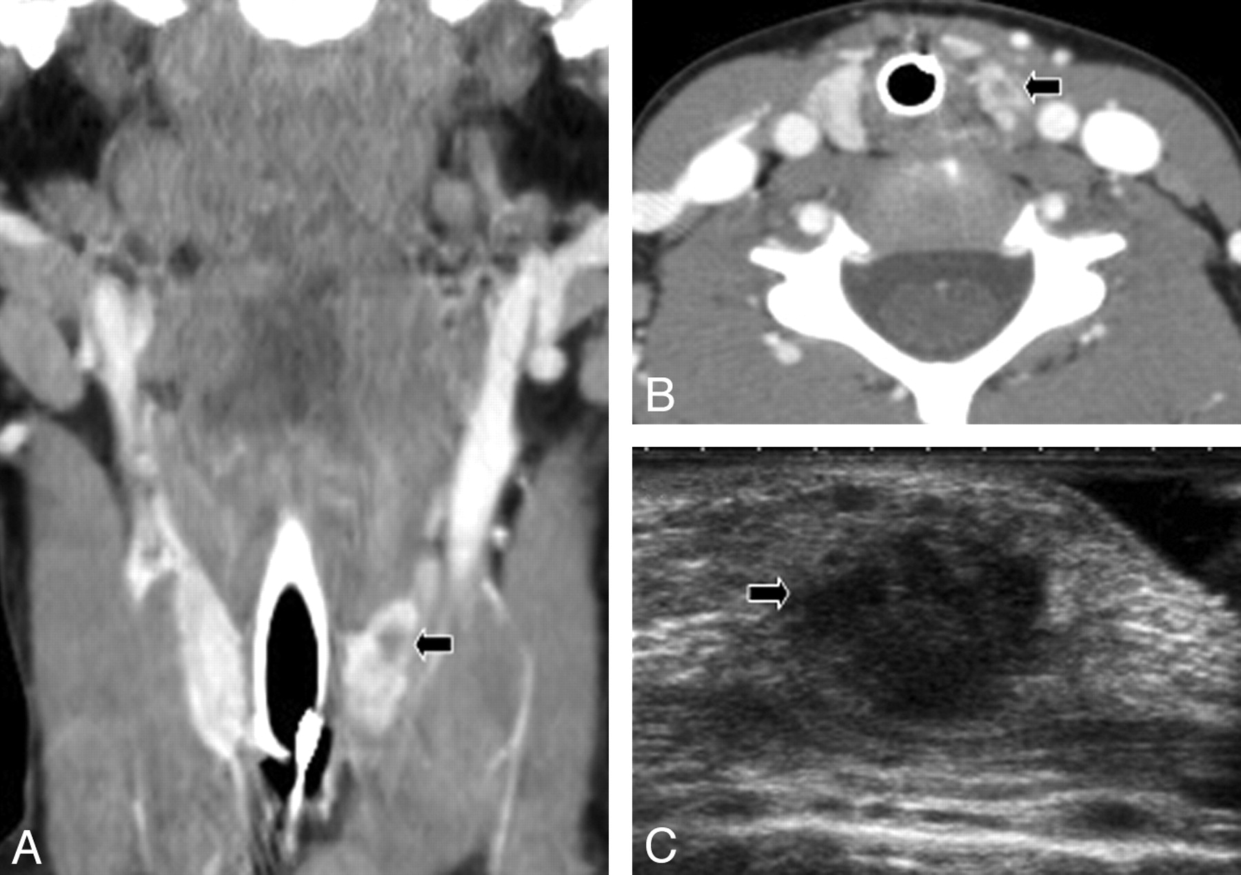
- Fig 1.
Infective lesion involving the upper pole of left thyroid lobe (arrow) associated with a third/fourth brachial remnant. Contrast-enhanced coronal CT reformation of the neck (A), contrast-enhanced coronal axial CT scan at the level of the thyroid gland (B), and a sonogram of the left thyroid lobe (C) show the phlegmonous lesion. Pharyngoscopy (not shown) revealed an opening at the apex of the left piriform sinus.
- Fig 2.
Extensive left-sided neck infection with abscess (thin black arrow) formation and involvement of the left thyroid lobe (thick arrow). Contrast-enhanced coronal T1 fat-saturated MR image of the neck (A), contrast-enhanced axial T1 fat-saturated MR image (B), and contrast-enhanced axial CT scan at the level of the thyroid gland (C) demonstrate the lesion well.
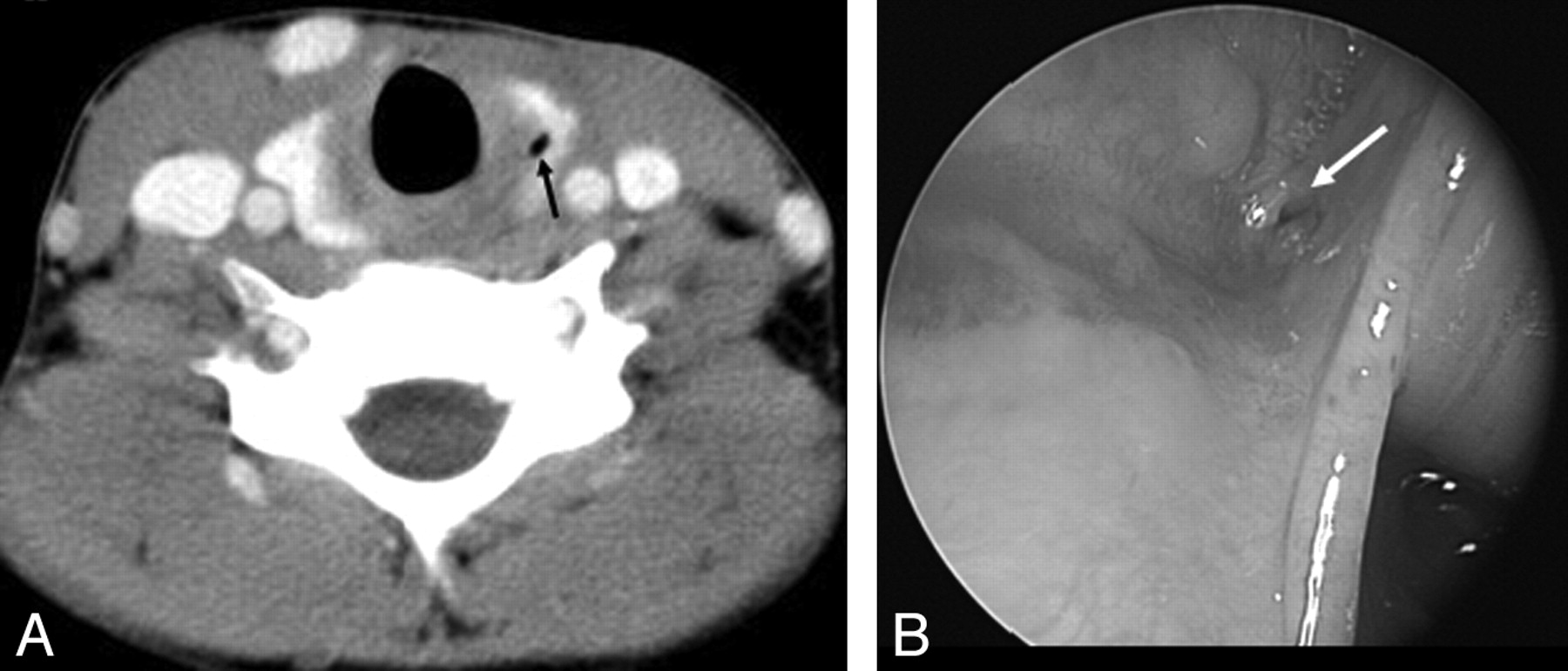
- Fig 3.
A, Contrast-enhanced axial CT scan at the level of the thyroid gland shows a small air pocket within the left lobe of the thyroid gland (black arrow), which is thought to be characteristic of a branchial sinus remnant. B, Pharyngoscopy photograph shows the opening (white arrow) of the branchial pouch sinus in the apex of the pyriform fossa.
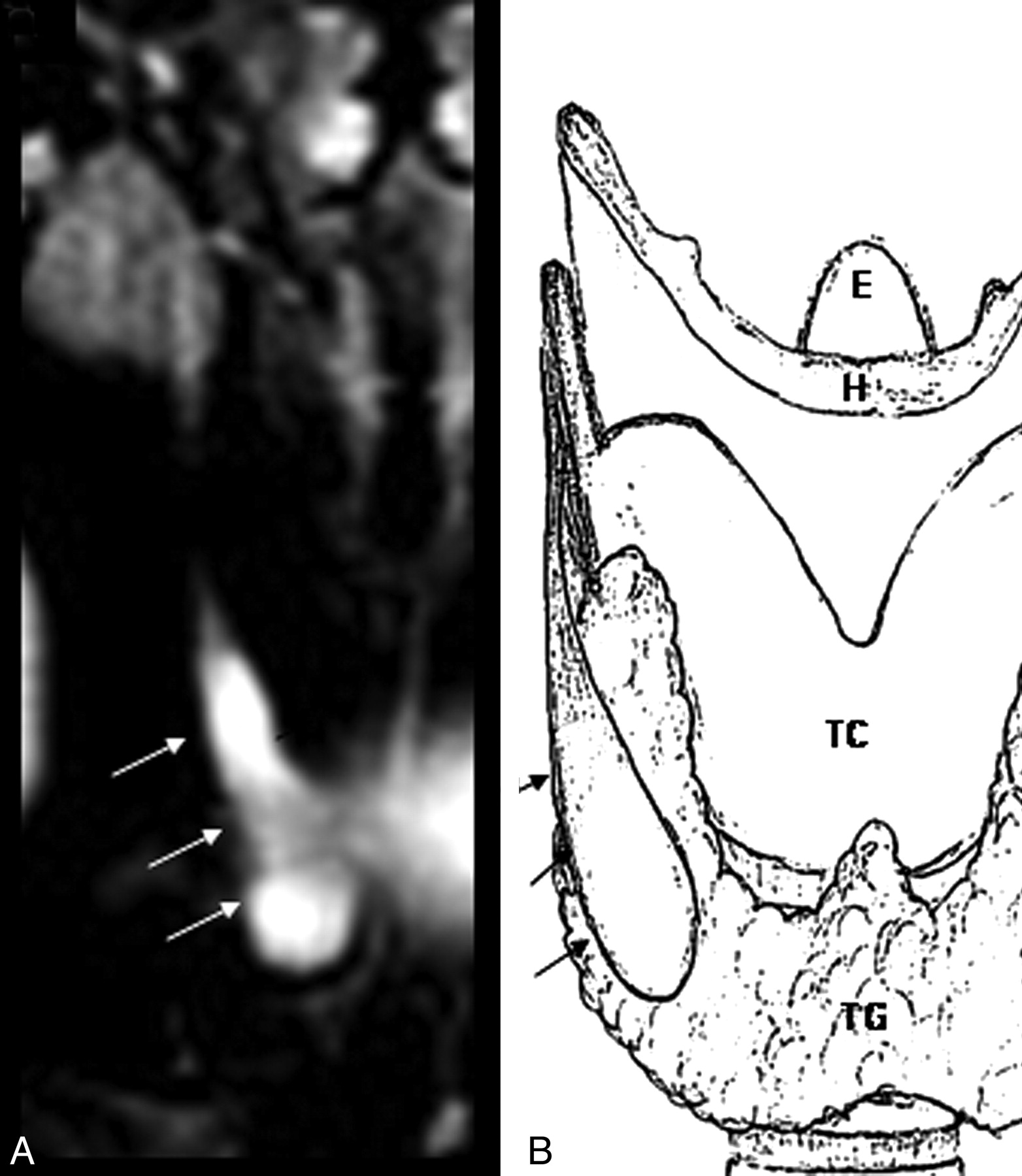
- Fig 4.
Noninfected third branchial cleft cyst on the right side of the neck following the course of the embryonal thymopharyngeal duct (arrows). A, Coronal T2 fat-saturated image. B, Schematic representation of the course of the thymopharyngeal duct.


Tables
Demographics, clinical presentation, pharyngoscopy findings, and imaging
Sl No. Age (mo) Sex Presentation Side Abscess Thyroid Inv Piriform S Imaging 01 60 M Infl neck mass L – + +L CT/MRI 02 108 F Infl neck mass L + + +L US/CT/MRI 03 120 F Noninfl neck mass, cyst R – Adj +R MRI 04 20 F Infl neck mass L + + +L US/CT 05 17 F Infl neck mass L + + +L CT 06 90 M Infl neck mass L + + +L CT/MRI 07 9 M Infl neck mass L – + – CT 08 117 F Noninfl neck mass, cyst R – Adj +R US/CT 09 125 F Infl neck mass L + + +L US/CT 10 127 M Infl neck mass L – + +L CT/MRI 11 50 M Infl neck mass, cyst R – Adj +R US/CT 12 99 M Infl neck mass L + + +L US/CT 13 32 M Infl neck mass L + + +L CT/MRI 14 23 F Infl neck mass L + + +L US/barium 15 182 F Infl neck mass L + + +L CT 16 104 F Infl neck mass L – + +L US/CT 17 31 F Infl neck mass L – + +L CT 18 41 M Infl neck mass L – + +L US/CT 19 145 F Infl neck mass L – + – US/nuclear 20 191 F Infl neck mass R + + +L + R US/CT -
+ indicates present; –, absent.
-


