Advertisement

Main menu

  • Home
  • Content
    • Current Issue
    • Accepted Manuscripts
    • Article Preview
    • Past Issue Archive
    • Video Articles
    • AJNR Case Collection
    • Case of the Week Archive
    • Case of the Month Archive
    • Classic Case Archive
  • Special Collections
    • AJNR Awards
    • Low-Field MRI
    • Alzheimer Disease
    • ASNR Foundation Special Collection
    • Photon-Counting CT
    • View All
  • Multimedia
    • AJNR Podcasts
    • AJNR SCANtastic
    • Trainee Corner
    • MRI Safety Corner
    • Imaging Protocols
  • For Authors
    • Submit a Manuscript
    • Submit a Video Article
    • Submit an eLetter to the Editor/Response
    • Manuscript Submission Guidelines
    • Statistical Tips
    • Fast Publishing of Accepted Manuscripts
    • Graphical Abstract Preparation
    • Imaging Protocol Submission
    • Author Policies
  • About Us
    • About AJNR
    • Editorial Board
    • Editorial Board Alumni
  • More
    • Become a Reviewer/Academy of Reviewers
    • Subscribers
    • Permissions
    • Alerts
    • Feedback
    • Advertisers
    • ASNR Home

User menu

  • Alerts
  • Log in

Search

  • Advanced search
American Journal of Neuroradiology

American Journal of Neuroradiology

ASHNR American Society of Functional Neuroradiology ASHNR American Society of Pediatric Neuroradiology ASSR
  • Alerts
  • Log in

Advanced Search

  • Home
  • Content
    • Current Issue
    • Accepted Manuscripts
    • Article Preview
    • Past Issue Archive
    • Video Articles
    • AJNR Case Collection
    • Case of the Week Archive
    • Case of the Month Archive
    • Classic Case Archive
  • Special Collections
    • AJNR Awards
    • Low-Field MRI
    • Alzheimer Disease
    • ASNR Foundation Special Collection
    • Photon-Counting CT
    • View All
  • Multimedia
    • AJNR Podcasts
    • AJNR SCANtastic
    • Trainee Corner
    • MRI Safety Corner
    • Imaging Protocols
  • For Authors
    • Submit a Manuscript
    • Submit a Video Article
    • Submit an eLetter to the Editor/Response
    • Manuscript Submission Guidelines
    • Statistical Tips
    • Fast Publishing of Accepted Manuscripts
    • Graphical Abstract Preparation
    • Imaging Protocol Submission
    • Author Policies
  • About Us
    • About AJNR
    • Editorial Board
    • Editorial Board Alumni
  • More
    • Become a Reviewer/Academy of Reviewers
    • Subscribers
    • Permissions
    • Alerts
    • Feedback
    • Advertisers
    • ASNR Home
  • Follow AJNR on Twitter
  • Visit AJNR on Facebook
  • Follow AJNR on Instagram
  • Join AJNR on LinkedIn
  • RSS Feeds

AJNR Awards, New Junior Editors, and more. Read the latest AJNR updates

Research ArticleFunctional
Open Access

Tactile Sensory and Pain Networks in the Human Spinal Cord and Brain Stem Mapped by Means of Functional MR Imaging

N.F. Ghazni, C.M. Cahill and P.W. Stroman
American Journal of Neuroradiology April 2010, 31 (4) 661-667; DOI: https://doi.org/10.3174/ajnr.A1909
N.F. Ghazni
  • Find this author on Google Scholar
  • Find this author on PubMed
  • Search for this author on this site
C.M. Cahill
  • Find this author on Google Scholar
  • Find this author on PubMed
  • Search for this author on this site
P.W. Stroman
  • Find this author on Google Scholar
  • Find this author on PubMed
  • Search for this author on this site
  • Article
  • Figures & Data
  • Info & Metrics
  • Responses
  • References
  • PDF
Loading

Article Figures & Data

Figures

  • Fig 1.
    • Download figure
    • Open in new tab
    • Download powerpoint
    Fig 1.

    A, Average numbers of voxels for the 2 g and 15 g stimuli across 8 healthy volunteers. Regions of interest include areas in the brain stem and cervical spinal cord. The only significance was found in the thalamus where there were more active voxels with the 15 g than 2 g stimulus. B, A box and whiskers plot of mean pain intensity and unpleasantness scores with SD across 8 healthy volunteers. Also included in the plot are the range of values for intensity and unpleasantness. The diamond corresponds to the median for the range of data and is indicated as a numeric value to the right of bars. The pain intensity and unpleasantness ratings for the 15 g filament were significantly higher than for the 2 g filament. Significance (*) was determined by using a 2-tailed, paired, Student t test (P < .05).

  • Fig 2.
    • Download figure
    • Open in new tab
    • Download powerpoint
    Fig 2.

    Combined data showing location of neuronal activity in touch (2 g and 15 g filament) and brush stimuli from 8 healthy volunteers superimposed onto transverse anatomic drawings. The figure shows significant areas of activity (T > 2.5) across the group. The T-value correlation map on the right indicates the corresponding color for each T-value. The asterisks indicate specific areas referred to in the text.

  • Fig 3.
    • Download figure
    • Open in new tab
    • Download powerpoint
    Fig 3.

    Positive and negative signal intensity responses with 2 g or 15 g von Frey filaments. Each row shows 8 consecutive transverse sections spanning the C6 level of the cord. Respective T-values (−7.0 < T < 7.0) are represented with colors as indicated at the bottom of the figure.
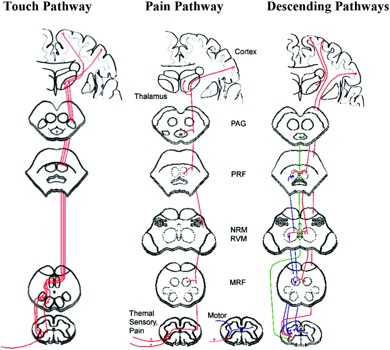
  • Fig 4.
    • Download figure
    • Open in new tab
    • Download powerpoint
    Fig 4.

    Schematic diagram showing ascending and descending pathways. Ascending touch pathways include the dorsal column–medial lemniscus. Touch sensation transmits via heavily myelinated Aβ fibers that ascend in the ipsilateral dorsal columns to the gracile or cuneate nuclei in the brain stem. Ascending pain pathways include the anterolateral pathway, which is a combination of the spinothalamic, spinoreticular, and spinomesencephalic pathways. Pain sensation transmits via thinly myelinated Aδ fibers and unmyelinated C primary afferent fibers. Descending fibers modulate touch and pain sensation.54 Red highlights descending pathways from the cortex, blue shows descending modulation from the reticular formation, and green highlights descending modulation from the raphe nuclei. PRF = pontine reticular formation; NRM = nucleus raphe magnus, RVM = rostral ventromedial medulla, MRF = medullary reticular formation. Pathways are drawn according to selected publications.34,53–55

PreviousNext
Back to top

In this issue

American Journal of Neuroradiology: 31 (4)
American Journal of Neuroradiology
Vol. 31, Issue 4
1 Apr 2010
  • Table of Contents
  • Index by author
Advertisement
Print
Download PDF
Email Article

Thank you for your interest in spreading the word on American Journal of Neuroradiology.

NOTE: We only request your email address so that the person you are recommending the page to knows that you wanted them to see it, and that it is not junk mail. We do not capture any email address.

Enter multiple addresses on separate lines or separate them with commas.
Tactile Sensory and Pain Networks in the Human Spinal Cord and Brain Stem Mapped by Means of Functional MR Imaging
(Your Name) has sent you a message from American Journal of Neuroradiology
(Your Name) thought you would like to see the American Journal of Neuroradiology web site.
CAPTCHA
This question is for testing whether or not you are a human visitor and to prevent automated spam submissions.
Cite this article
N.F. Ghazni, C.M. Cahill, P.W. Stroman
Tactile Sensory and Pain Networks in the Human Spinal Cord and Brain Stem Mapped by Means of Functional MR Imaging
American Journal of Neuroradiology Apr 2010, 31 (4) 661-667; DOI: 10.3174/ajnr.A1909

Citation Manager Formats

  • BibTeX
  • Bookends
  • EasyBib
  • EndNote (tagged)
  • EndNote 8 (xml)
  • Medlars
  • Mendeley
  • Papers
  • RefWorks Tagged
  • Ref Manager
  • RIS
  • Zotero
0 Responses
Respond to this article
Share
Bookmark this article
Tactile Sensory and Pain Networks in the Human Spinal Cord and Brain Stem Mapped by Means of Functional MR Imaging
N.F. Ghazni, C.M. Cahill, P.W. Stroman
American Journal of Neuroradiology Apr 2010, 31 (4) 661-667; DOI: 10.3174/ajnr.A1909
del.icio.us logo Twitter logo Facebook logo Mendeley logo
  • Tweet Widget
  • Facebook Like
  • Google Plus One
Purchase

Jump to section

  • Article
    • Abstract
    • Abbreviations
    • Materials and Methods
    • Results
    • Discussion
    • Conclusions
    • Acknowledgments
    • Footnotes
    • References
  • Figures & Data
  • Info & Metrics
  • Responses
  • References
  • PDF

Related Articles

  • No related articles found.
  • PubMed
  • Google Scholar

Cited By...

  • Simultaneous cortical, subcortical, and brainstem mapping of sensory activation
  • Functional magnetic resonance imaging of the lumbosacral cord during a lower extremity motor task
  • Differential fMRI Activation Patterns to Noxious Heat and Tactile Stimuli in the Primate Spinal Cord
  • Crossref (50)
  • Google Scholar

This article has been cited by the following articles in journals that are participating in Crossref Cited-by Linking.

  • Neuroimaging of the periaqueductal gray: State of the field
    Clas Linnman, Eric A. Moulton, Gabi Barmettler, Lino Becerra, David Borsook
    NeuroImage 2012 60 1
  • Pain imaging in health and disease — how far have we come?
    Petra Schweinhardt, M. Catherine Bushnell
    Journal of Clinical Investigation 2010 120 11
  • The current state-of-the-art of spinal cord imaging: Applications
    C.A. Wheeler-Kingshott, P.W. Stroman, J.M. Schwab, M. Bacon, R. Bosma, J. Brooks, D.W. Cadotte, T. Carlstedt, O. Ciccarelli, J. Cohen-Adad, A. Curt, N. Evangelou, M.G. Fehlings, M. Filippi, B.J. Kelley, S. Kollias, A. Mackay, C.A. Porro, S. Smith, S.M. Strittmatter, P. Summers, A.J. Thompson, I. Tracey
    NeuroImage 2014 84
  • Assessment of physiological noise modelling methods for functional imaging of the spinal cord
    Yazhuo Kong, Mark Jenkinson, Jesper Andersson, Irene Tracey, Jonathan C.W. Brooks
    NeuroImage 2012 60 2
  • Ten Key Insights into the Use of Spinal Cord fMRI
    Jocelyn M. Powers, Gabriela Ioachim, Patrick W. Stroman
    Brain Sciences 2018 8 9
  • Combined T2*-weighted measurements of the human brain and cervical spinal cord with a dynamic shim update
    Jürgen Finsterbusch, Christian Sprenger, Christian Büchel
    NeuroImage 2013 79
  • Central mechanisms of itch
    Hideki Mochizuki, Ryusuke Kakigi
    Clinical Neurophysiology 2015 126 9
  • Using Functional Magnetic Resonance Imaging to Determine if Cerebral Hemodynamic Responses to Pain Change Following Thoracic Spine Thrust Manipulation in Healthy Individuals
    Cheryl Sparks, Joshua A. Cleland, James M. Elliott, Michael Zagardo, Wen-Ching Liu
    Journal of Orthopaedic & Sports Physical Therapy 2013 43 5
  • Should systematic reviews assess the risk of bias from sham–placebo acupuncture control procedures?
    Ian Appleyard, Thomas Lundeberg, Nicola Robinson
    European Journal of Integrative Medicine 2014 6 2
  • Activation of Bilateral Secondary Somatosensory Cortex With Right Hand Touch Stimulation: A Meta-Analysis of Functional Neuroimaging Studies
    Gemma Lamp, Peter Goodin, Susan Palmer, Essie Low, Ayla Barutchu, Leeanne M. Carey
    Frontiers in Neurology 2019 9

More in this TOC Section

  • Glutaric Aciduria Type 1: DK vs. Conventional MRI
  • Kurtosis and Epileptogenic Tubers: A Pilot Study
  • Multiparametric MRI in PEDS Pontine Glioma
Show more FUNCTIONAL

Similar Articles

Advertisement

Indexed Content

  • Current Issue
  • Accepted Manuscripts
  • Article Preview
  • Past Issues
  • Editorials
  • Editor's Choice
  • Fellows' Journal Club
  • Letters to the Editor
  • Video Articles

Cases

  • Case Collection
  • Archive - Case of the Week
  • Archive - Case of the Month
  • Archive - Classic Case

More from AJNR

  • Trainee Corner
  • Imaging Protocols
  • MRI Safety Corner
  • Book Reviews

Multimedia

  • AJNR Podcasts
  • AJNR Scantastics

Resources

  • Turnaround Time
  • Submit a Manuscript
  • Submit a Video Article
  • Submit an eLetter to the Editor/Response
  • Manuscript Submission Guidelines
  • Statistical Tips
  • Fast Publishing of Accepted Manuscripts
  • Graphical Abstract Preparation
  • Imaging Protocol Submission
  • Evidence-Based Medicine Level Guide
  • Publishing Checklists
  • Author Policies
  • Become a Reviewer/Academy of Reviewers
  • News and Updates

About Us

  • About AJNR
  • Editorial Board
  • Editorial Board Alumni
  • Alerts
  • Permissions
  • Not an AJNR Subscriber? Join Now
  • Advertise with Us
  • Librarian Resources
  • Feedback
  • Terms and Conditions
  • AJNR Editorial Board Alumni

American Society of Neuroradiology

  • Not an ASNR Member? Join Now

© 2025 by the American Society of Neuroradiology All rights, including for text and data mining, AI training, and similar technologies, are reserved.
Print ISSN: 0195-6108 Online ISSN: 1936-959X

Powered by HighWire