
Article Figures & Data
Figures
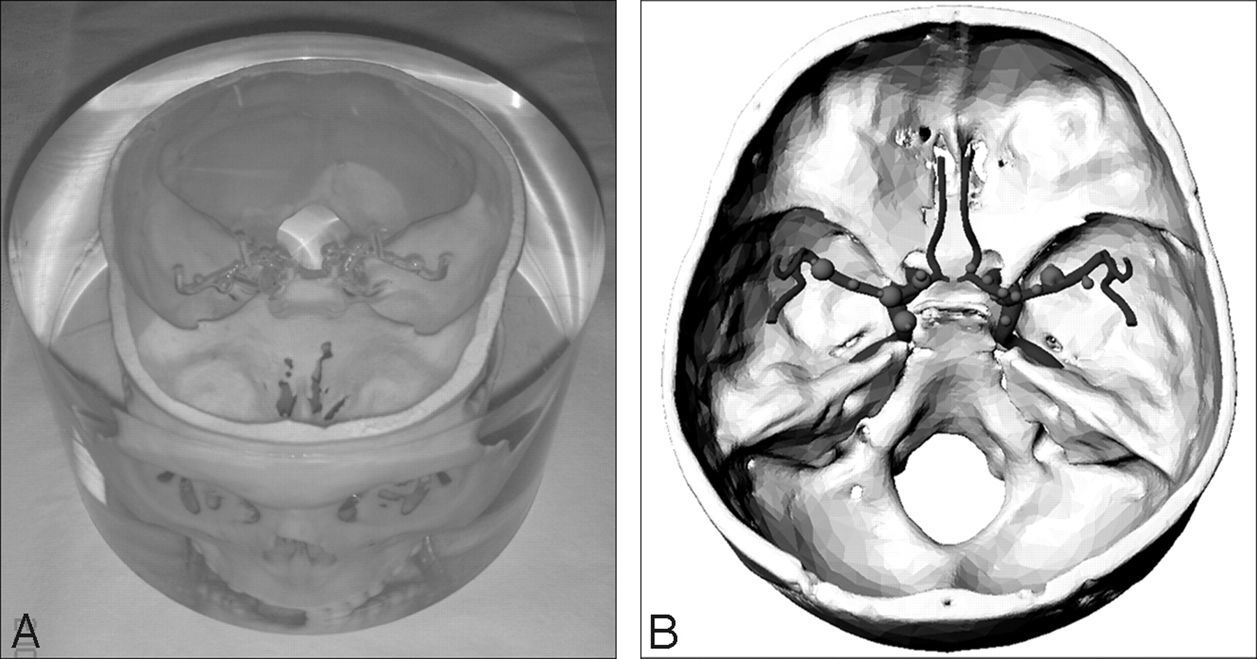
- Fig 1.
Photograph (A) and schematic drawing (B) of the anthropomorphic vascular phantom used in this study.
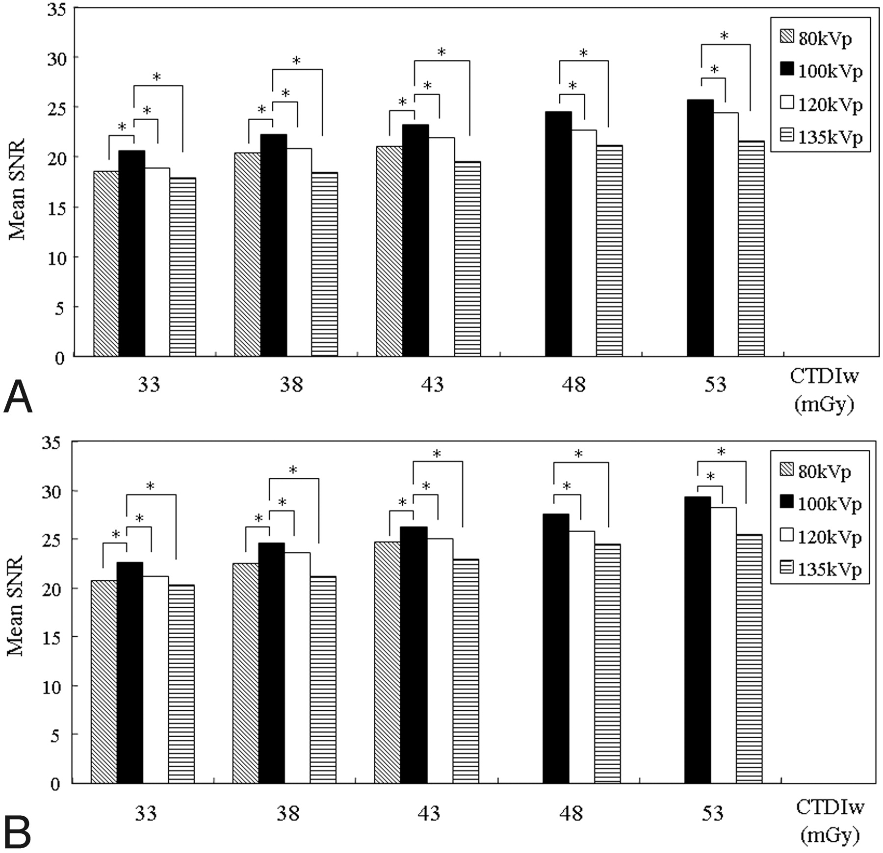
- Fig 2.
Graphs show the mean SNRs obtained at contrast material concentrations of 15 (A) and 20 mg I/mL (B). Asterisks show statistically significant differences at identical CTDIWs compared with the values of the standard CT image at 100 kV(p). At identical dose levels, the mean SNRs at 100 kV(p) from contrast material concentrations of 15 and 20 mg I/mL are significantly higher than those at 80, 120, and 135 kV(p) (P < .05).
- Fig 3.
Graphs show the mean CNRs obtained at contrast material concentrations of 15 (A) and 20 mg I/mL (B). Asterisks show statistically significant differences at identical CTDIWs, compared with the values of the standard CT image at 100 kV(p). At identical dose levels, the mean CNRs at 100 kV(p) from contrast material concentrations of 15 and 20 mg I/mL are significantly higher than those at 80, 120, and 135 kV(p) (P < .05).
- Fig 4.
CT angiograms at a concentration of 20 mg I/mL and at identical dose levels (CTDIw of 38 mGy). A, Tube voltage is 80 kV(p). B, 100 kV(p). C, 120 kV(p). D, 135 kV(p). For the mean qualitative image score with regard to the depiction of the simulated intracranial aneurysms (arrows) and aneurysmal blebs (arrowheads) on CT angiograms, 100 kV(p) is significantly superior to 80 and 135 kV(p) and is slightly superior to 120 kV(p).


Tables
kV(p) mA CTDIw (mGy) 80 360 33 420 38 470 43 100 210 33 240 38 270 43 300 48 330 53 120 140 33 160 38 180 43 200 48 220 53 135 110 33 120 38 130 43 140 48 150 53 CTDIw (mGy) 15 mg I/mL 20 mg I/mL 80 kV(p) 100 kV(p) 120 kV(p) 135 kVp 80 kV(p) 100 kV(p) 120 kV(p) 135 kV(p) 33 34.8 ± 0.18 24.2 ± 0.24 21.1 ± 0.18 20.0 ± 0.20 35.3 ± 0.14 24.9 ± 0.03 21.7 ± 0.08 20.0 ± 0.16 38 31.4 ± 0.26 22.3 ± 0.34 19.4 ± 0.27 19.3 ± 0.12 32.9 ± 0.28 23.0 ± 0.07 19.8 ± 0.14 19.2 ± 0.02 43 29.3 ± 0.13 21.5 ± 0.14 18.6 ± 0.08 18.0 ± 0.15 29.7 ± 0.28 21.6 ± 0.28 18.6 ± 0.07 17.7 ± 0.02 48 20.2 ± 0.16 17.7 ± 0.19 16.9 ± 0.16 20.5 ± 0.28 17.9 ± 0.01 16.6 ± 0.02 53 19.4 ± 0.11 16.6 ± 0.21 16.3 ± 0.13 19.4 ± 0.13 16.5 ± 0.16 15.9 ± 0.02 -
a Data are the mean ± SD.
-
- Table 3:
Mean qualitative image scores for the depiction of simulated aneurysms and blebsa
CTDIw (mGy) 15 mg I/mL 20 mg I/mL 80 kV(p) 100 kV(p) 120 kV(p) 135 kV(p) 80 kV(p) 100 kV(p) 120 kV(p) 135 kV(p) 33 1.86 ± 0.54b 2.86 ± 0.66 2.29 ± 0.72b 1.79 ± 0.58b 3.07 ± 0.62b 3.57 ± 0.65 3.36 ± 0.63 2.71 ± 0.47b 38 2.07 ± 0.73b 3.07 ± 0.62 2.57 ± 0.85b 2.21 ± 0.80b 3.29 ± 0.61b 3.79 ± 0.58 3.57 ± 0.51 2.92 ± 0.61b 43 2.57 ± 0.65b 3.14 ± 0.66 2.85 ± 0.77b 2.28 ± 0.82b 3.50 ± 0.65b 3.92 ± 0.48 3.85 ± 0.66 3.07 ± 0.62b 48 3.36 ± 0.63 3.29 ± 0.61 2.71 ± 0.91b 4.07 ± 0.48 4.00 ± 0.55 3.29 ± 0.61b 53 3.71 ± 0.61 3.57 ± 0.51 2.86 ± 0.77b 4.43 ± 0.65 4.21 ± 0.70 3.57 ± 0.51b -
a Data are the mean ± SD.
-
b Statistically significant difference at identical CTDIws, compared with the value of the standard CT image at 100 kV(p).
-
CTDIw (mGy) 15 mg I/mL 20 mg I/mL 80 kV(p) 100 kV(p) 120 kV(p) 135 kV(p) 80 kV(p) 100 kV(p) 120 kV(p) 135 kV(p) 33 2.45 ± 0.16 2.70 ± 0.11 2.63 ± 0.14 2.65 ± 0.17 2.63 ± 0.14 2.73 ± 0.17 2.73 ± 0.16 2.65 ± 0.18 38 2.68 ± 0.13 2.81 ± 0.16 2.70 ± 0.18 2.64 ± 0.21 2.69 ± 0.21 2.89 ± 0.29 2.70 ± 0.14 2.69 ± 0.20 43 2.75 ± 0.21 2.80 ± 0.08 2.85 ± 0.13 2.70 ± 0.22 2.85 ± 0.18 2.93 ± 0.14 2.88 ± 0.17 2.73 ± 0.17 48 2.88 ± 0.10 2.90 ± 0.13 2.73 ± 0.17 2.96 ± 0.06 2.95 ± 0.09 2.89 ± 0.26 53 2.88 ± 0.12 2.89 ± 0.10 2.76 ± 0.20 2.96 ± 0.04 2.93 ± 0.16 2.86 ± 0.19 -
a Data are the mean ± SD.
-


