
Article Figures & Data
Figures
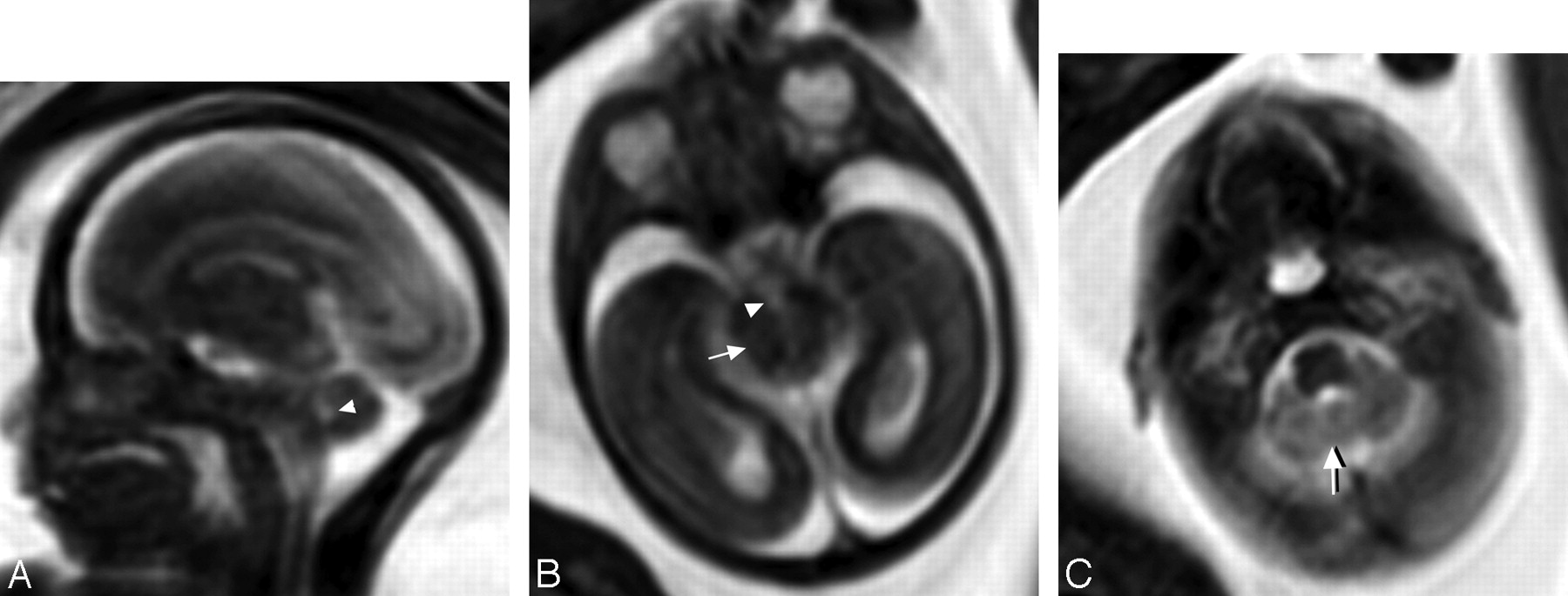
- Fig 1.
Female fetus at 20 weeks of gestation at risk of JSRD with normal brain in fetal MR imaging and a normal postnatal outcome. A, Prenatal midline sagittal MR image shows a normal fourth ventricle (arrowhead). The midline cerebellum appears intact and the CM is not dilated. B, Prenatal axial MR image through the level of the lower midbrain shows that the AP diameter of the IP fossa (arrowhead) is smaller than that of the midbrain (isthmus) (arrow). C, Prenatal axial MR image at the level of the pons shows a normal-sized fourth ventricle. Midline vermian tissues appear between the 2 cerebellar hemispheres (arrow). It is difficult to identify the normal superior cerebellar peduncles because of their small size and their downward oblique orientation
- Fig 2.
Female fetus at 22 weeks of gestation shows abnormal brain in MR imaging suggestive of JSRD. A, Prenatal axial MR image at the level of the pontomesencephalic junction shows the interpeduncular cistern (black arrow) deeper and wider than normal. The midbrain isthmus is abnormally narrowed in its AP diameter. The thick horizontal superior cerebellar peduncles (white arrow) represent the roots of the tooth for the MTS diagnostic of JSRD. There is dilation of the fourth ventricle (arrowhead) with a deformed anteriorly convex floor. B, Prenatal axial MR image at the level of the pons shows a midline sagittal CSF-containing cleft (arrow) separating the 2 cerebellar hemispheres denoting absent vermian tissues. C, Prenatal midsagittal MR image shows dilated CM, dilated fourth ventricle with rounding of its roof (arrowhead), and migration of the cerebellar hemispheres to the midline due to a hypoplastic/dysgenetic vermis. D, Prenatal coronal MR image shows the cerebellar hemispheres separated in the midline by sagittal cleft-containing CSF (arrowhead) caused by dysgenesis/agenesis of the vermis. E, Postnatal axial T1-weighted MR image at the age of 4 months documents JSRD diagnosis by showing a mild MTS (arrowhead).
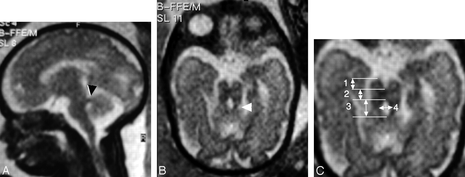
- Fig 3.
Male fetus at 30 weeks of gestation with abnormal brain MR imaging findings suggestive of JSRD. A, Prenatal sagittal MR image shows thick superior cerebellar peduncles at right angles to the posterior surface of the brain stem (arrowhead) suggesting JSRD. Note that the CM is not dilated. B, Prenatal axial MR image at the pontomesencephalic junction shows MTS (arrowhead). C, Measurements obtained in axial prenatal MR image at the lower midbrain/pontomesencephalic junction: the AP diameter of the interpeduncular fossa (no. 1), AP diameter of midbrain/isthmus (no. 2), as well as the AP (no. 3) and transverse (no. 4) diameters of the fourth ventricle. Note that the AP diameter of the interpeduncular fossa is almost equal to that of the narrow isthmus and the AP diameter of the superior aspect of the fourth ventricle is longer than its transverse diameter


