
Article Figures & Data
Figures
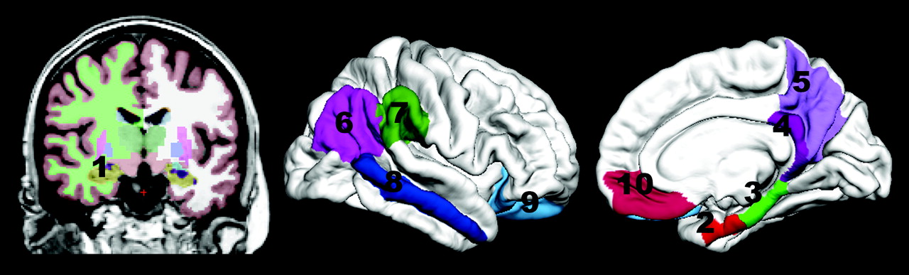
- Fig 1.
The regions of interest used are the following: 1) hippocampus and 2) entorhinal, 3) parahippocampal, 4) retrosplenial, 5) precuneus, 6) inferior parietal, 7) supramarginal, 8) middle temporal, 9) lateral orbitofrontal, and 10) medial orbitofrontal cortices.
- Fig 2.
Comparison of ROC curves for using 1 versus a combination of 2 and all 3 variables shown to be unique predictors of NC-versus-AD classification. Yellow is the predicted probability based on hippocampal volume alone (AUC = 0.900, SE = 0.033). Blue is the predicted probability based on hippocampal volume and t-tau/Aβ42 ratio (AUC = 0.950, SE = 0.022). Red is the predicted probability based on hippocampal volume, t-tau/Aβ42 ratio, and retrosplenial cortical thickness (AUC = 0.961, SE = 0.018).
- Fig 3.
The regression plots for 2-year change in scores in the MCI group significantly (P < .05) predicted from MR imaging morphometry and PET metabolism variables. A, CDR change predicted from retrosplenial cortical thickness. B and C, MMSE change predicted from retrosplenial cortical metabolism (B) and retrosplenial cortical thickness (C). D, Delayed logical memory change predicted from hippocampal volume.


Tables
NC (n = 42; 16F/26M) MCI (n = 73, 25F/48M) AD (n = 38, 16F/22M) M SD Range M SD Range M SD Range Age 75.5 (5.4) 62.2–84.7 74.5 (7.0) 55.5–88.9 76.2 (7.5) 58.8–88.1 Education 16.0 (3.2) 8–20 16.0 (2.9) 8–20 14.3 (3.6) 4–20 MMSE 29.1 (1.0) 26–30 27.0 (1.7) 24–30 23.8 (2.0) 20–26 MMSE_c −0.2 (1.6) −4–3 −1.3 (2.8) −13–4 −5.2 (5.8) −22–4 CDR 0.0 (0.0) 0–0 0.5 (0.0) 0.5–0.5 0.8 (0.3) 0.5–1.0 CDR_c 0.2 (0.7) −0.5–3.5 1.2 (1.6) −1.5–4.5 4.0 (3.1) 0–11 LM-del 12.0 (3.6) 6–22 4.1 (2.7) 0–8 1.1 (2.0) 0–8 LM-del_c 1.2 (4.1) −10–8 0 (3.3) −6–10 −0.7 (1.1) −4–1 a The numbers refer to baseline data, with the exception of MMSE_c, CDR_c, and LM-del_c, which refer to change across 2 years (baseline score subtracted from score at 2-year follow-up). MMSE and LM-del change scores were available for 36 NC, 51 MCI, and 25 AD subjects. CDR-SB change scores were available for 34 NC, 49 MCI, and 25 AD.
- Table 2:
Results from logistic regression analyses for each method predicting NC versus AD
Method Step Measure B P Odds Ratio % Corr. Class. R2a MRI 1 Hippocampus −2.306 .000 .100 NC: 83.3 .601 AD: 81.6 All: 82.5 2 Hippocampus −2.291 .000 .101 NC: 88.1 .665 Retrosplenial cortex −1.202 .014. .301 AD: 78.9 All: 83.8 3 Hippocampus −1.581 .011 .206 NC: 85.7 .714 Entorhinal cortex −1.314 .026 .269 AD: 84.2 Retrosplenial cortex −1.230 .024 .292 All: 85.0 PET 1 Entorhinal cortex −1.627 .000 .197 NC: 85.7 .461 AD: 73.7 All: 80.0 2 Entorhinal cortex −2.142 .000 .117 NC: 81.0 .506 Lateral orbitofrontal cortex .675 .048 1.964 AD: 76.3 All: 78.8 3 Entorhinal cortex −2.094 .000 .123 NC: 88.1 .620 Retrosplenial cortex −1.866 .003 .155 AD: 76.3 Lateral orbitofrontal cortex 1.701 .002 5.481 All: 82.5 CSF 1 t-τ:Aβ42 2.775 .000 16.036 NC: 85.7 .523 AD: 76.3 All: 81.2 a R2 is Nagelkerke R2.
- Table 3:
Results from the multimodal logistic regression analyses predicting NC versus ADa
Step Measure B P Odds Ratio % Corr. Class R2 1 MRI hippocampus −2.306 .000 .100 NC: 83.3 .601 AD: 81.6 All: 82.5 2 MRI hippocampus −2.029 .000 .132 NC: 88.1 .733 t-τ:Aβ42 2.141 .001 8.509 AD: 81.6 All: 85.0 3 MRI hippocampus −1.861 .002 .155 NC: 90.5 .778 MRI retrosplenial −1.239 .028 .290 AD: 86.8 t-τ:Aβ42 2.411 .002 11.140 All: 88.8 NC vs MCI 1 MR hippocampus −1.360 .000 .257 NC: 54.8 .312 MCI: 80.8 All: 71.3 2 MR hippocampus −1.124 .000 .325 NC: 64.3 .399 t-τ:Aβ42 1.422 .006 4.146 MCI: 87.7 All: 79.1 a The variables explaining unique variance within each method, as listed in Table 2, were included in the set of predictor variables, i.e. for MR: hippocampal volume, retrosplenial, and entorhinal thickness; for PET: entorhinal, retrosplenial, and lateral orbitofrontal metabolism; and for CSF: the ratio of T-tau to Abeta 42. R2 is Nagelkerke R2.
- Table 4:
Correlations between the variables included in the regression models predicting NC/AD classification and the change in CDR-SB (n = 49) and MMSE (n = 51) scores across 2 years in the MCI groupa
CDR-SB Change MMSE Change LM-Del Change MRI hippocampus −.29 .29 .41b MRI entorhinal −.17 .23 .34 MRI retrosplenial −.43b .42b .35 PET entorhinal −.30 .38b .28 PET retrosplenial −.22 .47b .11 PET lat. orbitofrontal −.02 .27 −.05 T-τ:Aβ42 .02 .08 −.23 a The variables explaining the unique variance within each method, as listed in Table 2, were included in the set of predictor variables (ie, for MR imaging, hippocampal volume and retrosplenial and entorhinal thickness; for PET, entorhinal, retrosplenial, and lateral orbitofrontal metabolism; and for CSF, the ratio of t-τ:Aβ42).
b P < .05, corrected for 7 comparisons.
In this issue


Jump to section
Related Articles
Cited By...
- AD plasma biomarkers are stable for an extended period at -20{degrees}C: implications for resource-constrained environments
- A reusable benchmark of brain-age prediction from M/EEG resting-state signals
- Structural Covariance of the Default Network in Healthy and Pathological Aging
- MRI and cerebrospinal fluid biomarkers for predicting progression to Alzheimer's disease in patients with mild cognitive impairment: a diagnostic accuracy study
- Application of the National Institute on Aging-Alzheimer's Association AD criteria to ADNI
- Clinical Utility and Analytical Challenges in Measurement of Cerebrospinal Fluid Amyloid-{beta}1-42 and {tau} Proteins as Alzheimer Disease Biomarkers
- Can MRI screen for CSF biomarkers in neurodegenerative disease?
- Imaging Approaches for Dementia
- White matter imaging contributes to the multimodal diagnosis of frontotemporal lobar degeneration
- The Mitochondria-Targeted Antioxidant MitoQ Prevents Loss of Spatial Memory Retention and Early Neuropathology in a Transgenic Mouse Model of Alzheimer's Disease
- Comparing predictors of conversion and decline in mild cognitive impairment