
Article Figures & Data
Figures
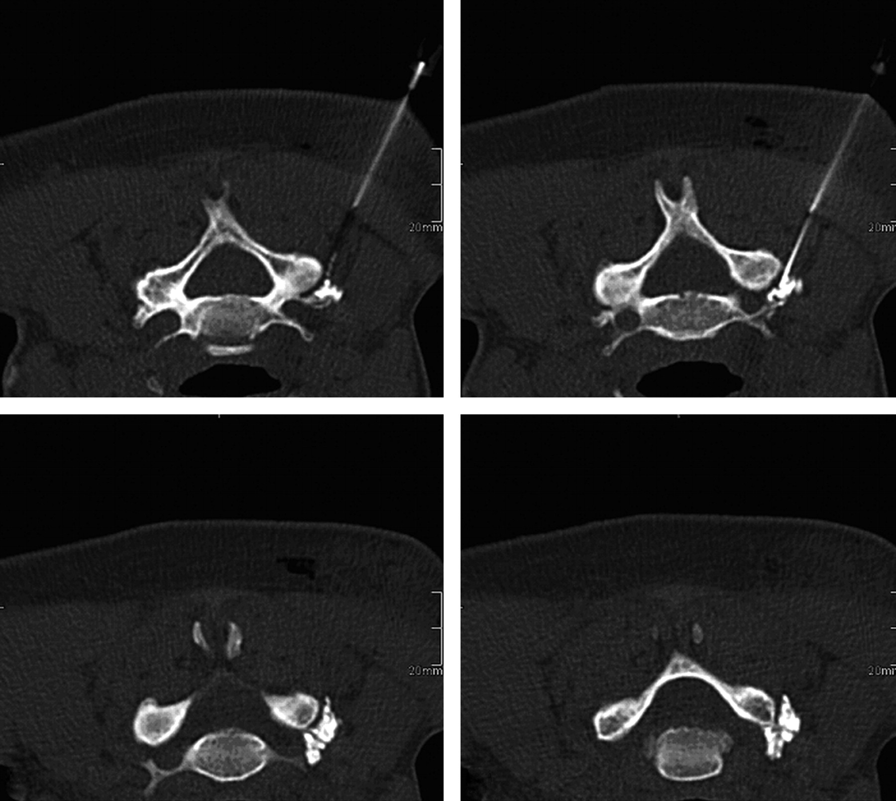
- Fig 1.
Extraforaminal contrast distribution after CSNRB of the right C6 nerve root.
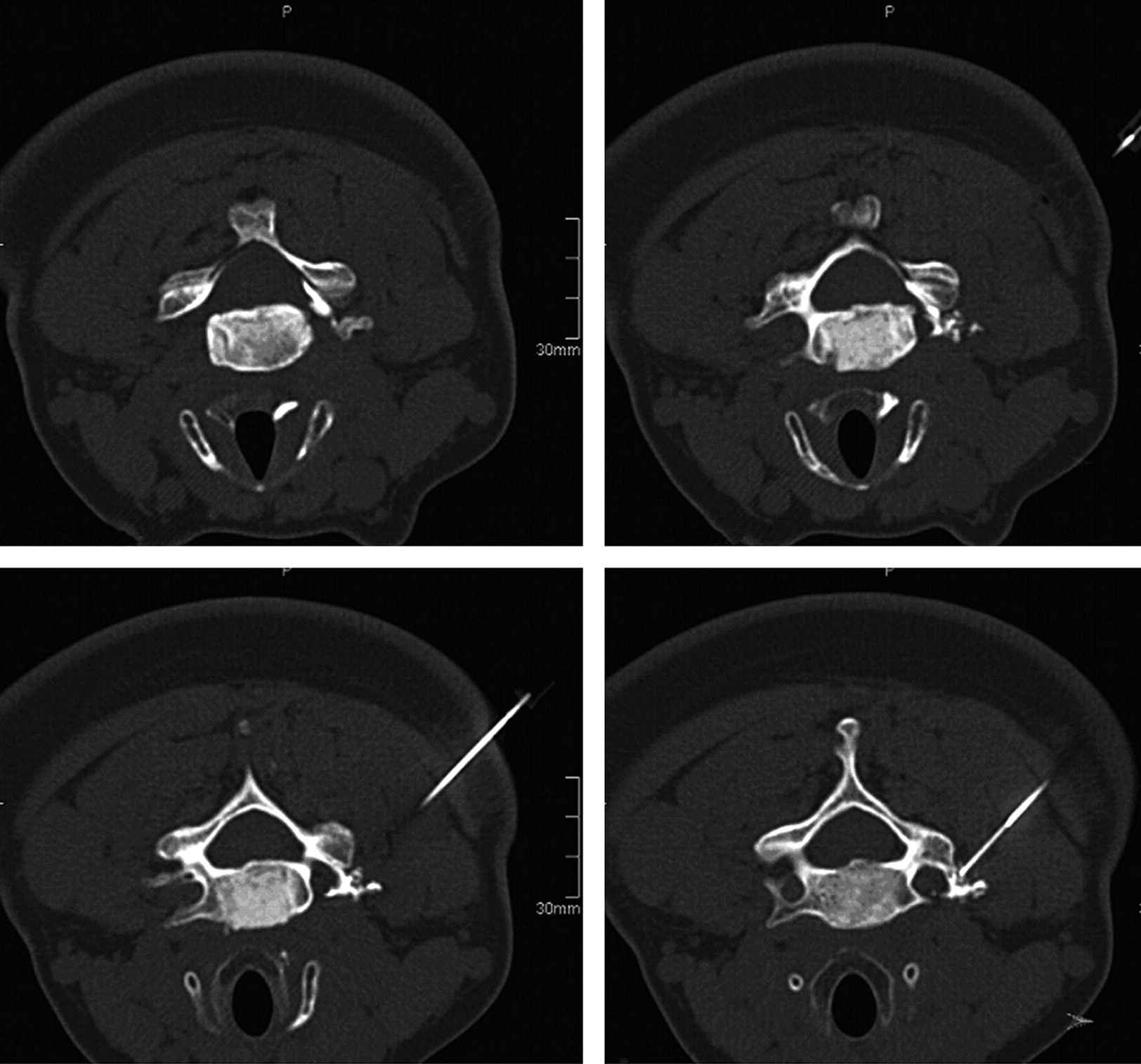
- Fig 2.
Extra- and intraforaminal contrast distribution after CSNRB of the right C6 nerve root.
- Fig 3.
Schematic illustration of the needle tip and entrance of the foramen. 1 indicates the facet joint; 2, the uncinate process; 3, the line reaching from the anterior part of the uncinate process to the anterior part of the zygapophyseal joint; 4, the area in which the needle tip in the dorsal-approach technique is expected; and 5, the area in which the nerve root and nerve supplying the artery are expected.


Tables
Number of different nerve root blocksa
Nerve Root All Indications Diagnostic Indication Therapeutic Indication No. + − No. + − No. + − C3 1 1 0 1 1 0 0 0 0 C4 4 3 1 3 2 1 1 1 0 C5 3 1 2 3 1 2 0 0 0 C6 22 16 6 14 9 5 8 7 1 C7 13 10 2 12 9 3 1 1 0 C8 10 9 1 5 4 1 5 5 0 Σ 53 40 13 38 26 12 15 14 1 -
a + indicates evaluated as a positive block; −, evaluated as a negative block.
-
In this issue


Jump to section
Related Articles
Cited By...
- CT-Fluoroscopic Cervical Transforaminal Epidural Steroid Injections: Extraforaminal Needle Tip Position Decreases Risk of Intravascular Injection
- Ultrasound-Guided Lower Cervical Nerve Root Injectate Volumes Associated With Dorsal Root Ganglion and Epidural Spread
- Incidence of Inadvertent Intravascular Injection during CT Fluoroscopy-Guided Epidural Steroid Injections
- Lateral Decubitus Positioning for Cervical Nerve Root Block Using CT Image Guidance Minimizes Effective Radiation Dose and Procedural Time
- Safety and Efficacy of CT-Guided Transforaminal Cervical Epidural Steroid Injections Using a Posterior Approach