Advertisement
  • Main menu
  • User menu
  • Search
  • English ▼
    • English
    • Afrikaans
    • Albanian
    • Amharic
    • Arabic
    • Armenian
    • Azerbaijani
    • Basque
    • Belarusian
    • Bengali
    • Bosnian
    • Bulgarian
    • Catalan
    • Cebuano
    • Chichewa
    • Chinese (Simplified)
    • Chinese (Traditional)
    • Corsican
    • Croatian
    • Czech
    • Danish
    • Dutch
    • Esperanto
    • Estonian
    • Filipino
    • Finnish
    • French
    • Frisian
    • Galician
    • Georgian
    • German
    • Greek
    • Gujarati
    • Haitian Creole
    • Hausa
    • Hawaiian
    • Hebrew
    • Hindi
    • Hmong
    • Hungarian
    • Icelandic
    • Igbo
    • Indonesian
    • Irish
    • Italian
    • Japanese
    • Javanese
    • Kannada
    • Kazakh
    • Khmer
    • Korean
    • Kurdish (Kurmanji)
    • Kyrgyz
    • Lao
    • Latin
    • Latvian
    • Lithuanian
    • Luxembourgish
    • Macedonian
    • Malagasy
    • Malay
    • Malayalam
    • Maltese
    • Maori
    • Marathi
    • Mongolian
    • Myanmar (Burmese)
    • Nepali
    • Norwegian
    • Pashto
    • Persian
    • Polish
    • Portuguese
    • Punjabi
    • Romanian
    • Russian
    • Samoan
    • Scottish Gaelic
    • Serbian
    • Sesotho
    • Shona
    • Sindhi
    • Sinhala
    • Slovak
    • Slovenian
    • Somali
    • Spanish
    • Sudanese
    • Swahili
    • Swedish
    • Tajik
    • Tamil
    • Telugu
    • Thai
    • Turkish
    • Ukrainian
    • Urdu
    • Uzbek
    • Vietnamese
    • Welsh
    • Xhosa
    • Yiddish
    • Yoruba
    • Zulu

Main menu

  • Home
  • Content
    • Current Issue
    • Accepted Manuscripts
    • Article Preview
    • Past Issue Archive
    • Video Articles
    • AJNR Case Collection
    • Case of the Week Archive
    • Case of the Month Archive
    • Classic Case Archive
  • Special Collections
    • Advancing NeuroMRI with High-Relaxivity Contrast Agents
    • AJNR Awards
    • Low-Field MRI
    • Alzheimer Disease
    • ASNR Foundation Special Collection
    • Photon-Counting CT
    • View All
  • Multimedia
    • AJNR Podcasts
    • AJNR SCANtastic
    • Trainee Corner
    • MRI Safety Corner
    • Imaging Protocols
  • For Authors
    • Submit a Manuscript
    • Submit a Video Article
    • Submit an eLetter to the Editor/Response
    • Manuscript Submission Guidelines
    • Statistical Tips
    • Fast Publishing of Accepted Manuscripts
    • Graphical Abstract Preparation
    • Imaging Protocol Submission
    • Author Policies
  • About Us
    • About AJNR
    • Editorial Board
    • Editorial Board Alumni
  • More
    • Become a Reviewer/Academy of Reviewers
    • Subscribers
    • Permissions
    • Alerts
    • Feedback
    • Advertisers
    • ASNR Home

User menu

  • Alerts
  • Log in

Search

  • Advanced search
American Journal of Neuroradiology

American Journal of Neuroradiology

ASHNR American Society of Functional Neuroradiology ASHNR American Society of Pediatric Neuroradiology ASSR
  • Alerts
  • Log in

Advanced Search

English ▼
  • English
  • Afrikaans
  • Albanian
  • Amharic
  • Arabic
  • Armenian
  • Azerbaijani
  • Basque
  • Belarusian
  • Bengali
  • Bosnian
  • Bulgarian
  • Catalan
  • Cebuano
  • Chichewa
  • Chinese (Simplified)
  • Chinese (Traditional)
  • Corsican
  • Croatian
  • Czech
  • Danish
  • Dutch
  • Esperanto
  • Estonian
  • Filipino
  • Finnish
  • French
  • Frisian
  • Galician
  • Georgian
  • German
  • Greek
  • Gujarati
  • Haitian Creole
  • Hausa
  • Hawaiian
  • Hebrew
  • Hindi
  • Hmong
  • Hungarian
  • Icelandic
  • Igbo
  • Indonesian
  • Irish
  • Italian
  • Japanese
  • Javanese
  • Kannada
  • Kazakh
  • Khmer
  • Korean
  • Kurdish (Kurmanji)
  • Kyrgyz
  • Lao
  • Latin
  • Latvian
  • Lithuanian
  • Luxembourgish
  • Macedonian
  • Malagasy
  • Malay
  • Malayalam
  • Maltese
  • Maori
  • Marathi
  • Mongolian
  • Myanmar (Burmese)
  • Nepali
  • Norwegian
  • Pashto
  • Persian
  • Polish
  • Portuguese
  • Punjabi
  • Romanian
  • Russian
  • Samoan
  • Scottish Gaelic
  • Serbian
  • Sesotho
  • Shona
  • Sindhi
  • Sinhala
  • Slovak
  • Slovenian
  • Somali
  • Spanish
  • Sudanese
  • Swahili
  • Swedish
  • Tajik
  • Tamil
  • Telugu
  • Thai
  • Turkish
  • Ukrainian
  • Urdu
  • Uzbek
  • Vietnamese
  • Welsh
  • Xhosa
  • Yiddish
  • Yoruba
  • Zulu
  • Home
  • Content
    • Current Issue
    • Accepted Manuscripts
    • Article Preview
    • Past Issue Archive
    • Video Articles
    • AJNR Case Collection
    • Case of the Week Archive
    • Case of the Month Archive
    • Classic Case Archive
  • Special Collections
    • Advancing NeuroMRI with High-Relaxivity Contrast Agents
    • AJNR Awards
    • Low-Field MRI
    • Alzheimer Disease
    • ASNR Foundation Special Collection
    • Photon-Counting CT
    • View All
  • Multimedia
    • AJNR Podcasts
    • AJNR SCANtastic
    • Trainee Corner
    • MRI Safety Corner
    • Imaging Protocols
  • For Authors
    • Submit a Manuscript
    • Submit a Video Article
    • Submit an eLetter to the Editor/Response
    • Manuscript Submission Guidelines
    • Statistical Tips
    • Fast Publishing of Accepted Manuscripts
    • Graphical Abstract Preparation
    • Imaging Protocol Submission
    • Author Policies
  • About Us
    • About AJNR
    • Editorial Board
    • Editorial Board Alumni
  • More
    • Become a Reviewer/Academy of Reviewers
    • Subscribers
    • Permissions
    • Alerts
    • Feedback
    • Advertisers
    • ASNR Home
  • Follow AJNR on Twitter
  • Visit AJNR on Facebook
  • Follow AJNR on Instagram
  • Join AJNR on LinkedIn
  • RSS Feeds

AJNR Awards, New Junior Editors, and more. Read the latest AJNR updates


Improved Turnaround Times | Median time to first decision: 12 days

Research ArticleFunctional

Magnetization Transfer MR Imaging Demonstrates Degeneration of the Subcortical and Cortical Gray Matter in Huntington Disease

A. Ginestroni, M. Battaglini, S. Diciotti, R. Della Nave, L.N. Mazzoni, C. Tessa, M. Giannelli, S. Piacentini, N. De Stefano and M. Mascalchi
American Journal of Neuroradiology November 2010, 31 (10) 1807-1812; DOI: https://doi.org/10.3174/ajnr.A2225
A. Ginestroni
  • Find this author on Google Scholar
  • Find this author on PubMed
  • Search for this author on this site
M. Battaglini
  • Find this author on Google Scholar
  • Find this author on PubMed
  • Search for this author on this site
S. Diciotti
  • Find this author on Google Scholar
  • Find this author on PubMed
  • Search for this author on this site
R. Della Nave
  • Find this author on Google Scholar
  • Find this author on PubMed
  • Search for this author on this site
L.N. Mazzoni
  • Find this author on Google Scholar
  • Find this author on PubMed
  • Search for this author on this site
C. Tessa
  • Find this author on Google Scholar
  • Find this author on PubMed
  • Search for this author on this site
M. Giannelli
  • Find this author on Google Scholar
  • Find this author on PubMed
  • Search for this author on this site
S. Piacentini
  • Find this author on Google Scholar
  • Find this author on PubMed
  • Search for this author on this site
N. De Stefano
  • Find this author on Google Scholar
  • Find this author on PubMed
  • Search for this author on this site
M. Mascalchi
  • Find this author on Google Scholar
  • Find this author on PubMed
  • Search for this author on this site
  • Article
  • Figures & Data
  • Info & Metrics
  • Responses
  • References
  • PDF
Loading

Article Figures & Data

Figures

  • Tables
  • Fig 1.
    • Download figure
    • Open in new tab
    • Download powerpoint
    Fig 1.

    A–H, Example of the FIRST segmentation of the striata and thalami (light blue indicates the caudate; light green, the pallidus; yellow ochre, the putamen; lilac, the thalamus) in HD gene carrier in clinical stage I (A–D), which was manually edited on the basis of the structure contour identified in the subject's heavily T1-weighted images (E–H). A–D, The automatic definition of the cortical GM ribbon performed by the in-house-developed software is also displayed with different colors corresponding to the 4 cerebral lobes (yellow indicates frontal; green, parietal-insular; dark blue, temporal; red, occipital).
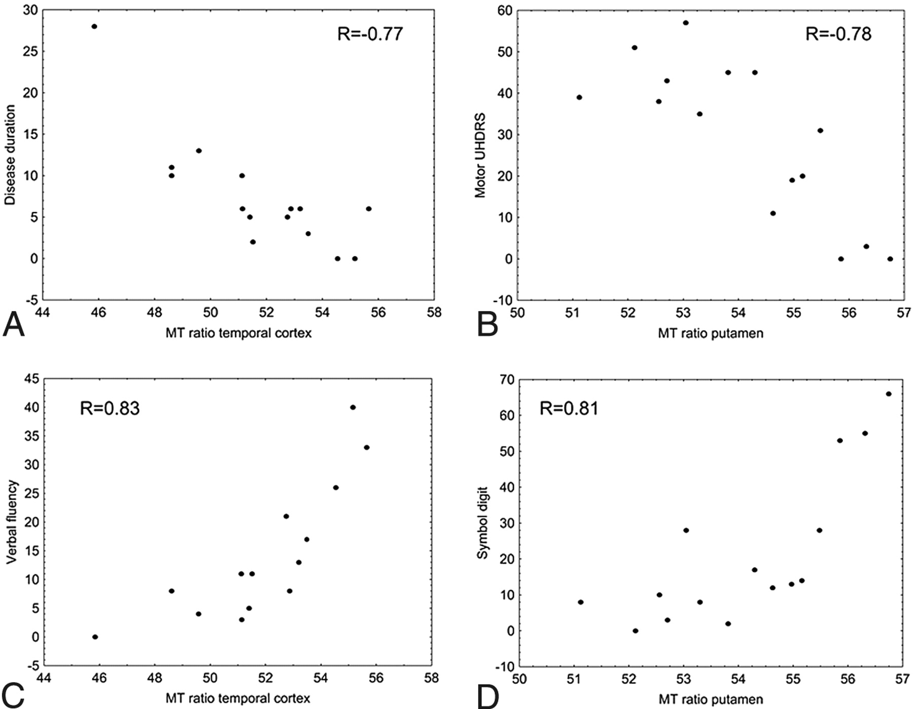
  • Fig 2.
    • Download figure
    • Open in new tab
    • Download powerpoint
    Fig 2.

    Plots showing correlation between clinical variables (disease duration, motor UHDRS, Verbal Fluency test, and Symbol Digit test) and MT ratio measurements in the temporal lobe cortex (A,C) and putamen (B,D).

Tables

  • Figures
    • View popup
    Table 1:

    Genetic and clinical features in 15 HD gene carriers

    MeanRange
    Age (yr)56 ± 1234–75
    CAG repeat size44 ± 340–50
    Sex (M:F)8:7
    Disease duration (yr)6.9 ± 6.60–28
    UHDRS motor score33 ± 180–57
    Verbal Fluency test13 ± 113–40
    Symbol Digit test19 ± 190–66
    Stroop Color Word Interference Test25 ± 343–120
    • View popup
    Table 2:

    Volumes and MT ratios of the striatal nuclei and thalami in 15 healthy controls and 15 HD carriers

    Mean Volume (mm3)Mean MT Ratio
    ControlsHD carriersP ValueaControlsHD CarriersP Valuea
    Caudate nuclei6221 ± 8063048 ± 1834<.00155.0 ± 0.951.8 ± 3.2<.01
    Putamen7222 ± 8864374 ± 1590<.00155.4 ± 0.754.1 ± 1.6NS
    Globi pallidi3061 ± 3211513 ± 553<.00156.9 ± 0.855.4 ± 1.5.02
    Thalami10551 ± 11979802 ± 1908NS58.6 ± 0.756.8 ± 1.9<.01
    • a Mann-Whitney U test with Bonferroni correction.

    • View popup
    Table 3:

    Mean normalized volumes and MT ratios of the cerebral cortical GM in 15 healthy controls and 15 HD carriers

    LobesMean Normalized Volume (mm3)Mean MT Ratio
    ControlsHD CarriersP ValueaControlsHD CarriersP Valuea
    Frontal202 ± 16179 ± 19<.0155.1 ± 0.950.7 ± 3.5<.001
    Parietal161 ± 15135 ± 20<.0154.5 ± 0.851.4 ± 2.9<.01
    Temporal143 ± 11128 ± 13.0254.9 ± 0.851.7 ± 2.7<.01
    Occipital91 ± 1072 ± 10<.0153.2 ± 1.348.2 ± 3.4<.01
    • a Mann-Whitney U test with Bonferroni correction.

    • View popup
    Table 4:

    Spearman rank correlation coefficients between the clinical-genetic variables and volumes and MT ratios in 15 HD carriers

    No. of TripletsDisease DurationMotor UHDRSVerbal FluencySymbol DigitStroop
    RPRPRPRPRPRP
    Volumes
        NWMV0.46NS−0.22NS−0.44NS0.44NS0.65<.010.54.04
        NGMV−0.16NS−0.50NS−0.67<.010.65<.010.41NS0.40NS
        Frontal cortex−0.40NS−0.60.02−0.59.020.65<.010.33NS0.31NS
        Parietal cortex−0.38NS−0.67<.01−0.60.020.74<.010.37NS0.37NS
        Temporal cortex−0.32NS−0.45NS−0.62.010.60.020.34NS0.45NS
        Occipital cortex−0.07NS−0.36NS−0.66<.010.77<.0010.61.010.68<.01
        Caudate nuclei−0.16NS−0.55.03−0.80<.0010.65<.010.53.040.46NS
        Putamen−0.24NS−0.59.02−0.78<.0010.59.020.50NS0.49NS
        Globi pallidi−0.14NS−0.63.01−0.79<.0010.51.04960.46NS0.37NS
        Thalami−0.24NS−0.34NS−0.71<.010.57.030.51NS0.62.01
    MT ratios
        WM−0.14NS−0.72<.01−0.81<.0010.73<.010.64.010.64.01
        GM−0.25NS−0.68<.01−0.81<.0010.88<.00010.57.040.58.03
        Frontal cortex−0.34NS−0.65<.01−0.55.030.61.020.26NS0.23NS
        Parietal cortex−0.26NS−0.66<.01−0.64.010.63.010.34NS0.30NS
        Temporal cortex−0.05NS−0.77<.001−0.77<.0010.83<.0010.58.020.49NS
        Occipital cortex−0.36NS−0.61.02−0.71<0.010.74<.010.39NS0.41NS
        Caudate nuclei0.02NS−0.60.02−0.23NS0.35NS0.21NS0.10NS
        Putamen0.24NS−0.58.02−0.78<.0010.72<.010.81<.0010.72<.01
        Globi pallidi0.05NS−0.29NS0.55.030.46NS0.51NS0.48NS
        Thalami0.12NS−0.56.03−0.30NS0.30NS0.22NS0.09NS
    • Note:—Bold indicates statistically significant data.

PreviousNext
Back to top

In this issue

American Journal of Neuroradiology: 31 (10)
American Journal of Neuroradiology
Vol. 31, Issue 10
1 Nov 2010
  • Table of Contents
  • Index by author
Advertisement
Print
Download PDF
Email Article
Cite this article
0 Responses
Respond to this article
Share
Bookmark this article
Magnetization Transfer MR Imaging Demonstrates Degeneration of the Subcortical and Cortical Gray Matter in Huntington Disease
A. Ginestroni, M. Battaglini, S. Diciotti, R. Della Nave, L.N. Mazzoni, C. Tessa, M. Giannelli, S. Piacentini, N. De Stefano, M. Mascalchi
American Journal of Neuroradiology Nov 2010, 31 (10) 1807-1812; DOI: 10.3174/ajnr.A2225
del.icio.us logo Twitter logo Facebook logo Mendeley logo
  • Tweet Widget
Purchase

Jump to section

  • Article
    • Abstract
    • Abbreviations
    • Materials and Methods
    • Results
    • Discussion
    • Footnotes
    • References
  • Figures & Data
  • Info & Metrics
  • Responses
  • References
  • PDF

Related Articles

  • No related articles found.
  • PubMed
  • Google Scholar

Cited By...

  • Magnetization Transfer Imaging in Premanifest and Manifest Huntington Disease: A 2-Year Follow-Up
  • Magnetization Transfer Imaging in Premanifest and Manifest Huntington Disease
  • Magnetization Transfer Imaging Demonstrates a Distributed Pattern of Microstructural Changes of the Cerebral Cortex in Amyotrophic Lateral Sclerosis
  • Crossref
  • Google Scholar

This article has not yet been cited by articles in journals that are participating in Crossref Cited-by Linking.

More in this TOC Section

  • Kurtosis and Epileptogenic Tubers: A Pilot Study
  • Glutaric Aciduria Type 1: DK vs. Conventional MRI
  • Multiparametric MRI in PEDS Pontine Glioma
Show more Functional

Similar Articles

Advertisement

Indexed Content

  • Current Issue
  • Accepted Manuscripts
  • Article Preview
  • Past Issues
  • Editorials
  • Editor's Choice
  • Fellows' Journal Club
  • Letters to the Editor
  • Video Articles

Cases

  • Case Collection
  • Archive - Case of the Week
  • Archive - Case of the Month
  • Archive - Classic Case

More from AJNR

  • Trainee Corner
  • Imaging Protocols
  • MRI Safety Corner
  • Book Reviews

Multimedia

  • AJNR Podcasts
  • AJNR Scantastics

Resources

  • Turnaround Time
  • Submit a Manuscript
  • Submit a Video Article
  • Submit an eLetter to the Editor/Response
  • Manuscript Submission Guidelines
  • Statistical Tips
  • Fast Publishing of Accepted Manuscripts
  • Graphical Abstract Preparation
  • Imaging Protocol Submission
  • Evidence-Based Medicine Level Guide
  • Publishing Checklists
  • Author Policies
  • Become a Reviewer/Academy of Reviewers
  • News and Updates

About Us

  • About AJNR
  • Editorial Board
  • Editorial Board Alumni
  • Alerts
  • Permissions
  • Not an AJNR Subscriber? Join Now
  • Advertise with Us
  • Librarian Resources
  • Feedback
  • Terms and Conditions
  • AJNR Editorial Board Alumni

American Society of Neuroradiology

  • Not an ASNR Member? Join Now

© 2025 by the American Society of Neuroradiology All rights, including for text and data mining, AI training, and similar technologies, are reserved.
Print ISSN: 0195-6108 Online ISSN: 1936-959X

Powered by HighWire
Email this Article

Thank you for your interest in spreading the word on American Journal of Neuroradiology.

NOTE: We only request your email address so that the person you are recommending the page to knows that you wanted them to see it, and that it is not junk mail. We do not capture any email address.

Enter multiple addresses on separate lines or separate them with commas.
Magnetization Transfer MR Imaging Demonstrates Degeneration of the Subcortical and Cortical Gray Matter in Huntington Disease
(Your Name) has sent you a message from American Journal of Neuroradiology
(Your Name) thought you would like to see the American Journal of Neuroradiology web site.
CAPTCHA
This question is for testing whether or not you are a human visitor and to prevent automated spam submissions.
Cite this article
A. Ginestroni, M. Battaglini, S. Diciotti, R. Della Nave, L.N. Mazzoni, C. Tessa, M. Giannelli, S. Piacentini, N. De Stefano, M. Mascalchi
Magnetization Transfer MR Imaging Demonstrates Degeneration of the Subcortical and Cortical Gray Matter in Huntington Disease
American Journal of Neuroradiology Nov 2010, 31 (10) 1807-1812; DOI: 10.3174/ajnr.A2225

Citation Manager Formats

  • BibTeX
  • Bookends
  • EasyBib
  • EndNote (tagged)
  • EndNote 8 (xml)
  • Medlars
  • Mendeley
  • Papers
  • RefWorks Tagged
  • Ref Manager
  • RIS
  • Zotero

We use cookies on this site to enhance your user experience. By clicking any link on this page you are giving your consent for us to set cookies.