Advertisement
  • Main menu
  • User menu
  • Search
  • English ▼
    • English
    • Afrikaans
    • Albanian
    • Amharic
    • Arabic
    • Armenian
    • Azerbaijani
    • Basque
    • Belarusian
    • Bengali
    • Bosnian
    • Bulgarian
    • Catalan
    • Cebuano
    • Chichewa
    • Chinese (Simplified)
    • Chinese (Traditional)
    • Corsican
    • Croatian
    • Czech
    • Danish
    • Dutch
    • Esperanto
    • Estonian
    • Filipino
    • Finnish
    • French
    • Frisian
    • Galician
    • Georgian
    • German
    • Greek
    • Gujarati
    • Haitian Creole
    • Hausa
    • Hawaiian
    • Hebrew
    • Hindi
    • Hmong
    • Hungarian
    • Icelandic
    • Igbo
    • Indonesian
    • Irish
    • Italian
    • Japanese
    • Javanese
    • Kannada
    • Kazakh
    • Khmer
    • Korean
    • Kurdish (Kurmanji)
    • Kyrgyz
    • Lao
    • Latin
    • Latvian
    • Lithuanian
    • Luxembourgish
    • Macedonian
    • Malagasy
    • Malay
    • Malayalam
    • Maltese
    • Maori
    • Marathi
    • Mongolian
    • Myanmar (Burmese)
    • Nepali
    • Norwegian
    • Pashto
    • Persian
    • Polish
    • Portuguese
    • Punjabi
    • Romanian
    • Russian
    • Samoan
    • Scottish Gaelic
    • Serbian
    • Sesotho
    • Shona
    • Sindhi
    • Sinhala
    • Slovak
    • Slovenian
    • Somali
    • Spanish
    • Sudanese
    • Swahili
    • Swedish
    • Tajik
    • Tamil
    • Telugu
    • Thai
    • Turkish
    • Ukrainian
    • Urdu
    • Uzbek
    • Vietnamese
    • Welsh
    • Xhosa
    • Yiddish
    • Yoruba
    • Zulu

Main menu

  • Home
  • Content
    • Current Issue
    • Accepted Manuscripts
    • Article Preview
    • Past Issue Archive
    • Video Articles
    • AJNR Case Collection
    • Case of the Week Archive
    • Case of the Month Archive
    • Classic Case Archive
  • Special Collections
    • AJNR Awards
    • Low-Field MRI
    • Alzheimer Disease
    • ASNR Foundation Special Collection
    • Photon-Counting CT
    • View All
  • Multimedia
    • AJNR Podcasts
    • AJNR SCANtastic
    • Trainee Corner
    • MRI Safety Corner
    • Imaging Protocols
  • For Authors
    • Submit a Manuscript
    • Submit a Video Article
    • Submit an eLetter to the Editor/Response
    • Manuscript Submission Guidelines
    • Statistical Tips
    • Fast Publishing of Accepted Manuscripts
    • Graphical Abstract Preparation
    • Imaging Protocol Submission
    • Author Policies
  • About Us
    • About AJNR
    • Editorial Board
    • Editorial Board Alumni
  • More
    • Become a Reviewer/Academy of Reviewers
    • Subscribers
    • Permissions
    • Alerts
    • Feedback
    • Advertisers
    • ASNR Home

User menu

  • Alerts
  • Log in

Search

  • Advanced search
American Journal of Neuroradiology

American Journal of Neuroradiology

ASHNR American Society of Functional Neuroradiology ASHNR American Society of Pediatric Neuroradiology ASSR
  • Alerts
  • Log in

Advanced Search

English ▼
  • English
  • Afrikaans
  • Albanian
  • Amharic
  • Arabic
  • Armenian
  • Azerbaijani
  • Basque
  • Belarusian
  • Bengali
  • Bosnian
  • Bulgarian
  • Catalan
  • Cebuano
  • Chichewa
  • Chinese (Simplified)
  • Chinese (Traditional)
  • Corsican
  • Croatian
  • Czech
  • Danish
  • Dutch
  • Esperanto
  • Estonian
  • Filipino
  • Finnish
  • French
  • Frisian
  • Galician
  • Georgian
  • German
  • Greek
  • Gujarati
  • Haitian Creole
  • Hausa
  • Hawaiian
  • Hebrew
  • Hindi
  • Hmong
  • Hungarian
  • Icelandic
  • Igbo
  • Indonesian
  • Irish
  • Italian
  • Japanese
  • Javanese
  • Kannada
  • Kazakh
  • Khmer
  • Korean
  • Kurdish (Kurmanji)
  • Kyrgyz
  • Lao
  • Latin
  • Latvian
  • Lithuanian
  • Luxembourgish
  • Macedonian
  • Malagasy
  • Malay
  • Malayalam
  • Maltese
  • Maori
  • Marathi
  • Mongolian
  • Myanmar (Burmese)
  • Nepali
  • Norwegian
  • Pashto
  • Persian
  • Polish
  • Portuguese
  • Punjabi
  • Romanian
  • Russian
  • Samoan
  • Scottish Gaelic
  • Serbian
  • Sesotho
  • Shona
  • Sindhi
  • Sinhala
  • Slovak
  • Slovenian
  • Somali
  • Spanish
  • Sudanese
  • Swahili
  • Swedish
  • Tajik
  • Tamil
  • Telugu
  • Thai
  • Turkish
  • Ukrainian
  • Urdu
  • Uzbek
  • Vietnamese
  • Welsh
  • Xhosa
  • Yiddish
  • Yoruba
  • Zulu
  • Home
  • Content
    • Current Issue
    • Accepted Manuscripts
    • Article Preview
    • Past Issue Archive
    • Video Articles
    • AJNR Case Collection
    • Case of the Week Archive
    • Case of the Month Archive
    • Classic Case Archive
  • Special Collections
    • AJNR Awards
    • Low-Field MRI
    • Alzheimer Disease
    • ASNR Foundation Special Collection
    • Photon-Counting CT
    • View All
  • Multimedia
    • AJNR Podcasts
    • AJNR SCANtastic
    • Trainee Corner
    • MRI Safety Corner
    • Imaging Protocols
  • For Authors
    • Submit a Manuscript
    • Submit a Video Article
    • Submit an eLetter to the Editor/Response
    • Manuscript Submission Guidelines
    • Statistical Tips
    • Fast Publishing of Accepted Manuscripts
    • Graphical Abstract Preparation
    • Imaging Protocol Submission
    • Author Policies
  • About Us
    • About AJNR
    • Editorial Board
    • Editorial Board Alumni
  • More
    • Become a Reviewer/Academy of Reviewers
    • Subscribers
    • Permissions
    • Alerts
    • Feedback
    • Advertisers
    • ASNR Home
  • Follow AJNR on Twitter
  • Visit AJNR on Facebook
  • Follow AJNR on Instagram
  • Join AJNR on LinkedIn
  • RSS Feeds

AJNR Awards, New Junior Editors, and more. Read the latest AJNR updates

Research ArticleBrain
Open Access

Diffusion-Weighted MR Imaging Derived Apparent Diffusion Coefficient Is Predictive of Clinical Outcome in Primary Central Nervous System Lymphoma

R.F. Barajas, J.L. Rubenstein, J.S. Chang, J. Hwang and S. Cha
American Journal of Neuroradiology January 2010, 31 (1) 60-66; DOI: https://doi.org/10.3174/ajnr.A1750
R.F. Barajas Jr
  • Find this author on Google Scholar
  • Find this author on PubMed
  • Search for this author on this site
J.L. Rubenstein
  • Find this author on Google Scholar
  • Find this author on PubMed
  • Search for this author on this site
J.S. Chang
  • Find this author on Google Scholar
  • Find this author on PubMed
  • Search for this author on this site
J. Hwang
  • Find this author on Google Scholar
  • Find this author on PubMed
  • Search for this author on this site
S. Cha
  • Find this author on Google Scholar
  • Find this author on PubMed
  • Search for this author on this site
  • Article
  • Figures & Data
  • Info & Metrics
  • Responses
  • References
  • PDF
Loading

Article Figures & Data

Figures

  • Tables
  • Fig 1.
    • Download figure
    • Open in new tab
    • Download powerpoint
    Fig 1.

    Comparison of diffusion-weighted imaging (DWI) and cellular density between high and low apparent diffusion coefficient (ADC) groups (A and F). Contrast-enhanced T1-weighted image with regions of interest surrounding enhancing regions that were pathologically diagnosed as primary central nervous system non-Hodgkin lymphoma. Arrow indicates enhancing region subjected to stereotactic biopsy. DWI (B and G), black and white ADC map (C and H), color ADC map (D and I), and biopsy specimens (E and J) from patient 2 (low ADC group) and patient 17 (high ADC group) (hematoxylin-eosin, original magnification ×100).
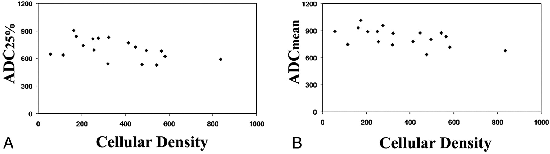
  • Fig 2.
    • Download figure
    • Open in new tab
    • Download powerpoint
    Fig 2.

    Scatterplots of cellular density measurements versus 25th percentile (ADC25%, A) and mean ADC (ADCmean, B) values within contrast-enhancing tumor regions for all 18 subjects included in this study, demonstrating statistically significant inverse correlations (ADC25%, R = −0.47, P = .05; ADCmean, R = −0.54, P = .02).
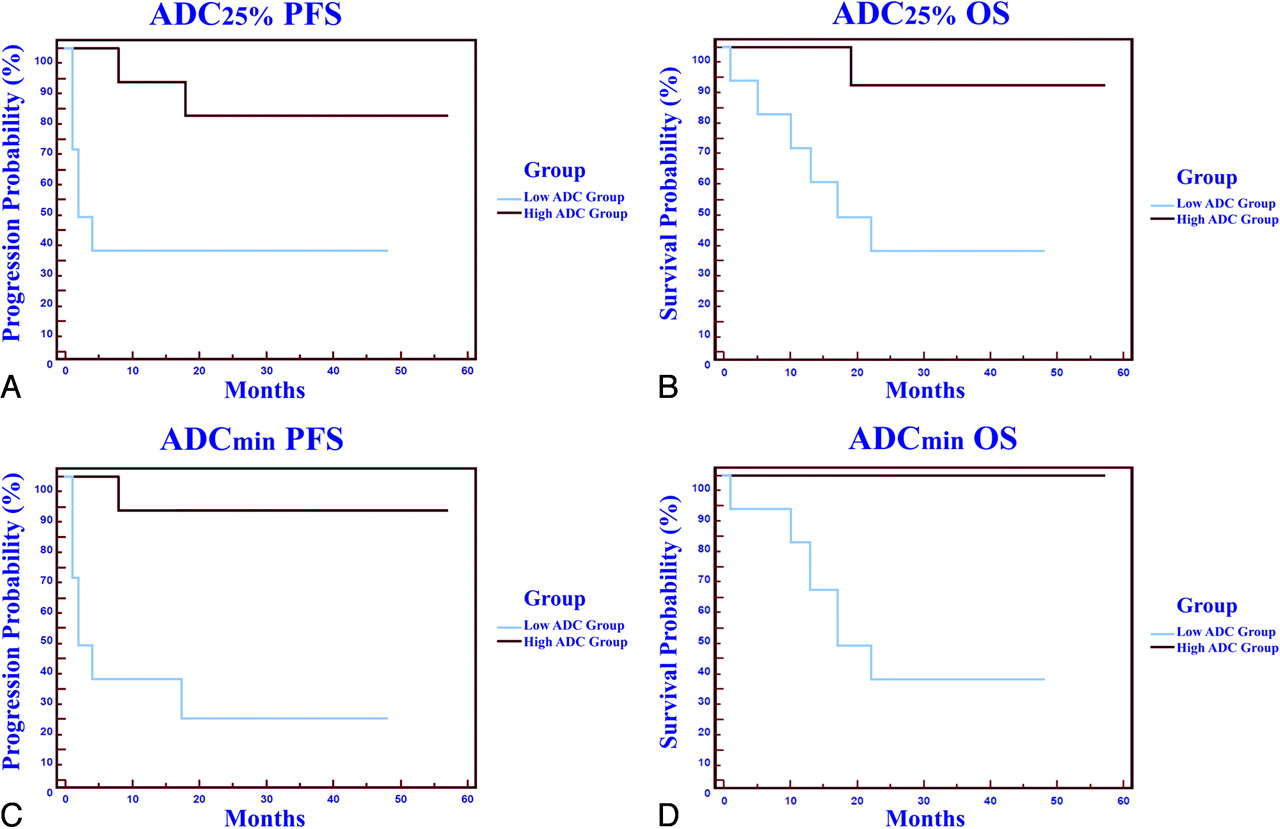
  • Fig 3.
    • Download figure
    • Open in new tab
    • Download powerpoint
    Fig 3.

    A and B, Patient outcome as a function of ADC25% stratification into low and high groups. Kaplan Meier analysis (A) of progression-free survival (PFS) for patients stratified into the low ADC25% group (<692, blue line) with a mean progression time of 9.4 months versus those stratified into high ADC25% group (>692, red line) with a mean progression time of 30.0 months (P = .02, logrank test). Kaplan Meier plot (B) of overall survival (OS) for patients stratified into the low ADC25% group (<692, blue line) with a mean survival of 15.8 months versus those stratified into high ADC25% group (>692, red line) with a mean survival of 30.9 months (P = .01, logrank test). C and D, Patient outcome as a function of minimum ADC value shows a statistically significant difference in PFS and OS between low and high groups (P < .05).

Tables

  • Figures
    • View popup
    Table 1:

    Clinical, imaging, and pathologic patient population characteristicsa

    Patient No.ADC GroupAge (yr)/SexNo. Enhancing LesionsResponse to TxADC25%ADCminADCmeanOverall Cellularity
    1Low45/FMPPD534156637High
    2Low82/FSGPD541167745High
    3Low57/MMPPD646191894High
    4Low61/MMPPR528214877High
    5Low62/FMPPD589280681High
    6Low54/MMPPD638338749Low
    7Low70/MSGPR623367719High
    8Low54/FSGCR688456805High
    9Low26/MSGCR692398835High
    10High52/FMPCR693294779Low
    11High43/FSGCR725371876High
    12High81/MMPCR822411959Low
    13High53/FMPCR831414873Mixed
    14High68/MMPCR8424791016Low
    15High61/FMPCR772510781Low
    16High53/MMPCR741525891Low
    17High56/FMPCR815609893Mixed
    18High67 mol/LSGCR906614933Low
    • Note:—ADC indicates apparent diffusion coefficient; MP, multiple; SG, single; Tx, treatment; PD, progressive disease; PR, partial response; CR, complete response; ADC25%, 25th percentile ADC value; ADCmin, minimum ADC value; ADCmean, mean ADC value.

    • a Patients are stratified on the basis of the median 25th percentile value.

    • View popup
    Table 2:

    Comparison of prognostic factors, cellular density, and clinical outcomea

    Age (yr)MMSEKPSDays to TxMonths to F/U MRINo. Tx to F/U MRIChange CE VolumeMonths to ProgressionMonths to Death
    Low ADC56.7 (15.6)25.2 (6.11)62.2 (6.67)13.8 (14.6)8.3 (14)2.00 (1.41)2.50 (2.88)9.4 (12.0)15.8 (9.87)
    High ADC59.3 (11.3)25.7 (4.50)66.7 (5.00)13.4 (11.7)4.0 (8.0)1.70 (0.70)3.92 (2.39)30.0 (18.0)30.9 (17.0)
    Pvalue.70.83.13.94.39.530.37<.01.03
    • Note:—MMSE indicates pretherapeutic Mini-Mental State Examination score; KPS, Karnovsky performance status; Days to Tx, mean number of days between initial diagnosis by MR imaging and initiation of methotrexate-based chemotherapy; Months to F/U MRI, mean number of months between pre- and intratherapeutic MR imaging; No. Tx to F/U MRI, mean number of methotrexate treatments between pre- and intratherapeutic MR imaging; Change CE, difference in enhancing volume between pre-and posttherapeutic imaging series; Months to Progression, mean number of months to progression based on MR imaging if event occurred; Months to Death, mean number of months to death if event occurred.

    • a All data are presented as mean (± SD). Patients are stratified based on median 25th percentile value.

    • View popup
    Table 3:

    Comparison of MR imaging findingsa

    ADCminChange ADCminADC25%Change ADC25%ADCmeanChange ADCmean
    Low ADC258 (109)−18.7 (219)608 (62)84.7 (114)771 (87.7)116 (106)
    High ADC469 (107)−120 (224)794 (67)−55.5 (84.7)889 (77.0)−42 (74)
    P value<.01.31<.01.04<.01.01
    • Note:—Change ADCmin indicates the difference between pre- and posttherapeutic ADCmin values (negative values signify net decrease in value); Change ADC25%, difference between pre- and posttherapeutic ADC25% values; Change ADCmean, difference between pre- and posttherapeutic ADCmean values.

    • a All data are presented as mean (SD). All ADC values are reported as 100 × 10−6 mm2/s.

PreviousNext
Back to top

In this issue

American Journal of Neuroradiology: 31 (1)
American Journal of Neuroradiology
Vol. 31, Issue 1
1 Jan 2010
  • Table of Contents
  • Index by author
Advertisement
Print
Download PDF
Email Article
Cite this article
0 Responses
Respond to this article
Share
Bookmark this article
Diffusion-Weighted MR Imaging Derived Apparent Diffusion Coefficient Is Predictive of Clinical Outcome in Primary Central Nervous System Lymphoma
R.F. Barajas, J.L. Rubenstein, J.S. Chang, J. Hwang, S. Cha
American Journal of Neuroradiology Jan 2010, 31 (1) 60-66; DOI: 10.3174/ajnr.A1750
del.icio.us logo Twitter logo Facebook logo Mendeley logo
  • Tweet Widget
Purchase

Jump to section

  • Article
    • Abstract
    • Materials and Methods
    • Results
    • Discussion
    • Conclusions
    • Acknowledgment
    • Footnotes
    • References
  • Figures & Data
  • Info & Metrics
  • Responses
  • References
  • PDF

Related Articles

  • No related articles found.
  • PubMed
  • Google Scholar

Cited By...

  • Hypovascular Cellular Tumor in Primary Central Nervous System Lymphoma is Associated with Treatment Resistance: Tumor Habitat Analysis Using Physiologic MRI
  • Simultaneous Multislice for Accelerating Diffusion MRI in Clinical Neuroradiology Protocols
  • Correlation Between Minimum Apparent Diffusion Coefficient (ADCmin) and Tumor Cellularity: A Meta-analysis
  • Diffusion-Weighted Imaging for Predicting and Monitoring Primary Central Nervous System Lymphoma Treatment Response
  • Differentiating Hemangioblastomas from Brain Metastases Using Diffusion-Weighted Imaging and Dynamic Susceptibility Contrast-Enhanced Perfusion-Weighted MR Imaging
  • Improved Brain Tumor Classification by Sodium MR Imaging: Prediction of IDH Mutation Status and Tumor Progression
  • The Combined Performance of ADC, CSF CXC Chemokine Ligand 13, and CSF Interleukin 10 in the Diagnosis of Central Nervous System Lymphoma
  • Potential Role of Preoperative Conventional MRI Including Diffusion Measurements in Assessing Epidermal Growth Factor Receptor Gene Amplification Status in Patients with Glioblastoma
  • "Dazed and diffused": making sense of diffusion abnormalities in neurologic pathologies
  • Rituximab, Methotrexate, Procarbazine, and Vincristine Followed by Consolidation Reduced-Dose Whole-Brain Radiotherapy and Cytarabine in Newly Diagnosed Primary CNS Lymphoma: Final Results and Long-Term Outcome
  • How I treat CNS lymphomas
  • The role of pre-treatment diffusion-weighted MRI in predicting long-term outcome of colorectal liver metastasis
  • Atypical Imaging Features of Epstein-Barr Virus-Positive Primary Central Nervous System Lymphomas in Patients without AIDS
  • MR Imaging of Neoplastic Central Nervous System Lesions: Review and Recommendations for Current Practice
  • Immunochemotherapy with Intensive Consolidation for Primary CNS Lymphoma: A Pilot Study and Prognostic Assessment by Diffusion-Weighted MRI
  • Central Nervous System Lymphoma: Characteristic Findings on Traditional and Advanced Imaging
  • Crossref
  • Google Scholar

This article has not yet been cited by articles in journals that are participating in Crossref Cited-by Linking.

More in this TOC Section

  • Progression of Microstructural Damage in Spinocerebellar Ataxia Type 2: A Longitudinal DTI Study
  • SWI or T2*: Which MRI Sequence to Use in the Detection of Cerebral Microbleeds? The Karolinska Imaging Dementia Study
  • Statin Therapy Does Not Affect the Radiographic and Clinical Profile of Patients with TIA and Minor Stroke
Show more Brain

Similar Articles

Advertisement

Indexed Content

  • Current Issue
  • Accepted Manuscripts
  • Article Preview
  • Past Issues
  • Editorials
  • Editor's Choice
  • Fellows' Journal Club
  • Letters to the Editor
  • Video Articles

Cases

  • Case Collection
  • Archive - Case of the Week
  • Archive - Case of the Month
  • Archive - Classic Case

More from AJNR

  • Trainee Corner
  • Imaging Protocols
  • MRI Safety Corner
  • Book Reviews

Multimedia

  • AJNR Podcasts
  • AJNR Scantastics

Resources

  • Turnaround Time
  • Submit a Manuscript
  • Submit a Video Article
  • Submit an eLetter to the Editor/Response
  • Manuscript Submission Guidelines
  • Statistical Tips
  • Fast Publishing of Accepted Manuscripts
  • Graphical Abstract Preparation
  • Imaging Protocol Submission
  • Evidence-Based Medicine Level Guide
  • Publishing Checklists
  • Author Policies
  • Become a Reviewer/Academy of Reviewers
  • News and Updates

About Us

  • About AJNR
  • Editorial Board
  • Editorial Board Alumni
  • Alerts
  • Permissions
  • Not an AJNR Subscriber? Join Now
  • Advertise with Us
  • Librarian Resources
  • Feedback
  • Terms and Conditions
  • AJNR Editorial Board Alumni

American Society of Neuroradiology

  • Not an ASNR Member? Join Now

© 2025 by the American Society of Neuroradiology All rights, including for text and data mining, AI training, and similar technologies, are reserved.
Print ISSN: 0195-6108 Online ISSN: 1936-959X

Powered by HighWire
Email this Article

Thank you for your interest in spreading the word on American Journal of Neuroradiology.

NOTE: We only request your email address so that the person you are recommending the page to knows that you wanted them to see it, and that it is not junk mail. We do not capture any email address.

Enter multiple addresses on separate lines or separate them with commas.
Diffusion-Weighted MR Imaging Derived Apparent Diffusion Coefficient Is Predictive of Clinical Outcome in Primary Central Nervous System Lymphoma
(Your Name) has sent you a message from American Journal of Neuroradiology
(Your Name) thought you would like to see the American Journal of Neuroradiology web site.
CAPTCHA
This question is for testing whether or not you are a human visitor and to prevent automated spam submissions.
Cite this article
R.F. Barajas, J.L. Rubenstein, J.S. Chang, J. Hwang, S. Cha
Diffusion-Weighted MR Imaging Derived Apparent Diffusion Coefficient Is Predictive of Clinical Outcome in Primary Central Nervous System Lymphoma
American Journal of Neuroradiology Jan 2010, 31 (1) 60-66; DOI: 10.3174/ajnr.A1750

Citation Manager Formats

  • BibTeX
  • Bookends
  • EasyBib
  • EndNote (tagged)
  • EndNote 8 (xml)
  • Medlars
  • Mendeley
  • Papers
  • RefWorks Tagged
  • Ref Manager
  • RIS
  • Zotero

We use cookies on this site to enhance your user experience. By clicking any link on this page you are giving your consent for us to set cookies.