Advertisement

Main menu

  • Home
  • Content
    • Current Issue
    • Accepted Manuscripts
    • Article Preview
    • Past Issue Archive
    • Video Articles
    • AJNR Case Collection
    • Case of the Week Archive
    • Case of the Month Archive
    • Classic Case Archive
  • Special Collections
    • AJNR Awards
    • Low-Field MRI
    • Alzheimer Disease
    • ASNR Foundation Special Collection
    • Photon-Counting CT
    • View All
  • Multimedia
    • AJNR Podcasts
    • AJNR SCANtastic
    • Trainee Corner
    • MRI Safety Corner
    • Imaging Protocols
  • For Authors
    • Submit a Manuscript
    • Submit a Video Article
    • Submit an eLetter to the Editor/Response
    • Manuscript Submission Guidelines
    • Statistical Tips
    • Fast Publishing of Accepted Manuscripts
    • Graphical Abstract Preparation
    • Imaging Protocol Submission
    • Author Policies
  • About Us
    • About AJNR
    • Editorial Board
    • Editorial Board Alumni
  • More
    • Become a Reviewer/Academy of Reviewers
    • Subscribers
    • Permissions
    • Alerts
    • Feedback
    • Advertisers
    • ASNR Home

User menu

  • Alerts
  • Log in

Search

  • Advanced search
American Journal of Neuroradiology

American Journal of Neuroradiology

ASHNR American Society of Functional Neuroradiology ASHNR American Society of Pediatric Neuroradiology ASSR
  • Alerts
  • Log in

Advanced Search

  • Home
  • Content
    • Current Issue
    • Accepted Manuscripts
    • Article Preview
    • Past Issue Archive
    • Video Articles
    • AJNR Case Collection
    • Case of the Week Archive
    • Case of the Month Archive
    • Classic Case Archive
  • Special Collections
    • AJNR Awards
    • Low-Field MRI
    • Alzheimer Disease
    • ASNR Foundation Special Collection
    • Photon-Counting CT
    • View All
  • Multimedia
    • AJNR Podcasts
    • AJNR SCANtastic
    • Trainee Corner
    • MRI Safety Corner
    • Imaging Protocols
  • For Authors
    • Submit a Manuscript
    • Submit a Video Article
    • Submit an eLetter to the Editor/Response
    • Manuscript Submission Guidelines
    • Statistical Tips
    • Fast Publishing of Accepted Manuscripts
    • Graphical Abstract Preparation
    • Imaging Protocol Submission
    • Author Policies
  • About Us
    • About AJNR
    • Editorial Board
    • Editorial Board Alumni
  • More
    • Become a Reviewer/Academy of Reviewers
    • Subscribers
    • Permissions
    • Alerts
    • Feedback
    • Advertisers
    • ASNR Home
  • Follow AJNR on Twitter
  • Visit AJNR on Facebook
  • Follow AJNR on Instagram
  • Join AJNR on LinkedIn
  • RSS Feeds

AJNR Awards, New Junior Editors, and more. Read the latest AJNR updates

Research ArticleBrain

Demonstration of Cerebral Venous Variations in the Region of the Third Ventricle on Phase-Sensitive Imaging

S. Fujii, Y. Kanasaki, E. Matsusue, S. Kakite, T. Kminou and T. Ogawa
American Journal of Neuroradiology January 2010, 31 (1) 55-59; DOI: https://doi.org/10.3174/ajnr.A1752
S. Fujii
  • Find this author on Google Scholar
  • Find this author on PubMed
  • Search for this author on this site
Y. Kanasaki
  • Find this author on Google Scholar
  • Find this author on PubMed
  • Search for this author on this site
E. Matsusue
  • Find this author on Google Scholar
  • Find this author on PubMed
  • Search for this author on this site
S. Kakite
  • Find this author on Google Scholar
  • Find this author on PubMed
  • Search for this author on this site
T. Kminou
  • Find this author on Google Scholar
  • Find this author on PubMed
  • Search for this author on this site
T. Ogawa
  • Find this author on Google Scholar
  • Find this author on PubMed
  • Search for this author on this site
  • Article
  • Figures & Data
  • Info & Metrics
  • Responses
  • References
  • PDF
Loading

Abstract

BACKGROUND AND PURPOSE: Susceptibility-weighted (SW) MR imaging has enabled noninvasive visualization of the cerebral veins and has shed light on the nature of venous architecture. For successful surgery of the third ventricle, understanding of the anatomy of the subependymal veins of the lateral ventricle and their relationships to the foramen of Monro is required preoperatively. The purpose of this study was to evaluate the anatomic variations of the subependymal veins around the third ventricle by use of phase-sensitive imaging (PSI) on the basis of principles similar to those of SW MR imaging.

MATERIALS AND METHODS: Included in this study were 642 sides in 321 patients. The courses of the anterior septal vein (ASV), thalamostriate vein, and internal cerebral vein (ICV) were evaluated. We classified these into 4 types (IA, IB, IIA, IIB) on the basis of standard classic angiographic criteria. The classification is based on their relationship with the ASV-ICV junction and the presence of a venous angle or a false venous angle, according to the method in a previous study. Other venous variations were classified as type III.

RESULTS: A venous angle was formed in 519 (80.9%), whereas a false venous angle was formed in 123 (19.1%). The ASV-ICV junction was located at the venous angle (type IA) in 407 (63.4%) of 642 sides. In 235 sides (36.6%), the ASV-ICV junction was located posteriorly beyond the foramen of Monro (types IB, IIA, IIB, and III).

CONCLUSIONS: PSI is useful for understanding normal variations of the subependymal veins in the region of the third ventricle.

Susceptibility-weighted (SW) MR imaging is a 3D gradient-echo imaging technique based on the blood oxygen level–dependent (BOLD)-induced phase effects between venous blood and the surrounding brain parenchyma.1,2 The technique of SW MR imaging for visualization of the venous system is based on the BOLD effect, which exploits the fact that paramagnetic deoxyhemoglobin causes a local magnetic field inhomogeneity, resulting in both a reduction of T2* and a phase difference between the venous vessel and its surroundings. Thus, SW MR imaging enables noninvasive visualization of the cerebral veins and has shed light on the nature of venous architecture3–5 and cerebral venous malformations.6–8 Currently, SW MR imaging is used mainly to investigate brain parenchymal lesions such as multiple sclerosis,9 small hemorrhage,10 and tumors.6–8

Cerebral angiography initially played the dominant role in the evaluation of cerebral venous anatomy in patients with brain tumors, but its role is now greatly reduced following advances in tomographic imaging. In surgery of the third ventricle, however, successful surgery necessitates understanding of the anatomy of the subependymal veins of the lateral ventricle and their relationships to the foramen of Monro preoperatively.11 Therefore, radiologists should be familiar with the cerebral venous anatomy in the region of the third ventricle and have an appreciation of the venous variations in this area.

Phase-sensitive imaging (PSI), on the basis of principles similar to those of SW MR imaging, is obtained by multiplication of the phase and magnitude images by use of Windows-based software.12 In this study, we demonstrate cerebral venous variations in the region of the third ventricle on PSI.

Materials and Methods

Patient Population

Included in this retrospective study were 734 sides in 367 consecutive patients who underwent MR imaging examinations including PSI at our institution between November 2007 and October 2008. PSI images of the brain were obtained for screening of headache or evaluation of hemorrhage, infarction, brain tumor, or degenerative diseases. We excluded 46 sides in 23 patients because the third ventricular area was not included on PSI. Sixteen sides in 8 patients were excluded because of motion artifacts or susceptibility artifacts because of the nasal sinuses or dental prostheses. In addition, 30 sides in 15 patients were excluded because their images could not be evaluated due to susceptibility artifacts caused by an intraventricular or basal ganglion hemorrhage around the third ventricle. A total of 642 sides in 321 consecutive patients (age range, 0–90 years; mean age, 58 years) who underwent PSI on a 3T MR imaging system were enrolled in the study. However, there were no cases of a third ventricular lesion in the present study. The Institutional Review Board approved our study, which did not require informed consent.

MR Imaging Technique

We obtained all brain MR imaging with a 3T MR system (Signa Excite HD; GE Healthcare, Milwaukee, Wisconsin) using an 8-channel high-resolution brain phased array coil (Invivo Corp., Gainesville, Florida).

We acquired PSI with a 3D echo-spoiled gradient-echo sequence with flow compensation using the following imaging parameters: TR, 45 ms; TE, 30 ms; FA, 20°; FOV, 21 cm; matrix, 512 × 192; section thickness, 1.5 mm; acquisition time, 7 min 40 to 50 s. All images were obtained in the axial plane. We postprocessed the phase-sensitive images using a high-pass filter and then converted the images into negative phase masks that were multiplied 4 times into the corresponding magnitude images using research software (PSIRecon; GE Yokogawa Medical Systems, Tokyo, Japan). We used a minimal-intensity projection (PSI) to display the processed data and contiguous 10.5-mm-thick sections with 7-mm overlap in the transverse plane (Advantage workstation version 4.1; GE Healthcare).

Evaluation of the Venous Anatomy in the Region of the Third Ventricle

Two experienced neuroradiologists consensually assessed each side with regard to venous variation on PSI. The reviewers identified the anterior septal vein (ASV) and thalamostriate vein (TSV) as running the following courses. The ASV initially runs posteromedially on the anterior wall of the frontal horn and behind the genu of the corpus callosum. When it reaches the anteromedial corner of the frontal horn, it runs backwards along the septum pellucidum and continues backward along its lower border. It then follows the lateral border of the anterior column of the fornix, generally joining the internal cerebral vein (ICV) at the posterosuperior margin of the foramen of Monro.13

The TSV begins in the region of the anterior wall of the atrium or in the inferolateral wall of the body of the lateral ventricle, in the linea terminalis. It passes anteriorly and medially, beneath the stria terminalis, receiving several transverse caudate veins into which the longitudinal caudate veins drain, on its way toward the foramen of Monro. At the level of the foramen of Monro, the TSV runs around the anterior tubercle of the thalamus and receives the vein from the head of the caudate nucleus. It then passes medially, generally joining the ICV at the posterosuperior margin of the foramen of Monro.13

The characteristic U-shaped junction of the TSV and ICV adjacent to the posterior margin of the foramen of Monro is referred to as the venous angle (Fig 1). When the junction of the TSV and ICV lies beyond the posterior margin of the foramen of Monro, this variation is referred to as the false venous angle (Fig 1). We classified these into 4 types on the basis of their relationship with the ASV-ICV junction and the presence of a venous angle or a false venous angle, according to the method used in a previous study (Fig 2 ).11

Fig 1.
  • Download figure
  • Open in new tab
  • Download powerpoint
Fig 1.

Diagram demonstrating the venous angle and false venous angle (lv, lateral ventricles; asv, anterior septal vein; tsv, thalamostriate vein; icv, internal cerebral vein; fm, foramen of Monro; cc, corpus callosum; ln, lentiform nucleus; ch, head of caudate nuclei; th, thalamus).

Fig 2.
  • Download figure
  • Open in new tab
  • Download powerpoint
Fig 2.

Variations in location of the ASV-ICV junction relative to the foramen of Monro (fm). (Reprinted by permission of Ture U, Yasargil, MG, Al-Mefty O. The transcallosal-transforaminal approach to the third ventricle with regard to the venous variations in this region. J Neurosurg 1997;87:706–1511).

The 4 types were classified as follows:

  • Type IA: ASV-ICV junction was located at the venous angle, which lies adjacent to the posterior margin of the foramen of Monro. In the other types, the ASV-ICV junction was located beyond the posterior margin of the foramen of Monro, in 3 different locations.

  • Type IB: the ASV joined the mainstem of the ICV posteriorly beyond both the foramen of Monro and the venous angle.

  • Type IIA: the ASV joined the false venous angle, which lies beyond the foramen of Monro.

  • Type IIB: the ASV joined the mainstem of the ICV far beyond both the foramen of Monro and the false venous angle. We classified any other venous variations as type III.

Results

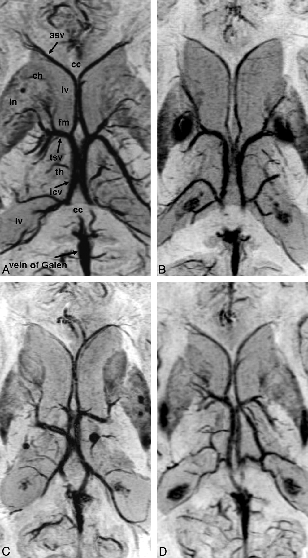
Figure 3 shows representative cases of the 4 types of cerebral venous variation in the region of the third ventricle. Data regarding the types of cerebral venous variation in the region of the third ventricle are summarized in the accompanying Table. In only 1 case of type III, the ASV joined the medial atrial vein (Fig 3).

Fig 3.
  • Download figure
  • Open in new tab
  • Download powerpoint
Fig 3.

Cerebral venous variations in the region of the third ventricle on PSI. Abbreviations are the same as in Fig 1. A, Type IA: the ASV-ICV junction is located at the venous angle, which lies adjacent to the posterior margin of the foramen of Monro. B, Type IIA (left): the ASV joined the false venous angle, which lies beyond the foramen of Monro. Type IB (right): the ASV joined the mainstem of the ICV posteriorly beyond both the foramen of Monro and the venous angle. C, Type IA (left), type IIB (right): the ASV joined the mainstem of the ICV far beyond both the foramen of Monro and the false venous angle. D, Type III (left): the ASV joined the medial atrial vein and the venous angle is formed. Type IA (right).

View this table:
  • View inline
  • View popup

Frequency of each type of cerebral venous variation in the region of the third ventricle

In 519 (80.9%) of 642 sides, the venous angle (TSV joins the ICV at the margin of the foramen of Monro) was formed. In 123 (19.1%) of 642 sides, a false venous angle was formed (Table; Fig 3).

In 407 (63.4%) of 642 sides, the ASV-ICV junction was located at the venous angle (type IA). In 235 (36.6%) of 642 sides, the ASV-ICV junction was located posteriorly, beyond the foramen of Monro (types IB, IIA, IIB, and III) (Fig 3).

Discussion

Surgery in the region of the third ventricle poses a considerable challenge, even for experienced neurosurgeons. The numerous publications describing various surgical approaches attest to the difficulties encountered when exploring this area.11 With use of the transcortical-transventricular and the anterior transcallosal approaches, access is gained to the anterior and middle portions of the third ventricle. The anterior transcallosal approach usually provides a direct and adequate pathway to the lateral ventricles, where the foramen of Monro serves as a natural entrance into the anterior and middle portions of the third ventricle, particularly when the foramen is dilated by tumor tissue. When the foramen of Monro is not dilated by a tumor, however, access to the midsuperior portion of the third ventricle is limited. The location of the ASV-ICV junction relative to the foramen of Monro is the key to this exposure. When the ASV-ICV junction is located posteriorly, beyond the foramen of Monro, the transforaminal exposure can be adapted. The foramen of Monro, ipsilateral to the more posteriorly located ASV-ICV junction, can be enlarged along the choroidal fissure as far as the junction to provide a wide access to the third ventricle.11 In our study, PSI demonstrated cerebral venous variations in the region of the third ventricle. Therefore, PSI may play an important role in the preoperative planning for lesions located around the third ventricle.

Various studies describe the deep venous system of the brain and venous variations in the region of the foramen of Monro.14–24 On cerebral angiography, the incidence of a false venous angle is reported to vary between 14% and 26%.16–18,20–22 In our study, we observed the presence of a false venous angle in 19.1% of sides, which is within the range reported for angiography.

According to an anatomic study,11 the frequency of a false venous angle (beyond the foramen of Monro) was 32.5%, and the posterior location of the ASV-ICV junction was 47.5%. The frequencies reported in the present study are low compared with this anatomic study. The reason for this discrepancy is not clear but may relate to the small population (40 hemispheres) in the anatomic study and the cases in the present PSI study for which accurate assessment was difficult because of the very short distance between the venous junction and the foramen of Monro.

MR venography is used in the evaluation of the venous anatomy24; however, we believe that SW MR imaging, including PSI, should be obtained for evaluation of the deep cerebral venous anatomy because it can depict the cerebral vein with high resolution,3–5 and it enables evaluation of brain parenchymal lesions, particularly a small hemorrhage.10 Therefore, we consider PSI to be a useful MR imaging technique that could be added to imaging protocols.

There were some limitations in our study. First, the potential existed for artifacts in PSI. Application of this imaging method may be difficult in areas with B0 inhomogeneity, restricting its use to anatomic brain regions free of large static field inhomogeneities caused by susceptibility differences, such as regions near the paranasal sinuses and skull base.5 Susceptibility artifacts because of hemorrhage are naturally stronger on PSI than that for other imaging sequences. Therefore, some patients were excluded from our study, but the proportion was low.

Second, the sequence parameters were optimized for evaluation of intraparenchymal lesions rather than for visualization of veins. Although we could evaluate the veins in most cases, further examination may be needed.

Third, surgical confirmation was not performed because third ventricular lesions were not included because of the absence of such a case. A prospective study is necessary for side-by-side comparison of the MR imaging results with the surgical findings.

Conclusions

PSI is useful for gaining an understanding of normal variations of the subependymal veins in the region of the third ventricle. PSI may play an important role in the preoperative planning of lesions in the third ventricle.

Acknowledgments

We thank Eijirou Yamashita, BS; Takuro Tanaka, BS; Naoki Iwata, BS; and Shota Sakimoto, BS, for technical support in obtaining the high-quality MR images used in this study.

References

  1. 1.↵
    1. Reichenbach JR,
    2. Haacke EM
    . High-resolution BOLD venographic imaging: a window into brain function. NMR Biomed 2001; 14: 453–67
    CrossRefPubMed
  2. 2.↵
    1. Rauscher A,
    2. Sedlacik J,
    3. Barth M,
    4. et al
    . Nonnvasive assessment of vascular architecture and function during modulated blood oxygenation using susceptibility weighted magnetic resonance imaging. Magn Reson Med 2005; 54: 87–95
    CrossRefPubMed
  3. 3.↵
    1. Reichenbach JR,
    2. Venkatesan R,
    3. Schillinger DJ,
    4. et al
    . Small vessels in the human brain: MR venography with deoxyhemoglobin as an intrinsic contrast agent. Radiology 1997; 204: 272–77
    PubMed
  4. 4.↵
    1. Lee BC,
    2. Vo KD,
    3. Kido DK,
    4. et al
    . MR high-resolution blood oxygenation level-dependent venography of occult (low-flow) vascular lesions. AJNR Am J Neuroradiol 1999; 20: 1239–42
    Abstract/FREE Full Text
  5. 5.↵
    1. Reichenbach JR,
    2. Barth M,
    3. Haacke EM,
    4. et al
    . High-resolution MR venography at 3.0 Tesla. J Comput Assist Tomogr 2000; 24: 949–57
    CrossRefPubMed
  6. 6.↵
    1. Sehgal V,
    2. Delproposto Z,
    3. Haacke EM,
    4. et al
    . Clinical applications of neuroimaging with susceptibility-weighted imaging. J Magn Reson Imaging 2005; 22: 439–50
    CrossRefPubMed
  7. 7.↵
    1. Thomas B,
    2. Somasundaram S,
    3. Thamburaj K,
    4. et al
    . Clinical applications of susceptibility weighted MR imaging of the brain—a pictorial review. Neuroradiology 2008; 50: 105–16
    CrossRefPubMed
  8. 8.↵
    1. Tong KA,
    2. Ashwal S,
    3. Obenaus A,
    4. et al
    . Susceptibility-weighted MR imaging: a review of clinical applications in children. AJNR Am J Neuroradiol 2008; 29: 9–17
    Abstract/FREE Full Text
  9. 9.↵
    1. Tan IL,
    2. van Schijndel RA,
    3. Pouwels PJ,
    4. et al
    . MR venography of multiple sclerosis. AJNR Am J Neuroradiol 2000; 21: 1039–42
    Abstract/FREE Full Text
  10. 10.↵
    1. Akter M,
    2. Hirai T,
    3. Hiai Y,
    4. et al
    . Detection of hemorrhagic hypointense foci in the brain on susceptibility-weighted imaging clinical and phantom studies. Acad Radiol 2007; 14: 1011–19
    CrossRefPubMed
  11. 11.↵
    1. Ture U,
    2. Yasargil MG,
    3. Al-Mefty O
    . The transcallosal-transforaminal approach to the third ventricle with regard to the venous variations in this region. J Neurosurg 1997; 87: 706–15
    CrossRefPubMed
  12. 12.↵
    1. Kakeda S,
    2. Korogi Y,
    3. Kamada K,
    4. et al
    . Signal intensity of the motor cortex on phase-weighted imaging at 3T. AJNR Am J Neuroradiol 2008; 29: 1171–75
    Abstract/FREE Full Text
  13. 13.↵
    1. Salamon G,
    2. Huang YP
    1. Huang YP
    . Deep cerebral veins. In: Salamon G, Huang YPeds. Radiologic Anatomy of the Brain. Berlin: Springer-Verlag; 1976: 210–61
  14. 14.↵
    1. Johanson C
    . The central veins and deep dural sinuses of the brain; an anatomical and angiographic study. Acta Radiol Suppl 1954; 107: 1–184
  15. 15.↵
    1. Krayenbuhl HA,
    2. Yasargil MG
    . Cerebral Angiography, 2nd ed. Philadelphia: Lippincott; 1968
  16. 16.↵
    1. Lang J
    . Surgical anatomy of the hypothalamus. Acta Neurochir 1985; 75: 5–22
    CrossRefPubMed
  17. 17.↵
    1. Mokrohisky JF,
    2. Paul RE,
    3. Lin PM,
    4. et al
    . The diagnostic importance of normal variants in deep cerebral phlebography. With special emphasis on the true and false “venous angles of the brain” and evaluation of venous angle measurement. Radiology 1956; 67: 34–47
    CrossRefPubMed
  18. 18.↵
    1. Ono M,
    2. Rhoton AL Jr.,
    3. Peace D,
    4. et al
    . Microsurgical anatomy of the deep venous system of the brain. Neurosurgery 1984; 15: 621–57
    CrossRefPubMed
  19. 19.↵
    1. Probst FP
    . Position of the “venous angle” in the median sagittal plane. A new topometric method. Acta Radiol (Diagn) 1970; 10: 271–88
    PubMed
  20. 20.↵
    1. Ring BA
    . Variations in the striate and other cerebral veins affecting measurements of the “venous angle”;. Acta Radiol 1959; 52: 433–47
    CrossRef
  21. 21.↵
    1. Newton TH,
    2. Potts DG
    1. Stein RL,
    2. Rosenbaum AE
    . Deep supratentorial veins. Section I. Normal deep cerebral venous system. In: Newton TH, Potts DGeds. Radiology of the Skull and Brain. Angiography. Vol 2. St. Louis: Mosby; 1974: 1903–98
  22. 22.↵
    1. Wolf BS,
    2. Huang YP
    . The subependymal veins of the lateral ventricles. AJR Am J Roentgenol 1964; 91: 406–26
  23. 23.↵
    1. Zimmer AE,
    2. Annes GP
    . The septal vein. An indicator of space occupying lesions in the anterior cranial fossa. Radiology 1966; 87: 813–23
    CrossRefPubMed
  24. 24.↵
    1. Cimsit NC,
    2. Ture U,
    3. Ekinci G,
    4. et al
    . Venous variations in the region of the third ventricle: the role of MR venography. Neuroradiology 2003; 45: 900–04
    CrossRefPubMed
  • Received April 9, 2009.
  • Accepted after revision May 21, 2009.
  • Copyright © American Society of Neuroradiology
View Abstract
PreviousNext
Back to top

In this issue

American Journal of Neuroradiology: 31 (1)
American Journal of Neuroradiology
Vol. 31, Issue 1
1 Jan 2010
  • Table of Contents
  • Index by author
Advertisement
Print
Download PDF
Email Article

Thank you for your interest in spreading the word on American Journal of Neuroradiology.

NOTE: We only request your email address so that the person you are recommending the page to knows that you wanted them to see it, and that it is not junk mail. We do not capture any email address.

Enter multiple addresses on separate lines or separate them with commas.
Demonstration of Cerebral Venous Variations in the Region of the Third Ventricle on Phase-Sensitive Imaging
(Your Name) has sent you a message from American Journal of Neuroradiology
(Your Name) thought you would like to see the American Journal of Neuroradiology web site.
CAPTCHA
This question is for testing whether or not you are a human visitor and to prevent automated spam submissions.
Cite this article
S. Fujii, Y. Kanasaki, E. Matsusue, S. Kakite, T. Kminou, T. Ogawa
Demonstration of Cerebral Venous Variations in the Region of the Third Ventricle on Phase-Sensitive Imaging
American Journal of Neuroradiology Jan 2010, 31 (1) 55-59; DOI: 10.3174/ajnr.A1752

Citation Manager Formats

  • BibTeX
  • Bookends
  • EasyBib
  • EndNote (tagged)
  • EndNote 8 (xml)
  • Medlars
  • Mendeley
  • Papers
  • RefWorks Tagged
  • Ref Manager
  • RIS
  • Zotero
0 Responses
Respond to this article
Share
Bookmark this article
Demonstration of Cerebral Venous Variations in the Region of the Third Ventricle on Phase-Sensitive Imaging
S. Fujii, Y. Kanasaki, E. Matsusue, S. Kakite, T. Kminou, T. Ogawa
American Journal of Neuroradiology Jan 2010, 31 (1) 55-59; DOI: 10.3174/ajnr.A1752
del.icio.us logo Twitter logo Facebook logo Mendeley logo
  • Tweet Widget
  • Facebook Like
  • Google Plus One
Purchase

Jump to section

  • Article
    • Abstract
    • Materials and Methods
    • Results
    • Discussion
    • Conclusions
    • Acknowledgments
    • References
  • Figures & Data
  • Info & Metrics
  • Responses
  • References
  • PDF

Related Articles

  • No related articles found.
  • PubMed
  • Google Scholar

Cited By...

  • The Internal Cerebral Vein: New Classification of Branching Patterns Based on CTA
  • Venous imaging-based biomarkers in acute ischaemic stroke
  • Variability of Cerebral Deep Venous System in Preterm and Term Neonates Evaluated on MR SWI Venography
  • Visualization of the Internal Cerebral Veins on MR Phase-Sensitive Imaging: Comparison with 3D Gadolinium-Enhanced MR Venography and Fast-Spoiled Gradient Recalled Imaging
  • Crossref (22)
  • Google Scholar

This article has been cited by the following articles in journals that are participating in Crossref Cited-by Linking.

  • Venous imaging-based biomarkers in acute ischaemic stroke
    Josep Munuera, Gerard Blasco, María Hernández-Pérez, Pepus Daunis-i-Estadella, Antoni Dávalos, David S Liebeskind, Max Wintermark, Andrew Demchuk, Bijoy K Menon, Götz Thomalla, Kambiz Nael, Salvador Pedraza, Josep Puig
    Journal of Neurology, Neurosurgery & Psychiatry 2017 88 1
  • Deep medullary vein engorgement and superficial medullary vein engorgement: two patterns of perinatal venous stroke
    Hedieh Khalatbari, Jason N. Wright, Gisele E. Ishak, Francisco A. Perez, Catherine M. Amlie-Lefond, Dennis W. W. Shaw
    Pediatric Radiology 2021 51 5
  • High incidence of asymptomatic cerebral microbleeds in patients with hemorrhagic onset-type moyamoya disease: a phase-sensitive MRI study and meta-analysis
    Ying Qin, Toshihide Ogawa, Shinya Fujii, Yuki Shinohara, Shin-ichiro Kitao, Fuminori Miyoshi, Marie Takasugi, Takashi Watanabe, Toshio Kaminou
    Acta Radiologica 2015 56 3
  • Time-resolved MR angiography of the intracranial venous system: an alternative MR venography technique
    Hasan Yiğit, Aynur Turan, Elif Ergün, Pınar Koşar, Uğur Koşar
    European Radiology 2012 22 5
  • Variability of Cerebral Deep Venous System in Preterm and Term Neonates Evaluated on MR SWI Venography
    D. Tortora, M. Severino, M. Malova, A. Parodi, G. Morana, L.A. Ramenghi, A. Rossi
    American Journal of Neuroradiology 2016 37 11
  • Susceptibility-Weighted Imaging of the Anatomic Variation of Thalamostriate Vein and Its Tributaries
    Xiao-fen Zhang, Jian-ce Li, Xin-dong Wen, Chuan-gen Ren, Ming Cai, Cheng-chun Chen, Heye Zhang
    PLOS ONE 2015 10 10
  • Evaluation of the anatomy and variants of internal cerebral veins with phase-sensitive MR imaging
    Jin Wang, Jiawei Wang, Jianzhong Sun, Xiangyang Gong
    Surgical and Radiologic Anatomy 2010 32 7
  • Susceptibility-weighted imaging of the venous networks around the brain stem
    Ming Cai, Xiao-Fen Zhang, Hui-Huang Qiao, Zhong-Xiao Lin, Chuan-Gen Ren, Jian-Ce Li, Cheng-Chun Chen, Nu Zhang
    Neuroradiology 2015 57 2
  • Comparison of increased venous contrast in ischemic stroke using phase-sensitive MR imaging with perfusion changes on flow-sensitive alternating inversion recovery at 3 Tesla
    Eijiro Yamashita, Yoshiko Kanasaki, Shinya Fujii, Takuro Tanaka, Yoshiharu Hirata, Toshihide Ogawa
    Acta Radiologica 2011 52 8
  • Visualization of Anatomic Variation of the Anterior Septal Vein on Susceptibility-Weighted Imaging
    Zhengzhen Chen, Huihuang Qiao, Yu Guo, Jiance Li, Huizhong Miao, Caiyun Wen, Xindong Wen, Xiaofen Zhang, Xindong Yang, Chengchun Chen, Yen-Yu Ian Shih
    PLOS ONE 2016 11 10

More in this TOC Section

  • Evaluating the Effects of White Matter Multiple Sclerosis Lesions on the Volume Estimation of 6 Brain Tissue Segmentation Methods
  • Quiet PROPELLER MRI Techniques Match the Quality of Conventional PROPELLER Brain Imaging Techniques
  • Predictors of Reperfusion in Patients with Acute Ischemic Stroke
Show more BRAIN

Similar Articles

Advertisement

Indexed Content

  • Current Issue
  • Accepted Manuscripts
  • Article Preview
  • Past Issues
  • Editorials
  • Editor's Choice
  • Fellows' Journal Club
  • Letters to the Editor
  • Video Articles

Cases

  • Case Collection
  • Archive - Case of the Week
  • Archive - Case of the Month
  • Archive - Classic Case

More from AJNR

  • Trainee Corner
  • Imaging Protocols
  • MRI Safety Corner

Multimedia

  • AJNR Podcasts
  • AJNR Scantastics

Resources

  • Turnaround Time
  • Submit a Manuscript
  • Submit a Video Article
  • Submit an eLetter to the Editor/Response
  • Manuscript Submission Guidelines
  • Statistical Tips
  • Fast Publishing of Accepted Manuscripts
  • Graphical Abstract Preparation
  • Imaging Protocol Submission
  • Evidence-Based Medicine Level Guide
  • Publishing Checklists
  • Author Policies
  • Become a Reviewer/Academy of Reviewers
  • News and Updates

About Us

  • About AJNR
  • Editorial Board
  • Editorial Board Alumni
  • Alerts
  • Permissions
  • Not an AJNR Subscriber? Join Now
  • Advertise with Us
  • Librarian Resources
  • Feedback
  • Terms and Conditions
  • AJNR Editorial Board Alumni

American Society of Neuroradiology

  • Not an ASNR Member? Join Now

© 2025 by the American Society of Neuroradiology All rights, including for text and data mining, AI training, and similar technologies, are reserved.
Print ISSN: 0195-6108 Online ISSN: 1936-959X

Powered by HighWire