Advertisement

Main menu

  • Home
  • Content
    • Current Issue
    • Accepted Manuscripts
    • Article Preview
    • Past Issue Archive
    • Video Articles
    • AJNR Case Collection
    • Case of the Week Archive
    • Case of the Month Archive
    • Classic Case Archive
  • Special Collections
    • Advancing NeuroMRI with High-Relaxivity Contrast Agents
    • AJNR Awards
    • Low-Field MRI
    • Alzheimer Disease
    • ASNR Foundation Special Collection
    • Photon-Counting CT
    • View All
  • Multimedia
    • AJNR Podcasts
    • AJNR SCANtastic
    • Trainee Corner
    • MRI Safety Corner
    • Imaging Protocols
  • For Authors
    • Submit a Manuscript
    • Submit a Video Article
    • Submit an eLetter to the Editor/Response
    • Manuscript Submission Guidelines
    • Statistical Tips
    • Fast Publishing of Accepted Manuscripts
    • Graphical Abstract Preparation
    • Imaging Protocol Submission
    • Author Policies
  • About Us
    • About AJNR
    • Editorial Board
    • Editorial Board Alumni
  • More
    • Become a Reviewer/Academy of Reviewers
    • Subscribers
    • Permissions
    • Alerts
    • Feedback
    • Advertisers
    • ASNR Home

User menu

  • Alerts
  • Log in

Search

  • Advanced search
American Journal of Neuroradiology

American Journal of Neuroradiology

ASHNR American Society of Functional Neuroradiology ASHNR American Society of Pediatric Neuroradiology ASSR
  • Alerts
  • Log in

Advanced Search

  • Home
  • Content
    • Current Issue
    • Accepted Manuscripts
    • Article Preview
    • Past Issue Archive
    • Video Articles
    • AJNR Case Collection
    • Case of the Week Archive
    • Case of the Month Archive
    • Classic Case Archive
  • Special Collections
    • Advancing NeuroMRI with High-Relaxivity Contrast Agents
    • AJNR Awards
    • Low-Field MRI
    • Alzheimer Disease
    • ASNR Foundation Special Collection
    • Photon-Counting CT
    • View All
  • Multimedia
    • AJNR Podcasts
    • AJNR SCANtastic
    • Trainee Corner
    • MRI Safety Corner
    • Imaging Protocols
  • For Authors
    • Submit a Manuscript
    • Submit a Video Article
    • Submit an eLetter to the Editor/Response
    • Manuscript Submission Guidelines
    • Statistical Tips
    • Fast Publishing of Accepted Manuscripts
    • Graphical Abstract Preparation
    • Imaging Protocol Submission
    • Author Policies
  • About Us
    • About AJNR
    • Editorial Board
    • Editorial Board Alumni
  • More
    • Become a Reviewer/Academy of Reviewers
    • Subscribers
    • Permissions
    • Alerts
    • Feedback
    • Advertisers
    • ASNR Home
  • Follow AJNR on Twitter
  • Visit AJNR on Facebook
  • Follow AJNR on Instagram
  • Join AJNR on LinkedIn
  • RSS Feeds

AJNR Awards, New Junior Editors, and more. Read the latest AJNR updates


Improved Turnaround Times | Median time to first decision: 12 days

Research ArticleBrain

Optimized Preload Leakage-Correction Methods to Improve the Diagnostic Accuracy of Dynamic Susceptibility-Weighted Contrast-Enhanced Perfusion MR Imaging in Posttreatment Gliomas

L.S. Hu, L.C. Baxter, D.S. Pinnaduwage, T.L. Paine, J.P. Karis, B.G. Feuerstein, K.M. Schmainda, A.C. Dueck, J. Debbins, K.A. Smith, P. Nakaji, J.M. Eschbacher, S.W. Coons and J.E. Heiserman
American Journal of Neuroradiology January 2010, 31 (1) 40-48; DOI: https://doi.org/10.3174/ajnr.A1787
L.S. Hu
  • Find this author on Google Scholar
  • Find this author on PubMed
  • Search for this author on this site
L.C. Baxter
  • Find this author on Google Scholar
  • Find this author on PubMed
  • Search for this author on this site
D.S. Pinnaduwage
  • Find this author on Google Scholar
  • Find this author on PubMed
  • Search for this author on this site
T.L. Paine
  • Find this author on Google Scholar
  • Find this author on PubMed
  • Search for this author on this site
J.P. Karis
  • Find this author on Google Scholar
  • Find this author on PubMed
  • Search for this author on this site
B.G. Feuerstein
  • Find this author on Google Scholar
  • Find this author on PubMed
  • Search for this author on this site
K.M. Schmainda
  • Find this author on Google Scholar
  • Find this author on PubMed
  • Search for this author on this site
A.C. Dueck
  • Find this author on Google Scholar
  • Find this author on PubMed
  • Search for this author on this site
J. Debbins
  • Find this author on Google Scholar
  • Find this author on PubMed
  • Search for this author on this site
K.A. Smith
  • Find this author on Google Scholar
  • Find this author on PubMed
  • Search for this author on this site
P. Nakaji
  • Find this author on Google Scholar
  • Find this author on PubMed
  • Search for this author on this site
J.M. Eschbacher
  • Find this author on Google Scholar
  • Find this author on PubMed
  • Search for this author on this site
S.W. Coons
  • Find this author on Google Scholar
  • Find this author on PubMed
  • Search for this author on this site
J.E. Heiserman
  • Find this author on Google Scholar
  • Find this author on PubMed
  • Search for this author on this site
  • Article
  • Figures & Data
  • Info & Metrics
  • Responses
  • References
  • PDF
Loading

Article Figures & Data

Figures

  • Tables
  • Fig 1.
    • Download figure
    • Open in new tab
    • Download powerpoint
    Fig 1.

    Diagram demonstrates the step-wise preload dosing (PLD) protocol. Circles depict the 6 sequential dynamic susceptibility-weighted contrast-enhanced (DSC)-acquisition 0.05-mmol/kg contrast injections, each separated by 3 minutes. Relative cerebral blood volume (rCBV) was calculated from each DSC acquisition. The PLD amount for each acquisition (ie, P2 through P6) equals the sum of all preceding contrast-injection amounts. The blue circle (P1) represents the control DSC acquisition with no PLD amount. MRI indicates MR imaging.

  • Fig 2.
    • Download figure
    • Open in new tab
    • Download powerpoint
    Fig 2.

    Graph shows cerebral blood volume (CBV) calculation methods. A, Uncorrected CBV calculation by trapezoidal integration without baseline subtraction (BLS). B, BLS integration excludes a triangular area (red) that estimates the T2/T2*WI residual effects. The remaining area under the curve (AUC) (blue lines) estimates the corrected CBV.
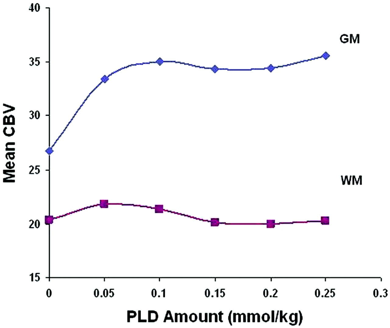
  • Fig 3.
    • Download figure
    • Open in new tab
    • Download powerpoint
    Fig 3.

    Graph shows CBV over an increasing PLD amount from normal-appearing gray matter (GM) and white matter (WM) regions of interest. CBV values are calculated by using BLS integration.

  • Fig 4.
    • Download figure
    • Open in new tab
    • Download powerpoint
    Fig 4.

    Graph shows distribution of rCBV values for tumor (red circles) and posttreatment radiation-effect (PTRE) (blue triangles) groups over the increasing PLD amounts without BLS (A) and with BLS (B). Gray boxes depict the range of overlapping rCBV values between groups under each condition. Asterisks in B denote conditions that demonstrated statistically significant increases in the rCBV receiver operator characteristic (ROC) AUC compared with the control acquisition (dotted circle). These are observed with BLS and PLD amounts of 0.05 (P = .045), 0.1 (P = .01), and 0.25 (P = .35) mmol/kg, with incubation times of 3, 6, and 15 minutes, respectively.

  • Fig 5.
    • Download figure
    • Open in new tab
    • Download powerpoint
    Fig 5.

    Graph shows the effects of PLD amount and the presence of BLS correction on DSC test accuracy (represented by ROC AUC). Asterisks (with P values) denote test conditions that demonstrate a statistically significant increase in rCBV ROC AUC compared with the control acquisition (red circle).

  • Fig 6.
    • Download figure
    • Open in new tab
    • Download powerpoint
    Fig 6.

    Graph shows that representative rCBV color maps are obtained with 2 different DSC protocols and are subsequently masked and overlaid on the same contrast-enhanced MR imaging lesion, with identical color scales that progress from blue (low) to red (high). A, Before leakage correction, the lesion inaccurately demonstrates an even mixture of tumor and PTRE voxels based on uncorrected rCBV. B, Following PLD-BLS (0.1 mmol/kg) correction, rCBV increase demonstrates a greater abundance of tumor voxels.

Tables

  • Figures
    • View popup
    Table 1:

    Patient demographic and clinical information

    SubjectAge (yr), SexPrimary Tumor (grade)RT Type, Dose, Timing (completed prior to imaging)Steroids at Imaging
    A31, MAA (III)60-Gy 3D conformal (9 mo)Yes
    25-Gy salvage IMRT (2 mo)
    B58, MGBM (IV)54-Gy 3D conformal (22 mo)Yes
    12-Gy gamma knife (5 mo)
    C36, MGBM (IV)60-Gy IMRT (3 mo)None
    D56, MGBM (IV)60-Gy IMRT (23 mo)Yes
    30-Gy IMRT (10 mo)
    E45, FGBM (IV)60-Gy 3D conformal (26 mo)None
    FaExcluded due to motion
    G50, MGBM (IV)37.5-Gy whole brain RT (8 mo)Yes
    30-Gy IMRT (5 mo)
    Hb38, MAna. GG (III)59.4-Gy IMRT (28.5 mo)Yes
    12-Gy gamma knife (15.5 mo)
    I59, MGBM (IV)59.4-Gy IMRT (13 mo)None
    JaExcluded due to motion
    K62, MGBM (IV)54-Gy 3D conformal (12 mo)None
    Lb38, MAna. GG (III)54-Gy IMRT (24 months)None
    12-Gy gamma knife (11 mo)
    M43, MGBM (IV)60-Gy 3D conformal (8 mo)Yes
    10-Gy gamma knife (7 mo)
    • Note:—RT indicates radiation therapy; AA, anaplastic astrocytoma; GBM, glioblastoma multiforme; Ana. GG, anaplastic ganglioglioma; IMRT, intensity modulated radiation therapy.

    • a Excluded from analysis due to motion during scanning.

    • b A single patient who underwent 2 separate surgeries 4.5 months apart.

    • View popup
    Table 2:

    Percentage of tumor and PTRE specimens that were correctly diagnosed by rCBV thresholds measured under various DSC conditions

    PLDBLSNo BLS
    rCBVaTumor No. (%) (Total = 21)bPTRE No. (%)(Total = 15)crCBVaTumor No. (%) (Total = 21)bPTRE No. (%) (Total = 15)c
    0.01.00–1.0613 (61.9)15 (100)0.93–0.9613 (61.9)15 (100)
    0.051.15–1.1617 (81.0)15 (100)1.09–1.1315 (71.4)15 (100)
    0.11.02–1.0319 (90.5)15 (100)1.14–1.1518 (85.7)13 (86.7)
    0.150.96–0.9819 (90.5)14 (93.3)1.01–1.0419 (90.5)13 (80)
    0.21.22–1.2217 (81)15 (100)1.35–1.3817 (81.0)12 (80.0)
    0.250.99–1.1219 (90.5)15 (100)1.19–1.2020 (95.2)13 (86.7)
    • Note:— PLD indicates preload dosing (mmol/kg); DCS, dynamic susceptibility-weighted contrast–enhanced MR imaging; BLS, baseline subtraction; rCBV, relative cerebral blood volume; PTRE, posttreatment radiation effect.

    • a Range of thresholds that maximized accuracy (average of sensitivity and specificity) to diagnose tumor and PTRE specimens under each test condition.

    • b Maximum number and percentage of specimens (out of a total of 21) correctly diagnosed by rCBV thresholds.

    • c Maximum number and percentage of specimens (out of a total of 15) correctly diagnosed by rCBV thresholds.

PreviousNext
Back to top

In this issue

American Journal of Neuroradiology: 31 (1)
American Journal of Neuroradiology
Vol. 31, Issue 1
1 Jan 2010
  • Table of Contents
  • Index by author
Advertisement
Print
Download PDF
Email Article

Thank you for your interest in spreading the word on American Journal of Neuroradiology.

NOTE: We only request your email address so that the person you are recommending the page to knows that you wanted them to see it, and that it is not junk mail. We do not capture any email address.

Enter multiple addresses on separate lines or separate them with commas.
Optimized Preload Leakage-Correction Methods to Improve the Diagnostic Accuracy of Dynamic Susceptibility-Weighted Contrast-Enhanced Perfusion MR Imaging in Posttreatment Gliomas
(Your Name) has sent you a message from American Journal of Neuroradiology
(Your Name) thought you would like to see the American Journal of Neuroradiology web site.
CAPTCHA
This question is for testing whether or not you are a human visitor and to prevent automated spam submissions.
Cite this article
L.S. Hu, L.C. Baxter, D.S. Pinnaduwage, T.L. Paine, J.P. Karis, B.G. Feuerstein, K.M. Schmainda, A.C. Dueck, J. Debbins, K.A. Smith, P. Nakaji, J.M. Eschbacher, S.W. Coons, J.E. Heiserman
Optimized Preload Leakage-Correction Methods to Improve the Diagnostic Accuracy of Dynamic Susceptibility-Weighted Contrast-Enhanced Perfusion MR Imaging in Posttreatment Gliomas
American Journal of Neuroradiology Jan 2010, 31 (1) 40-48; DOI: 10.3174/ajnr.A1787

Citation Manager Formats

  • BibTeX
  • Bookends
  • EasyBib
  • EndNote (tagged)
  • EndNote 8 (xml)
  • Medlars
  • Mendeley
  • Papers
  • RefWorks Tagged
  • Ref Manager
  • RIS
  • Zotero
0 Responses
Respond to this article
Share
Bookmark this article
Optimized Preload Leakage-Correction Methods to Improve the Diagnostic Accuracy of Dynamic Susceptibility-Weighted Contrast-Enhanced Perfusion MR Imaging in Posttreatment Gliomas
L.S. Hu, L.C. Baxter, D.S. Pinnaduwage, T.L. Paine, J.P. Karis, B.G. Feuerstein, K.M. Schmainda, A.C. Dueck, J. Debbins, K.A. Smith, P. Nakaji, J.M. Eschbacher, S.W. Coons, J.E. Heiserman
American Journal of Neuroradiology Jan 2010, 31 (1) 40-48; DOI: 10.3174/ajnr.A1787
del.icio.us logo Twitter logo Facebook logo Mendeley logo
  • Tweet Widget
  • Facebook Like
  • Google Plus One
Purchase

Jump to section

  • Article
    • Abstract
    • Materials and Methods
    • Results
    • Discussion
    • Conclusions
    • Acknowledgments
    • Footnotes
    • References
  • Figures & Data
  • Info & Metrics
  • Responses
  • References
  • PDF

Related Articles

  • No related articles found.
  • PubMed
  • Google Scholar

Cited By...

  • Identification of a Single-Dose, Low-Flip-Angle-Based CBV Threshold for Fractional Tumor Burden Mapping in Recurrent Glioblastoma
  • Arterial Spin-Labeling and DSC Perfusion Metrics Improve Agreement in Neuroradiologists Clinical Interpretations of Posttreatment High-Grade Glioma Surveillance MR Imaging--An Institutional Experience
  • Presurgical Identification of Primary Central Nervous System Lymphoma with Normalized Time-Intensity Curve: A Pilot Study of a New Method to Analyze DSC-PWI
  • Performance of Standardized Relative CBV for Quantifying Regional Histologic Tumor Burden in Recurrent High-Grade Glioma: Comparison against Normalized Relative CBV Using Image-Localized Stereotactic Biopsies
  • Perfusion MRI-Based Fractional Tumor Burden Differentiates between Tumor and Treatment Effect in Recurrent Glioblastomas and Informs Clinical Decision-Making
  • Moving Toward a Consensus DSC-MRI Protocol: Validation of a Low-Flip Angle Single-Dose Option as a Reference Standard for Brain Tumors
  • Optimization of Acquisition and Analysis Methods for Clinical Dynamic Susceptibility Contrast MRI Using a Population-Based Digital Reference Object
  • Effects of MRI Protocol Parameters, Preload Injection Dose, Fractionation Strategies, and Leakage Correction Algorithms on the Fidelity of Dynamic-Susceptibility Contrast MRI Estimates of Relative Cerebral Blood Volume in Gliomas
  • MRI Evaluation of Non-Necrotic T2-Hyperintense Foci in Pediatric Diffuse Intrinsic Pontine Glioma
  • Contrast Leakage Patterns from Dynamic Susceptibility Contrast Perfusion MRI in the Grading of Primary Pediatric Brain Tumors
  • Comparison of the Effect of Vessel Size Imaging and Cerebral Blood Volume Derived from Perfusion MR Imaging on Glioma Grading
  • Impact of Software Modeling on the Accuracy of Perfusion MRI in Glioma
  • The Added Prognostic Value of Preoperative Dynamic Contrast-Enhanced MRI Histogram Analysis in Patients with Glioblastoma: Analysis of Overall and Progression-Free Survival
  • Repeatability of Standardized and Normalized Relative CBV in Patients with Newly Diagnosed Glioblastoma
  • ASFNR Recommendations for Clinical Performance of MR Dynamic Susceptibility Contrast Perfusion Imaging of the Brain
  • Bayesian Estimation of Cerebral Perfusion Using Reduced-Contrast-Dose Dynamic Susceptibility Contrast Perfusion at 3T
  • Comparison of 18F-FET PET and Perfusion-Weighted MR Imaging: A PET/MR Imaging Hybrid Study in Patients with Brain Tumors
  • Arterial Spin-Labeling Assessment of Normalized Vascular Intratumoral Signal Intensity as a Predictor of Histologic Grade of Astrocytic Neoplasms
  • Diagnostic Accuracy of Dynamic Contrast-Enhanced MR Imaging Using a Phase-Derived Vascular Input Function in the Preoperative Grading of Gliomas
  • The Role of Preload and Leakage Correction in Gadolinium-Based Cerebral Blood Volume Estimation Determined by Comparison with MION as a Criterion Standard
  • Correlations between Perfusion MR Imaging Cerebral Blood Volume, Microvessel Quantification, and Clinical Outcome Using Stereotactic Analysis in Recurrent High-Grade Glioma
  • Multimodality Assessment of Brain Tumors and Tumor Recurrence
  • Crossref
  • Google Scholar

This article has not yet been cited by articles in journals that are participating in Crossref Cited-by Linking.

More in this TOC Section

  • Predictors of Reperfusion in Patients with Acute Ischemic Stroke
  • Enhanced Axonal Metabolism during Early Natalizumab Treatment in Relapsing-Remitting Multiple Sclerosis
  • Progression of Microstructural Damage in Spinocerebellar Ataxia Type 2: A Longitudinal DTI Study
Show more Brain

Similar Articles

Advertisement

Indexed Content

  • Current Issue
  • Accepted Manuscripts
  • Article Preview
  • Past Issues
  • Editorials
  • Editor's Choice
  • Fellows' Journal Club
  • Letters to the Editor
  • Video Articles

Cases

  • Case Collection
  • Archive - Case of the Week
  • Archive - Case of the Month
  • Archive - Classic Case

More from AJNR

  • Trainee Corner
  • Imaging Protocols
  • MRI Safety Corner
  • Book Reviews

Multimedia

  • AJNR Podcasts
  • AJNR Scantastics

Resources

  • Turnaround Time
  • Submit a Manuscript
  • Submit a Video Article
  • Submit an eLetter to the Editor/Response
  • Manuscript Submission Guidelines
  • Statistical Tips
  • Fast Publishing of Accepted Manuscripts
  • Graphical Abstract Preparation
  • Imaging Protocol Submission
  • Evidence-Based Medicine Level Guide
  • Publishing Checklists
  • Author Policies
  • Become a Reviewer/Academy of Reviewers
  • News and Updates

About Us

  • About AJNR
  • Editorial Board
  • Editorial Board Alumni
  • Alerts
  • Permissions
  • Not an AJNR Subscriber? Join Now
  • Advertise with Us
  • Librarian Resources
  • Feedback
  • Terms and Conditions
  • AJNR Editorial Board Alumni

American Society of Neuroradiology

  • Not an ASNR Member? Join Now

© 2025 by the American Society of Neuroradiology All rights, including for text and data mining, AI training, and similar technologies, are reserved.
Print ISSN: 0195-6108 Online ISSN: 1936-959X

Powered by HighWire