Advertisement

Main menu

  • Home
  • Content
    • Current Issue
    • Accepted Manuscripts
    • Article Preview
    • Past Issue Archive
    • Video Articles
    • AJNR Case Collection
    • Case of the Week Archive
    • Case of the Month Archive
    • Classic Case Archive
  • Special Collections
    • AJNR Awards
    • Low-Field MRI
    • Alzheimer Disease
    • ASNR Foundation Special Collection
    • Photon-Counting CT
    • View All
  • Multimedia
    • AJNR Podcasts
    • AJNR SCANtastic
    • Trainee Corner
    • MRI Safety Corner
    • Imaging Protocols
  • For Authors
    • Submit a Manuscript
    • Submit a Video Article
    • Submit an eLetter to the Editor/Response
    • Manuscript Submission Guidelines
    • Statistical Tips
    • Fast Publishing of Accepted Manuscripts
    • Graphical Abstract Preparation
    • Imaging Protocol Submission
    • Author Policies
  • About Us
    • About AJNR
    • Editorial Board
    • Editorial Board Alumni
  • More
    • Become a Reviewer/Academy of Reviewers
    • Subscribers
    • Permissions
    • Alerts
    • Feedback
    • Advertisers
    • ASNR Home

User menu

  • Alerts
  • Log in

Search

  • Advanced search
American Journal of Neuroradiology

American Journal of Neuroradiology

ASHNR American Society of Functional Neuroradiology ASHNR American Society of Pediatric Neuroradiology ASSR
  • Alerts
  • Log in

Advanced Search

  • Home
  • Content
    • Current Issue
    • Accepted Manuscripts
    • Article Preview
    • Past Issue Archive
    • Video Articles
    • AJNR Case Collection
    • Case of the Week Archive
    • Case of the Month Archive
    • Classic Case Archive
  • Special Collections
    • AJNR Awards
    • Low-Field MRI
    • Alzheimer Disease
    • ASNR Foundation Special Collection
    • Photon-Counting CT
    • View All
  • Multimedia
    • AJNR Podcasts
    • AJNR SCANtastic
    • Trainee Corner
    • MRI Safety Corner
    • Imaging Protocols
  • For Authors
    • Submit a Manuscript
    • Submit a Video Article
    • Submit an eLetter to the Editor/Response
    • Manuscript Submission Guidelines
    • Statistical Tips
    • Fast Publishing of Accepted Manuscripts
    • Graphical Abstract Preparation
    • Imaging Protocol Submission
    • Author Policies
  • About Us
    • About AJNR
    • Editorial Board
    • Editorial Board Alumni
  • More
    • Become a Reviewer/Academy of Reviewers
    • Subscribers
    • Permissions
    • Alerts
    • Feedback
    • Advertisers
    • ASNR Home
  • Follow AJNR on Twitter
  • Visit AJNR on Facebook
  • Follow AJNR on Instagram
  • Join AJNR on LinkedIn
  • RSS Feeds

AJNR Awards, New Junior Editors, and more. Read the latest AJNR updates

Research ArticlePediatric Neuroimaging

Maturation of the Olfactory Bulbs: MR Imaging Findings

J.F. Schneider and F. Floemer
American Journal of Neuroradiology June 2009, 30 (6) 1149-1152; DOI: https://doi.org/10.3174/ajnr.A1501
J.F. Schneider
  • Find this author on Google Scholar
  • Find this author on PubMed
  • Search for this author on this site
F. Floemer
  • Find this author on Google Scholar
  • Find this author on PubMed
  • Search for this author on this site
  • Article
  • Figures & Data
  • Info & Metrics
  • Responses
  • References
  • PDF
Loading

Article Figures & Data

Figures

  • Fig 1.
    • Download figure
    • Open in new tab
    • Download powerpoint
    Fig 1.

    Pattern 1 of OBs (black arrows) shows a continuous external neuronal layer and a central area of T2-hyperintense unmyelinated white matter.

  • Fig 2.
    • Download figure
    • Open in new tab
    • Download powerpoint
    Fig 2.

    Pattern 2 of OBs (black arrows) shows a thinning of the superior aspect and a still T2-hyperintense, not yet fully myelinated, central white matter area.

  • Fig 3.
    • Download figure
    • Open in new tab
    • Download powerpoint
    Fig 3.

    Pattern 3 of OBs (black arrows) shows a slightly J-shaped form with a more prominent lateral neuronal layer and a now fully myelinated central white matter area.

  • Fig 4.
    • Download figure
    • Open in new tab
    • Download powerpoint
    Fig 4.

    Demographics and age repartition according to the MR imaging pattern of OBs.
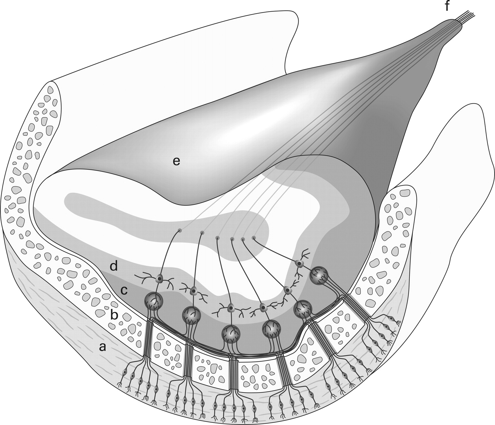
  • Fig 5.
    • Download figure
    • Open in new tab
    • Download powerpoint
    Fig 5.

    Schematic drawing with a coronal cut through the OB, demonstrating asymmetric neuronal layering in an adult OB. a, Nasal olfactory epithelium. b, Cribriform plate. c, Glomerular layer. d, Mitral cell layer. e, Olfactory bulb. f, Olfactory tract.

  • Fig 6.
    • Download figure
    • Open in new tab
    • Download powerpoint
    Fig 6.

    Serial MR imaging examinations (from top to bottom: ages 2 months, 8 months, and 18 months) in the same child show all 3 maturational steps.

PreviousNext
Back to top

In this issue

American Journal of Neuroradiology: 30 (6)
American Journal of Neuroradiology
Vol. 30, Issue 6
June 2009
  • Table of Contents
  • Index by author
Advertisement
Print
Download PDF
Email Article

Thank you for your interest in spreading the word on American Journal of Neuroradiology.

NOTE: We only request your email address so that the person you are recommending the page to knows that you wanted them to see it, and that it is not junk mail. We do not capture any email address.

Enter multiple addresses on separate lines or separate them with commas.
Maturation of the Olfactory Bulbs: MR Imaging Findings
(Your Name) has sent you a message from American Journal of Neuroradiology
(Your Name) thought you would like to see the American Journal of Neuroradiology web site.
CAPTCHA
This question is for testing whether or not you are a human visitor and to prevent automated spam submissions.
Cite this article
J.F. Schneider, F. Floemer
Maturation of the Olfactory Bulbs: MR Imaging Findings
American Journal of Neuroradiology Jun 2009, 30 (6) 1149-1152; DOI: 10.3174/ajnr.A1501

Citation Manager Formats

  • BibTeX
  • Bookends
  • EasyBib
  • EndNote (tagged)
  • EndNote 8 (xml)
  • Medlars
  • Mendeley
  • Papers
  • RefWorks Tagged
  • Ref Manager
  • RIS
  • Zotero
0 Responses
Respond to this article
Share
Bookmark this article
Maturation of the Olfactory Bulbs: MR Imaging Findings
J.F. Schneider, F. Floemer
American Journal of Neuroradiology Jun 2009, 30 (6) 1149-1152; DOI: 10.3174/ajnr.A1501
del.icio.us logo Twitter logo Facebook logo Mendeley logo
  • Tweet Widget
  • Facebook Like
  • Google Plus One
Purchase

Jump to section

  • Article
    • Abstract
    • Materials and Methods
    • Results
    • Discussion
    • References
  • Figures & Data
  • Info & Metrics
  • Responses
  • References
  • PDF

Related Articles

  • No related articles found.
  • PubMed
  • Google Scholar

Cited By...

  • Quantitative Analysis of the Olfactory System in COVID-19: An MR Imaging Study
  • Olfactory Bulb Signal Abnormality in Patients with COVID-19 Who Present with Neurologic Symptoms
  • Diagnostic Accuracy of MRI-Based Morphometric Parameters for Detecting Olfactory Nerve Dysfunction
  • MR Imaging-Based Evaluations of Olfactory Bulb Atrophy in Patients with Olfactory Dysfunction
  • Crossref (24)
  • Google Scholar

This article has been cited by the following articles in journals that are participating in Crossref Cited-by Linking.

  • Correlation between olfactory bulb volume and olfactory function in children and adolescents
    T. Hummel, M. Smitka, S. Puschmann, J. C. Gerber, B. Schaal, D. Buschhüter
    Experimental Brain Research 2011 214 2
  • Odor identification testing in children and young adults using the smell wheel
    E. Leslie Cameron, Richard L. Doty
    International Journal of Pediatric Otorhinolaryngology 2013 77 3
  • A Comparative Olfactory MRI, DTI and fMRI Study of COVID-19 Related Anosmia and Post Viral Olfactory Dysfunction
    Duzgun Yildirim, Sedat Giray Kandemirli, Deniz Esin Tekcan Sanli, Ozlem Akinci, Aytug Altundag
    Academic Radiology 2022 29 1
  • Olfactory Bulb Signal Abnormality in Patients with COVID-19 Who Present with Neurologic Symptoms
    S.B. Strauss, J.E. Lantos, L.A. Heier, D.R. Shatzkes, C.D. Phillips
    American Journal of Neuroradiology 2020 41 10
  • MR Imaging–Based Evaluations of Olfactory Bulb Atrophy in Patients with Olfactory Dysfunction
    M.S. Chung, W.R. Choi, H.-Y. Jeong, J.H. Lee, J.H. Kim
    American Journal of Neuroradiology 2018 39 3
  • Quantitative Analysis of the Olfactory System in COVID-19: An MR Imaging Study
    E. Altunisik, A.H. Baykan, S. Sahin, E. Aydin, S.M. Erturk
    American Journal of Neuroradiology 2021 42 12
  • Neuroradiological Basis of COVID‐19 Olfactory Dysfunction: A Systematic Review and Meta‐Analysis
    Claire Jing‐Wen Tan, Benjamin Kye Jyn Tan, Xin Yan Tan, Hui Ting Liu, Chong Boon Teo, Anna See, Shuhui Xu, Song Tar Toh, Si Wei Kheok, Tze Choong Charn, Neville Wei Yang Teo
    The Laryngoscope 2022 132 6
  • Imaging of lamination patterns of the adult human olfactory bulb and tract: In vitro comparison of standard- and high-resolution 3T MRI, and MR microscopy at 9.4T
    Hartmut P. Burmeister, Thomas Bitter, Patrick M. Heiler, Andrey Irintchev, Rosemarie Fröber, Matthias Dietzel, Pascal A. Baltzer, Lothar R. Schad, Jürgen R. Reichenbach, Hilmar Gudziol, Orlando Guntinas-Lichius, Werner A. Kaiser
    NeuroImage 2012 60 3
  • The Shape of the Olfactory Bulb Predicts Olfactory Function
    Xiaoguang Yan, Akshita Joshi, Yunpeng Zang, Francisca Assunção, Henrique M. Fernandes, Thomas Hummel
    Brain Sciences 2022 12 2
  • Olfactory bulb ventricles as a frequent finding—a myth or reality? Evaluation using high resolution 3 Tesla magnetic resonance imaging
    H.P. Burmeister, T. Bitter, P.A.T. Baltzer, M. Dietzel, O. Guntinas-Lichius, H. Gudziol, W.A. Kaiser
    Neuroscience 2011 172

More in this TOC Section

  • Neuroimaging Delineation and Progression of SLSMD
  • fetal brain development of 10 weeks gestation
  • CHARGE fetal MRI clival cleft
Show more Pediatric Neuroimaging

Similar Articles

Advertisement

Indexed Content

  • Current Issue
  • Accepted Manuscripts
  • Article Preview
  • Past Issues
  • Editorials
  • Editor's Choice
  • Fellows' Journal Club
  • Letters to the Editor
  • Video Articles

Cases

  • Case Collection
  • Archive - Case of the Week
  • Archive - Case of the Month
  • Archive - Classic Case

More from AJNR

  • Trainee Corner
  • Imaging Protocols
  • MRI Safety Corner
  • Book Reviews

Multimedia

  • AJNR Podcasts
  • AJNR Scantastics

Resources

  • Turnaround Time
  • Submit a Manuscript
  • Submit a Video Article
  • Submit an eLetter to the Editor/Response
  • Manuscript Submission Guidelines
  • Statistical Tips
  • Fast Publishing of Accepted Manuscripts
  • Graphical Abstract Preparation
  • Imaging Protocol Submission
  • Evidence-Based Medicine Level Guide
  • Publishing Checklists
  • Author Policies
  • Become a Reviewer/Academy of Reviewers
  • News and Updates

About Us

  • About AJNR
  • Editorial Board
  • Editorial Board Alumni
  • Alerts
  • Permissions
  • Not an AJNR Subscriber? Join Now
  • Advertise with Us
  • Librarian Resources
  • Feedback
  • Terms and Conditions
  • AJNR Editorial Board Alumni

American Society of Neuroradiology

  • Not an ASNR Member? Join Now

© 2025 by the American Society of Neuroradiology All rights, including for text and data mining, AI training, and similar technologies, are reserved.
Print ISSN: 0195-6108 Online ISSN: 1936-959X

Powered by HighWire