Advertisement

Main menu

  • Home
  • Content
    • Current Issue
    • Accepted Manuscripts
    • Article Preview
    • Past Issue Archive
    • Video Articles
    • AJNR Case Collection
    • Case of the Week Archive
    • Case of the Month Archive
    • Classic Case Archive
  • Special Collections
    • Advancing NeuroMRI with High-Relaxivity Contrast Agents
    • AJNR Awards
    • Low-Field MRI
    • Alzheimer Disease
    • ASNR Foundation Special Collection
    • Photon-Counting CT
    • View All
  • Multimedia
    • AJNR Podcasts
    • AJNR SCANtastic
    • Trainee Corner
    • MRI Safety Corner
    • Imaging Protocols
  • For Authors
    • Submit a Manuscript
    • Submit a Video Article
    • Submit an eLetter to the Editor/Response
    • Manuscript Submission Guidelines
    • Statistical Tips
    • Fast Publishing of Accepted Manuscripts
    • Graphical Abstract Preparation
    • Imaging Protocol Submission
    • Author Policies
  • About Us
    • About AJNR
    • Editorial Board
    • Editorial Board Alumni
  • More
    • Become a Reviewer/Academy of Reviewers
    • Subscribers
    • Permissions
    • Alerts
    • Feedback
    • Advertisers
    • ASNR Home

User menu

  • Alerts
  • Log in

Search

  • Advanced search
American Journal of Neuroradiology

American Journal of Neuroradiology

ASHNR American Society of Functional Neuroradiology ASHNR American Society of Pediatric Neuroradiology ASSR
  • Alerts
  • Log in

Advanced Search

  • Home
  • Content
    • Current Issue
    • Accepted Manuscripts
    • Article Preview
    • Past Issue Archive
    • Video Articles
    • AJNR Case Collection
    • Case of the Week Archive
    • Case of the Month Archive
    • Classic Case Archive
  • Special Collections
    • Advancing NeuroMRI with High-Relaxivity Contrast Agents
    • AJNR Awards
    • Low-Field MRI
    • Alzheimer Disease
    • ASNR Foundation Special Collection
    • Photon-Counting CT
    • View All
  • Multimedia
    • AJNR Podcasts
    • AJNR SCANtastic
    • Trainee Corner
    • MRI Safety Corner
    • Imaging Protocols
  • For Authors
    • Submit a Manuscript
    • Submit a Video Article
    • Submit an eLetter to the Editor/Response
    • Manuscript Submission Guidelines
    • Statistical Tips
    • Fast Publishing of Accepted Manuscripts
    • Graphical Abstract Preparation
    • Imaging Protocol Submission
    • Author Policies
  • About Us
    • About AJNR
    • Editorial Board
    • Editorial Board Alumni
  • More
    • Become a Reviewer/Academy of Reviewers
    • Subscribers
    • Permissions
    • Alerts
    • Feedback
    • Advertisers
    • ASNR Home
  • Follow AJNR on Twitter
  • Visit AJNR on Facebook
  • Follow AJNR on Instagram
  • Join AJNR on LinkedIn
  • RSS Feeds

AJNR Awards, New Junior Editors, and more. Read the latest AJNR updates


Improved Turnaround Times | Median time to first decision: 12 days

OtherBrain

Acquisition Time and Reproducibility of Continuous Arterial Spin-Labeling Perfusion Imaging at 3T

S. Gevers, C.B.L.M. Majoie, X.W. van den Tweel, C. Lavini and A.J. Nederveen
American Journal of Neuroradiology May 2009, 30 (5) 968-971; DOI: https://doi.org/10.3174/ajnr.A1454
S. Gevers
  • Find this author on Google Scholar
  • Find this author on PubMed
  • Search for this author on this site
C.B.L.M. Majoie
  • Find this author on Google Scholar
  • Find this author on PubMed
  • Search for this author on this site
X.W. van den Tweel
  • Find this author on Google Scholar
  • Find this author on PubMed
  • Search for this author on this site
C. Lavini
  • Find this author on Google Scholar
  • Find this author on PubMed
  • Search for this author on this site
A.J. Nederveen
  • Find this author on Google Scholar
  • Find this author on PubMed
  • Search for this author on this site
  • Article
  • Figures & Data
  • Info & Metrics
  • Responses
  • References
  • PDF
Loading

Article Figures & Data

Figures

  • Tables
  • Fig 1.
    • Download figure
    • Open in new tab
    • Download powerpoint
    Fig 1.

    Bland Altman plot of intrascan (A), intrasession (B), and intersession (C) WB-CBF differences plotted against mean WB-CBF. WB intrascan, intrasession, and intersession CBF differences are randomly distributed and are not dependent on mean WB-CBF. Dotted lines indicate that the mean WB-CBF difference ± 1.96 SD. 95% of the differences between repeated measurements are within 1.96 SD of the mean difference.
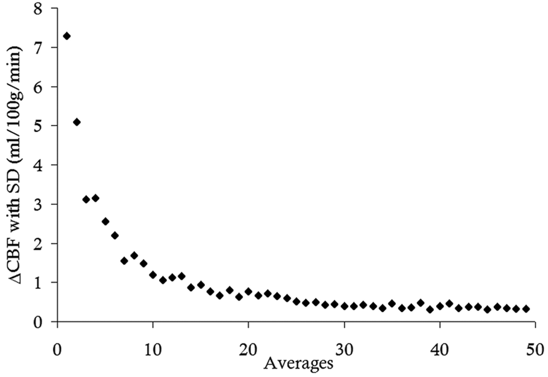
  • Fig 2.
    • Download figure
    • Open in new tab
    • Download powerpoint
    Fig 2.

    Absolute WB-CBF difference, as a function of the number of averages. Steady CBF values are reached after 20 averages.

  • Fig 3.
    • Download figure
    • Open in new tab
    • Download powerpoint
    Fig 3.

    Intrasession and intersession ICC as a function of the number of averages.
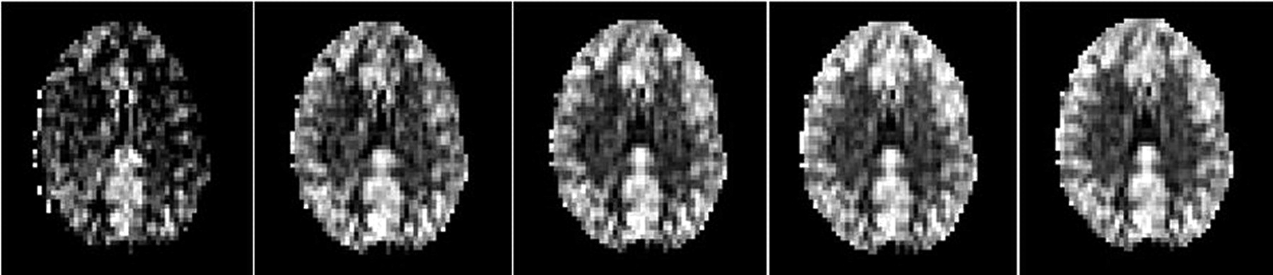
  • Fig 4.
    • Download figure
    • Open in new tab
    • Download powerpoint
    Fig 4.

    CASL sections obtained at 3T with 10, 20, 30, 40, and 50 averages, respectively (left to right); no clear differences are visible between perfusion images on the basis of 20 and 50 averages.

Tables

  • Figures
    • View popup
    Table 1:

    WB and vascular region CBF means and SDs

    Means ± SD (ml/100 g/min)WBRACALACARMCALMCARPCALPCA
    All sessions56.1 ± 11.160.2 ± 12.561.9 ± 13.563.8 ± 14.161.8 ± 13.854.8 ± 13.053.0 ± 14.2
        Male54.2 ± 9.2
        Female58.1 ± 12.6
    Mean Session I56.0 ± 12.160.4 ± 14.061.5 ± 14.563.0 ± 16.062.0 ± 14.054.2 ± 13.552.9 ± 13.4
    Mean Session II55.6 ± 9.559.8 ± 10.461.0 ± 11.663.2 ± 11.161.0 ± 13.255.2 ± 12.153.0 ± 14.2
    Mean Session III56.8 ± 12.060.3 ± 13.563.0 ± 14.765.4 ± 15.562.3 ± 14.755.2 ± 13.853.0 ± 15.5
    • Note:—CBF indicates cerebral blood flow; LACA, left anterior cerebral artery; LMCA, left middle cerebral artery; LPCA, left posterior cerebral artery; RACA, right anterior cerebral artery; RMCA, right middle cerebral artery; RPCA, right posterior cerebral artery; WB, whole brain.

    • View popup
    Table 2:

    Intrascan reproducibility on the basis of the measurement differences between the first 25 and the last 25 averages

    ReproducibilityWBRACALACARMCALMCARPCALPCA
    Intrascan
        RI (mL/100 g/min)10.212.413.112.213.512.811.6
        ICC (95% CI)0.88 (0.81–0.93)0.86 (0.78–0.92)0.87 (0.79–0.92)0.89 (0.83–0.93)0.86 (0.78–0.92)0.87 (0.79–0.92)0.91 (0.85–0.95)*
    • Note:—CI indicates confidence interval; ICC, intraclass correlation coefficient; RI, repeatability index.

    • * Log10 transformed data used in the calculation of the ICC.

    • View popup
    Table 3:

    Intrasession and intersession reproducibility on the basis of 20 averages

    Reproducibility 20 averagesWBRACALACARMCALMCARPCALPCA
    Intrasession
        RI (ml/100 g/min)10.713.313.713.412.614.114.7
        ICC (95% CI)0.85 (0.72–0.93)0.82 (0.65–0.91)0.85 (0.70–0.92)0.86 (0.73–0.93)0.87 (0.75–0.94)0.82 (0.66–0.91)0.79 (0.60–0.89)*
    Intersession
        RI (mL/100 g/min)16.818.020.721.719.417.516.0
        ICC (95% CI)0.61 (0.24–0.87)0.63 (0.27–0.88)0.62 (0.25–0.87)0.60 (0.22–0.87)0.68 (0.33–0.90)0.70 (0.37–0.91)0.78 (0.50–0.93)*
    • * Log10 transformed data used in the calculation of the ICC.

    • View popup
    Table 4:

    Intrasession and intersession reproducibility based on 50 averages

    Reproducibility 50 averagesWBRACALACARMCALMCARPCALPCA
    Intrasession
        RI (mL/100 g/min)10.713.714.113.212.214.111.1
        ICC (95% CI)0.88 (0.76–0.94)0.85 (0.70–0.92)0.86 (0.73–0.93)0.89 (0.78–0.95)0.90 (0.80–0.95)0.85 (0.71–0.93)0.90 (0.80–0.95)*
    Intersession
        RI (ml/100 g/min)14.214.016.619.517.013.812.4
        ICC (95% CI)0.78 (0.49–0.93)0.83 (0.59–0.95)0.79 (0.52–0.94)0.74 (0.43–0.92)0.80 (0.53–0.94)0.84 (0.62–0.95)0.89 (0,72–0,97)*
    • * Log10 transformed data used in the calculation of the ICC.

PreviousNext
Back to top

In this issue

American Journal of Neuroradiology: 30 (5)
American Journal of Neuroradiology
Vol. 30, Issue 5
May 2009
  • Table of Contents
  • Index by author
Advertisement
Print
Download PDF
Email Article

Thank you for your interest in spreading the word on American Journal of Neuroradiology.

NOTE: We only request your email address so that the person you are recommending the page to knows that you wanted them to see it, and that it is not junk mail. We do not capture any email address.

Enter multiple addresses on separate lines or separate them with commas.
Acquisition Time and Reproducibility of Continuous Arterial Spin-Labeling Perfusion Imaging at 3T
(Your Name) has sent you a message from American Journal of Neuroradiology
(Your Name) thought you would like to see the American Journal of Neuroradiology web site.
CAPTCHA
This question is for testing whether or not you are a human visitor and to prevent automated spam submissions.
Cite this article
S. Gevers, C.B.L.M. Majoie, X.W. van den Tweel, C. Lavini, A.J. Nederveen
Acquisition Time and Reproducibility of Continuous Arterial Spin-Labeling Perfusion Imaging at 3T
American Journal of Neuroradiology May 2009, 30 (5) 968-971; DOI: 10.3174/ajnr.A1454

Citation Manager Formats

  • BibTeX
  • Bookends
  • EasyBib
  • EndNote (tagged)
  • EndNote 8 (xml)
  • Medlars
  • Mendeley
  • Papers
  • RefWorks Tagged
  • Ref Manager
  • RIS
  • Zotero
0 Responses
Respond to this article
Share
Bookmark this article
Acquisition Time and Reproducibility of Continuous Arterial Spin-Labeling Perfusion Imaging at 3T
S. Gevers, C.B.L.M. Majoie, X.W. van den Tweel, C. Lavini, A.J. Nederveen
American Journal of Neuroradiology May 2009, 30 (5) 968-971; DOI: 10.3174/ajnr.A1454
del.icio.us logo Twitter logo Facebook logo Mendeley logo
  • Tweet Widget
  • Facebook Like
  • Google Plus One
Purchase

Jump to section

  • Article
    • Abstract
    • Technique
    • Discussion
    • Conclusions
    • Acknowledgments
    • References
  • Figures & Data
  • Info & Metrics
  • Responses
  • References
  • PDF

Related Articles

  • No related articles found.
  • PubMed
  • Google Scholar

Cited By...

  • Cerebral Perfusion Long Term after Therapeutic Occlusion of the Internal Carotid Artery in Patients Who Tolerated Angiographic Balloon Test Occlusion
  • Crossref
  • Google Scholar

This article has not yet been cited by articles in journals that are participating in Crossref Cited-by Linking.

More in this TOC Section

  • Progression of Microstructural Damage in Spinocerebellar Ataxia Type 2: A Longitudinal DTI Study
  • SWI or T2*: Which MRI Sequence to Use in the Detection of Cerebral Microbleeds? The Karolinska Imaging Dementia Study
  • Statin Therapy Does Not Affect the Radiographic and Clinical Profile of Patients with TIA and Minor Stroke
Show more Brain

Similar Articles

Advertisement

Indexed Content

  • Current Issue
  • Accepted Manuscripts
  • Article Preview
  • Past Issues
  • Editorials
  • Editor's Choice
  • Fellows' Journal Club
  • Letters to the Editor
  • Video Articles

Cases

  • Case Collection
  • Archive - Case of the Week
  • Archive - Case of the Month
  • Archive - Classic Case

More from AJNR

  • Trainee Corner
  • Imaging Protocols
  • MRI Safety Corner
  • Book Reviews

Multimedia

  • AJNR Podcasts
  • AJNR Scantastics

Resources

  • Turnaround Time
  • Submit a Manuscript
  • Submit a Video Article
  • Submit an eLetter to the Editor/Response
  • Manuscript Submission Guidelines
  • Statistical Tips
  • Fast Publishing of Accepted Manuscripts
  • Graphical Abstract Preparation
  • Imaging Protocol Submission
  • Evidence-Based Medicine Level Guide
  • Publishing Checklists
  • Author Policies
  • Become a Reviewer/Academy of Reviewers
  • News and Updates

About Us

  • About AJNR
  • Editorial Board
  • Editorial Board Alumni
  • Alerts
  • Permissions
  • Not an AJNR Subscriber? Join Now
  • Advertise with Us
  • Librarian Resources
  • Feedback
  • Terms and Conditions
  • AJNR Editorial Board Alumni

American Society of Neuroradiology

  • Not an ASNR Member? Join Now

© 2025 by the American Society of Neuroradiology All rights, including for text and data mining, AI training, and similar technologies, are reserved.
Print ISSN: 0195-6108 Online ISSN: 1936-959X

Powered by HighWire