Advertisement

Main menu

  • Home
  • Content
    • Current Issue
    • Accepted Manuscripts
    • Article Preview
    • Past Issue Archive
    • Video Articles
    • AJNR Case Collection
    • Case of the Week Archive
    • Case of the Month Archive
    • Classic Case Archive
  • Special Collections
    • AJNR Awards
    • Low-Field MRI
    • Alzheimer Disease
    • ASNR Foundation Special Collection
    • Photon-Counting CT
    • View All
  • Multimedia
    • AJNR Podcasts
    • AJNR SCANtastic
    • Trainee Corner
    • MRI Safety Corner
    • Imaging Protocols
  • For Authors
    • Submit a Manuscript
    • Submit a Video Article
    • Submit an eLetter to the Editor/Response
    • Manuscript Submission Guidelines
    • Statistical Tips
    • Fast Publishing of Accepted Manuscripts
    • Graphical Abstract Preparation
    • Imaging Protocol Submission
    • Author Policies
  • About Us
    • About AJNR
    • Editorial Board
    • Editorial Board Alumni
  • More
    • Become a Reviewer/Academy of Reviewers
    • Subscribers
    • Permissions
    • Alerts
    • Feedback
    • Advertisers
    • ASNR Home

User menu

  • Alerts
  • Log in

Search

  • Advanced search
American Journal of Neuroradiology

American Journal of Neuroradiology

ASHNR American Society of Functional Neuroradiology ASHNR American Society of Pediatric Neuroradiology ASSR
  • Alerts
  • Log in

Advanced Search

  • Home
  • Content
    • Current Issue
    • Accepted Manuscripts
    • Article Preview
    • Past Issue Archive
    • Video Articles
    • AJNR Case Collection
    • Case of the Week Archive
    • Case of the Month Archive
    • Classic Case Archive
  • Special Collections
    • AJNR Awards
    • Low-Field MRI
    • Alzheimer Disease
    • ASNR Foundation Special Collection
    • Photon-Counting CT
    • View All
  • Multimedia
    • AJNR Podcasts
    • AJNR SCANtastic
    • Trainee Corner
    • MRI Safety Corner
    • Imaging Protocols
  • For Authors
    • Submit a Manuscript
    • Submit a Video Article
    • Submit an eLetter to the Editor/Response
    • Manuscript Submission Guidelines
    • Statistical Tips
    • Fast Publishing of Accepted Manuscripts
    • Graphical Abstract Preparation
    • Imaging Protocol Submission
    • Author Policies
  • About Us
    • About AJNR
    • Editorial Board
    • Editorial Board Alumni
  • More
    • Become a Reviewer/Academy of Reviewers
    • Subscribers
    • Permissions
    • Alerts
    • Feedback
    • Advertisers
    • ASNR Home
  • Follow AJNR on Twitter
  • Visit AJNR on Facebook
  • Follow AJNR on Instagram
  • Join AJNR on LinkedIn
  • RSS Feeds

AJNR Awards, New Junior Editors, and more. Read the latest AJNR updates

LetterLetter

“Hot Cross Bun” Sign in Variant Creutzfeldt-Jakob Disease

J.P. Soares-Fernandes, M. Ribeiro and Á. Machado
American Journal of Neuroradiology March 2009, 30 (3) e37; DOI: https://doi.org/10.3174/ajnr.A1335
J.P. Soares-Fernandes
  • Find this author on Google Scholar
  • Find this author on PubMed
  • Search for this author on this site
M. Ribeiro
  • Find this author on Google Scholar
  • Find this author on PubMed
  • Search for this author on this site
Á. Machado
  • Find this author on Google Scholar
  • Find this author on PubMed
  • Search for this author on this site
  • Article
  • Figures & Data
  • Info & Metrics
  • Responses
  • References
  • PDF
Loading

The “hot cross bun” sign refers to pontine cruciform hyperintensity on long TR sequences, which can be observed in multiple-system atrophy, spinocerebellar atrophy types 2 and 3,1 and in parkinsonism secondary to vasculitis.2 It has not been previously demonstrated in variant Creutzfeldt-Jakob disease (vCJD), to our knowledge.

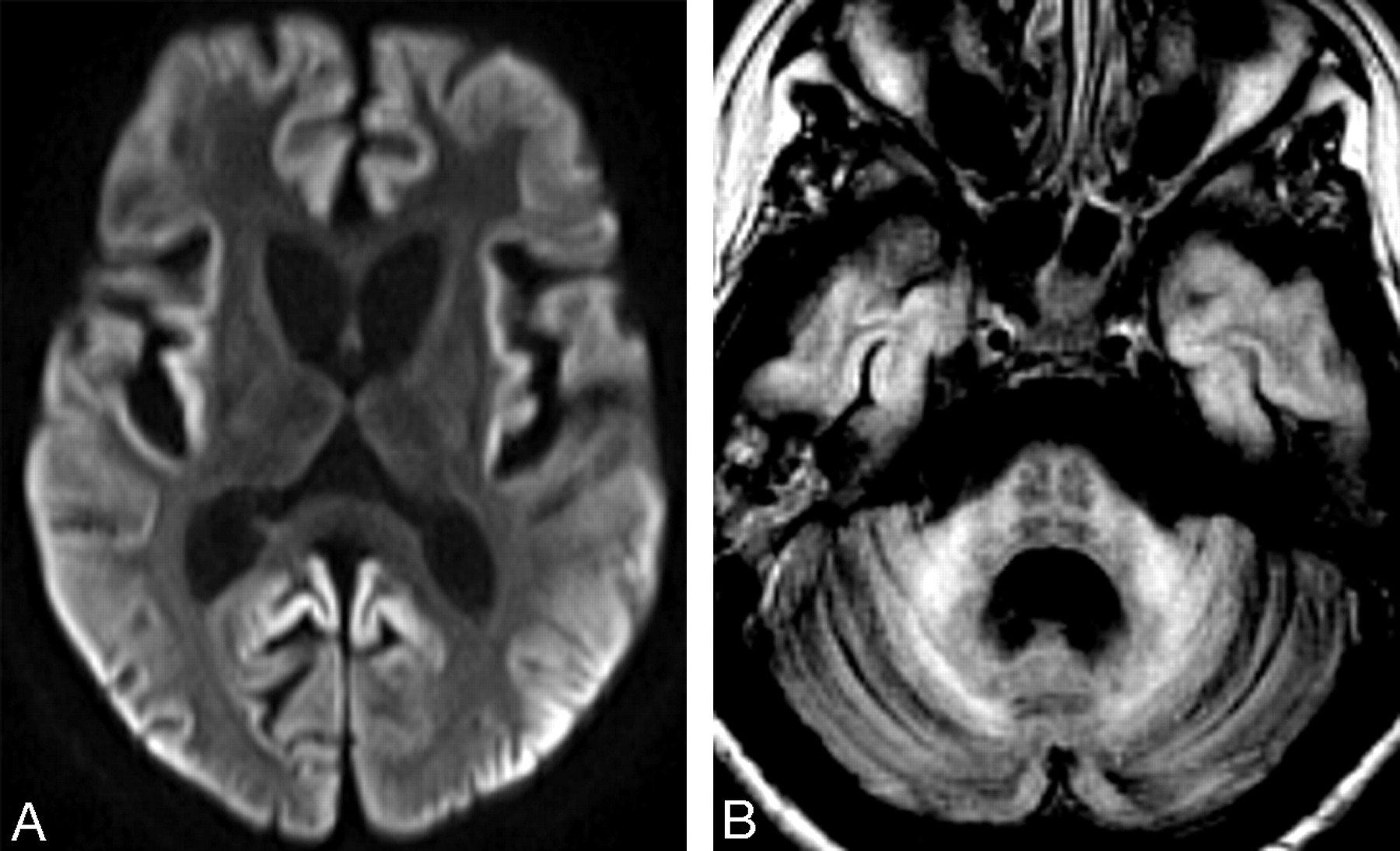
A 16-year-old girl, diagnosed with vCJD at age 14 years and described in detail elsewhere,3 had progressive clinical deterioration, leading to a persistent vegetative state. MR imaging at 18 months after onset of the disease showed global brain atrophy and widespread restricted cortical diffusion (Fig 1A). Pontine cruciform hyperintensity was seen on fluid-attenuated inversion recovery images (Fig 1B).

It is believed that the “hot cross bun” sign results from pontine nuclei neuronal loss and pontocerebellar tract degeneration, with preserved corticospinal tracts. In vCJD, cerebellar involvement is prominent, characterized by marked neuronal loss, astrocytosis, and florid plaques.4 In addition, spongiform changes have been detected in pontine nuclei.4 Secondary degeneration of pontocerebellar tracts is therefore likely to occur, thus supporting our observation. The presented case expands the differential diagnosis of neurodegenerative conditions in which the “hot cross bun” sign can be found.

  Fig 1.
  • Download figure
  • Open in new tab
  • Download powerpoint
Fig 1.

A, Axial diffusion-weighted image shows symmetric striatal and thalamic atrophy and widespread cortical high signal intensity. Apparent diffusion coefficient maps (not shown) revealed corresponding decreased cortical signal intensity, confirming restricted water diffusion. B, Axial fluid-attenuated inversion recovery image demonstrates pontine “hot cross bun” sign, atrophic and hyperintense middle cerebellar peduncles, global cerebellar atrophy, and cortical temporal lobe high signal intensity.

References

  1. ↵
    Bürk K, Skalej M, Dichgans J. Pontine MRI hyperintensities (“the cross sign”) are not pathognomonic for multiple system atrophy (MSA). Mov Disord 2001;16:535
    CrossRefPubMed
  2. ↵
    Muqit MM, Mort D, Miskiel KA, et al. “Hot cross bun” sign in a patient with parkinsonism secondary to presumed vasculitis. J Neurol Neurosurg Psychiatry 2001;71:565–66
    FREE Full Text
  3. ↵
    Machado A, Soares H, Antunes H, et al. Variant Creutzfeldt-Jakob [corrected] disease: the second case in Portugal and in the same geographical region [published erratum appears in J Neurol Neurosurg Psychiatry 2008;79:614]. J Neurol Neurosurg Psychiatry 2008;79:180–82
    Abstract/FREE Full Text
  4. ↵
    Ironside JW, Head MW, McCardle L, et al. Neuropathology of variant Creutzfeldt-Jakob disease. Acta Neurobiol Exp (Wars). 2002;62:175–82
    PubMed
  • Copyright © American Society of Neuroradiology
PreviousNext
Back to top

In this issue

American Journal of Neuroradiology: 30 (3)
American Journal of Neuroradiology
Vol. 30, Issue 3
March 2009
  • Table of Contents
  • Index by author
Advertisement
Print
Download PDF
Email Article

Thank you for your interest in spreading the word on American Journal of Neuroradiology.

NOTE: We only request your email address so that the person you are recommending the page to knows that you wanted them to see it, and that it is not junk mail. We do not capture any email address.

Enter multiple addresses on separate lines or separate them with commas.
“Hot Cross Bun” Sign in Variant Creutzfeldt-Jakob Disease
(Your Name) has sent you a message from American Journal of Neuroradiology
(Your Name) thought you would like to see the American Journal of Neuroradiology web site.
CAPTCHA
This question is for testing whether or not you are a human visitor and to prevent automated spam submissions.
Cite this article
J.P. Soares-Fernandes, M. Ribeiro, Á. Machado
“Hot Cross Bun” Sign in Variant Creutzfeldt-Jakob Disease
American Journal of Neuroradiology Mar 2009, 30 (3) e37; DOI: 10.3174/ajnr.A1335

Citation Manager Formats

  • BibTeX
  • Bookends
  • EasyBib
  • EndNote (tagged)
  • EndNote 8 (xml)
  • Medlars
  • Mendeley
  • Papers
  • RefWorks Tagged
  • Ref Manager
  • RIS
  • Zotero
0 Responses
Respond to this article
Share
Bookmark this article
“Hot Cross Bun” Sign in Variant Creutzfeldt-Jakob Disease
J.P. Soares-Fernandes, M. Ribeiro, Á. Machado
American Journal of Neuroradiology Mar 2009, 30 (3) e37; DOI: 10.3174/ajnr.A1335
del.icio.us logo Twitter logo Facebook logo Mendeley logo
  • Tweet Widget
  • Facebook Like
  • Google Plus One
Purchase

Jump to section

  • Article
    • References
  • Figures & Data
  • Info & Metrics
  • Responses
  • References
  • PDF

Related Articles

  • No related articles found.
  • PubMed
  • Google Scholar

Cited By...

  • Hot cross bun and bright middle cerebellar peduncle signs in cerebellar type multiple system atrophy
  • Crossref (25)
  • Google Scholar

This article has been cited by the following articles in journals that are participating in Crossref Cited-by Linking.

  • “Atypical” atypical parkinsonism: New genetic conditions presenting with features of progressive supranuclear palsy, corticobasal degeneration, or multiple system atrophy—A diagnostic guide
    Maria Stamelou, Niall P. Quinn, Kailash P. Bhatia
    Movement Disorders 2013 28 9
  • Significance of the hot-cross bun sign on T2*-weighted MRI for the diagnosis of multiple system atrophy
    Kazushi Deguchi, Kazuyo Ikeda, Kodai Kume, Tadayuki Takata, Yohei Kokudo, Masaki Kamada, Tetsuo Touge, Naomi Honjo, Tsutomu Masaki
    Journal of Neurology 2015 262 6
  • MRI Shrimp Sign in Cerebellar Progressive Multifocal Leukoencephalopathy: Description and Validation of a Novel Observation
    N. Adra, A.E. Goodheart, O. Rapalino, P. Caruso, S.S. Mukerji, R.G. González, N. Venna, J.D. Schmahmann
    American Journal of Neuroradiology 2021 42 6
  • “Hot cross bun” sign in multiple system atrophy with predominant cerebellar ataxia: A comparison between proton density-weighted imaging and T2-weighted imaging
    Seiko Kasahara, Yukio Miki, Mitsunori Kanagaki, Takayuki Kondo, Akira Yamamoto, Emiko Morimoto, Tomohisa Okada, Hidefumi Ito, Ryosuke Takahashi, Kaori Togashi
    European Journal of Radiology 2012 81 10
  • Various Diseases and Clinical Heterogeneity Are Associated With “Hot Cross Bun”
    Shuzhen Zhu, Hualing Li, Bin Deng, Jialing Zheng, Zifeng Huang, Zihan Chang, Yanjun Huang, Zhibo Wen, Yanran Liang, Mengjue Yu, Ling-Ling Chan, Eng-King Tan, Qing Wang
    Frontiers in Aging Neuroscience 2020 12
  • Differential Diagnosis of T2 Hyperintense Brainstem Lesions: Part 2. Diffuse Lesions
    Juan A. Guzmán-De-Villoria, Concepción Ferreiro-Argüelles, Pilar Fernández-García
    Seminars in Ultrasound, CT and MRI 2010 31 3
  • Differentiating multiple-system atrophy from Parkinson's disease
    N. Ramli, S.R. Nair, N.M. Ramli, S.Y. Lim
    Clinical Radiology 2015 70 5
  • ‘Hot cross bun’ sign in a case of cerebrotendinous xanthomatosis: a rare neuroimaging observation
    Rajendra Singh Jain, Raghavendra Bakki Sannegowda, Amit Agrawal, Deepika Hemrajani, Rahul Jain, Tarun Mathur
    BMJ Case Reports 2013 2013
  • Neuroimagen estructural y funcional en las enfermedades priónicas humanas
    S. Ortega-Cubero, M.R. Luquín, I. Domínguez, J. Arbizu, I. Pagola, M.M. Carmona-Abellán, M. Riverol
    Neurología 2013 28 5
  • ‘Hot cross bun’ sign with leptomeningeal metastases of breast cancer: a case report and review of the literature
    Zhenyu Pan, Guozi Yang, Tingting Yuan, Yongxiang Wang, Xiaochuan Pang, Yan Gao, Lihua Dong
    World Journal of Surgical Oncology 2015 13 1

More in this TOC Section

  • Brain AVM’s Nidus: What if We Hadn’t Understood Anything?
  • Letter to the Editor regarding “Automated Volumetric Software in Dementia: Help or Hindrance to the Neuroradiologist?”
  • Reply:
Show more LETTERS

Similar Articles

Advertisement

Indexed Content

  • Current Issue
  • Accepted Manuscripts
  • Article Preview
  • Past Issues
  • Editorials
  • Editor's Choice
  • Fellows' Journal Club
  • Letters to the Editor
  • Video Articles

Cases

  • Case Collection
  • Archive - Case of the Week
  • Archive - Case of the Month
  • Archive - Classic Case

More from AJNR

  • Trainee Corner
  • Imaging Protocols
  • MRI Safety Corner
  • Book Reviews

Multimedia

  • AJNR Podcasts
  • AJNR Scantastics

Resources

  • Turnaround Time
  • Submit a Manuscript
  • Submit a Video Article
  • Submit an eLetter to the Editor/Response
  • Manuscript Submission Guidelines
  • Statistical Tips
  • Fast Publishing of Accepted Manuscripts
  • Graphical Abstract Preparation
  • Imaging Protocol Submission
  • Evidence-Based Medicine Level Guide
  • Publishing Checklists
  • Author Policies
  • Become a Reviewer/Academy of Reviewers
  • News and Updates

About Us

  • About AJNR
  • Editorial Board
  • Editorial Board Alumni
  • Alerts
  • Permissions
  • Not an AJNR Subscriber? Join Now
  • Advertise with Us
  • Librarian Resources
  • Feedback
  • Terms and Conditions
  • AJNR Editorial Board Alumni

American Society of Neuroradiology

  • Not an ASNR Member? Join Now

© 2025 by the American Society of Neuroradiology All rights, including for text and data mining, AI training, and similar technologies, are reserved.
Print ISSN: 0195-6108 Online ISSN: 1936-959X

Powered by HighWire