
Article Figures & Data
Figures
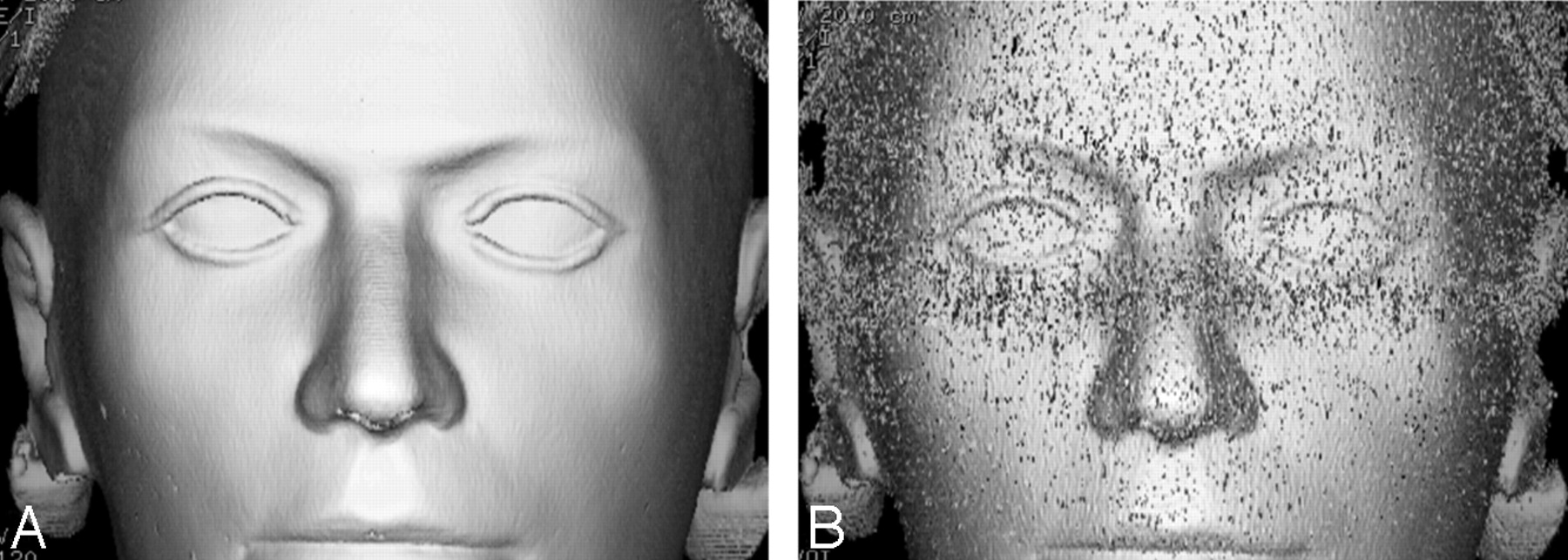
- Fig 1.
A and B, A surface-rendered image reconstructed on an Advantage Windows workstation (GE Healthcare) from a CT dataset performed with a CTDIvol of 65 mGy (A) shows no discernible noise, whereas with a CTDIvol of 9.5 mGy (B), delineation of facial contours is impaired due to image noise which might, according to our initial hypothesis, render surface registration impossible.
- Fig 2.
Screenshot from the navigation system display shows the information the ENT surgeon has available during the intervention. A low-dose dataset (CTDIvol of 3.1 mGy) was used.
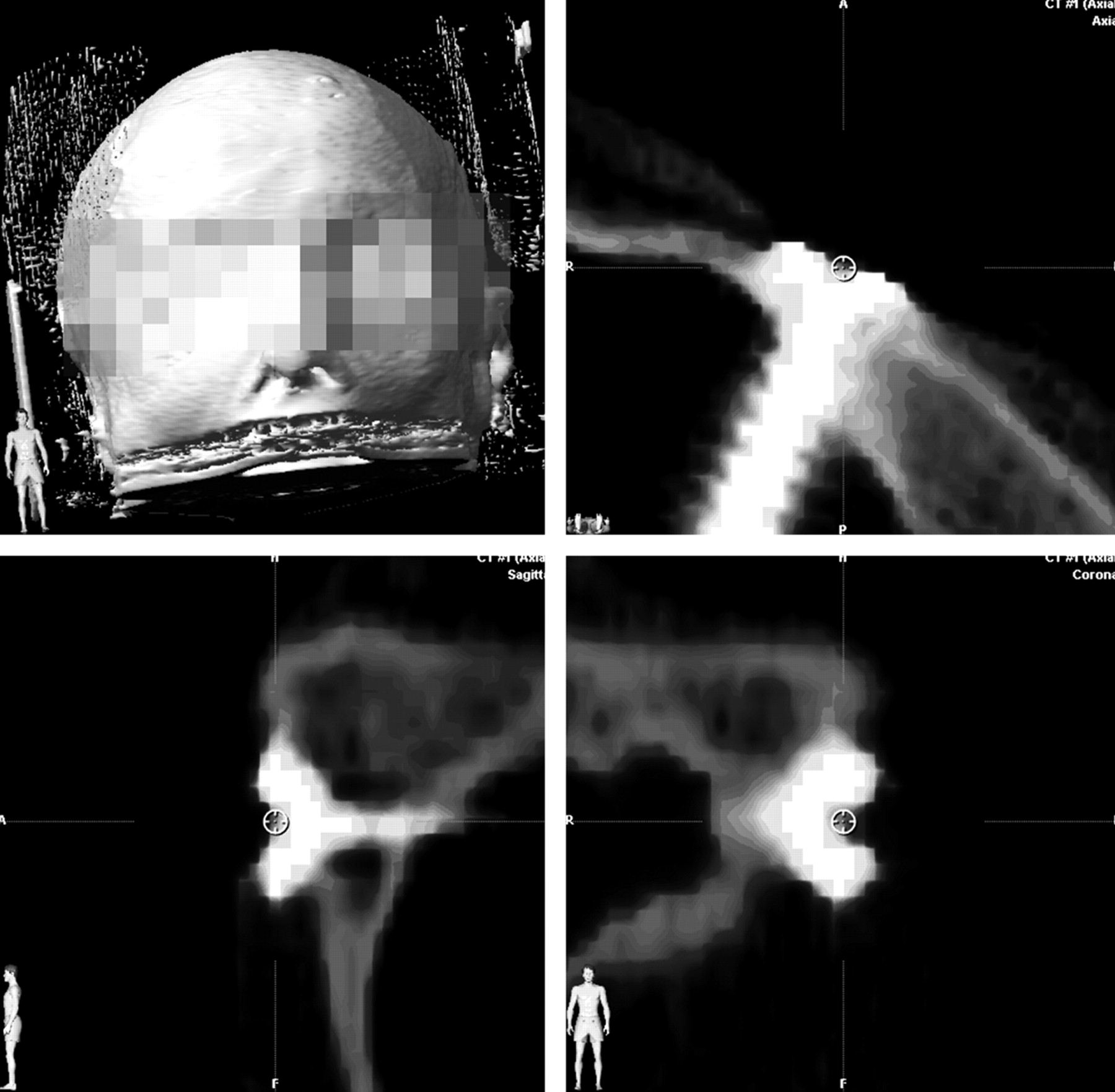
- Fig 3.
This screenshot from the planning software iPlan shows a CT dataset with coronal and sagittal reformations. The central screw head pit of the screw in the zygomatic bone is heavily magnified for positioning the marker circle used for the accuracy measurements.
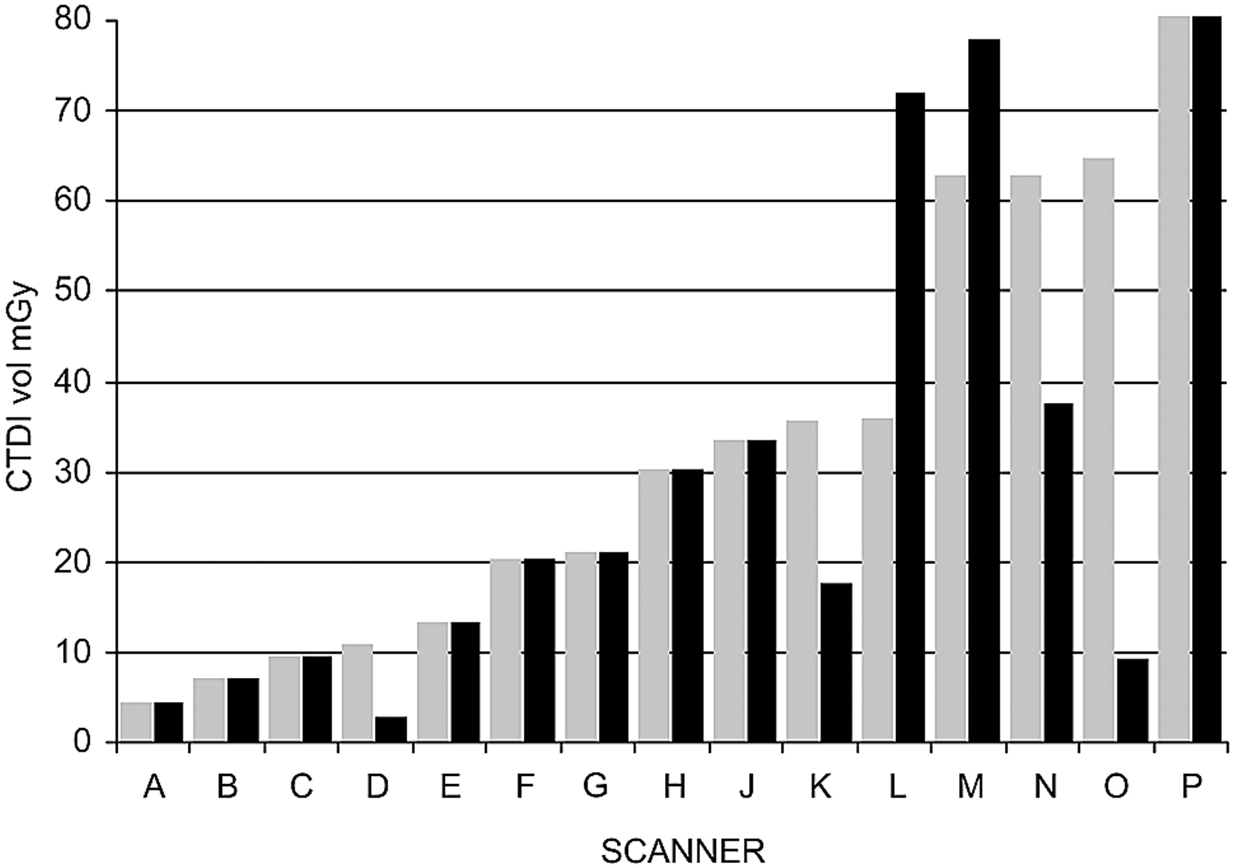
- Fig 4.
Results of the dose survey. Doses used for CAS-CT (gray) and diagnostic sinus CT (black) in 15 CT scanners. O is our institution before the CAS-CT protocol was adapted.
- Fig 5.
Scatterplot of accuracy measurements in millimeters for screw-marked points depicted on the y-axis. CAS-CT dose used in mGy depicted on the logarithmic x-axis. Each column represents the accuracy in mm for 7 screw-marked points on 4 cadaveric heads for registration with CAS-CT at a given dose. The regression line shows no clinically significant slope for accuracy with exponential growth of the radiation dose in the dose interval tested. Note that slight irregularities of the regression line due to the limited resolution of the software used for statistical analysis were straightened by using Photoshop (Adobe Systems, San Jose, Calif), without altering the content of the graphic.
- Fig 6.
Sinus CT in an edge-enhanced (bone) algorithm performed with a CTDIvol of 65 mGy (A) and with a CTDIvol of 9.5 mGy (B). The sevenfold dose difference is not appreciated well, though image B is obviously noisier than A.


Tables
Scanning parameters used in the experiments with phantom and cadaver heads
CTDIvol (mGy) kV mAs 65 120 210 9.5 120 30 6.3 120 20 4 100 20 3.1 120 10 2 100 10 1.1 80 10 Note:—CTDIvol indicates volumetric CT-dose index.
In this issue


Jump to section
Related Articles
Cited By...
- Influence of Ultra-Low-Dose and Iterative Reconstructions on the Visualization of Orbital Soft Tissues on Maxillofacial CT
- Dental Flat Panel Conebeam CT in the Evaluation of Patients with Inflammatory Sinonasal Disease: Diagnostic Efficacy and Radiation Dose Savings
- Radiation Dose Reduction in Paranasal Sinus CT Using Model-Based Iterative Reconstruction