
Article Figures & Data
Figures
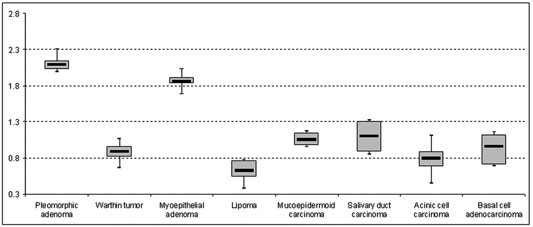
- Fig 1.
The boxplots demonstrate the mean value, 95% confidence interval (CI), and minimum and maximum values for each tumor entity with a number of patients >1. The central line of each boxplot indicates the mean value, whereas the range of the box displays the 95% CI. The whiskers represent the minimal and maximal values (unit of the vertical axis: × 10−3 mm2/s).
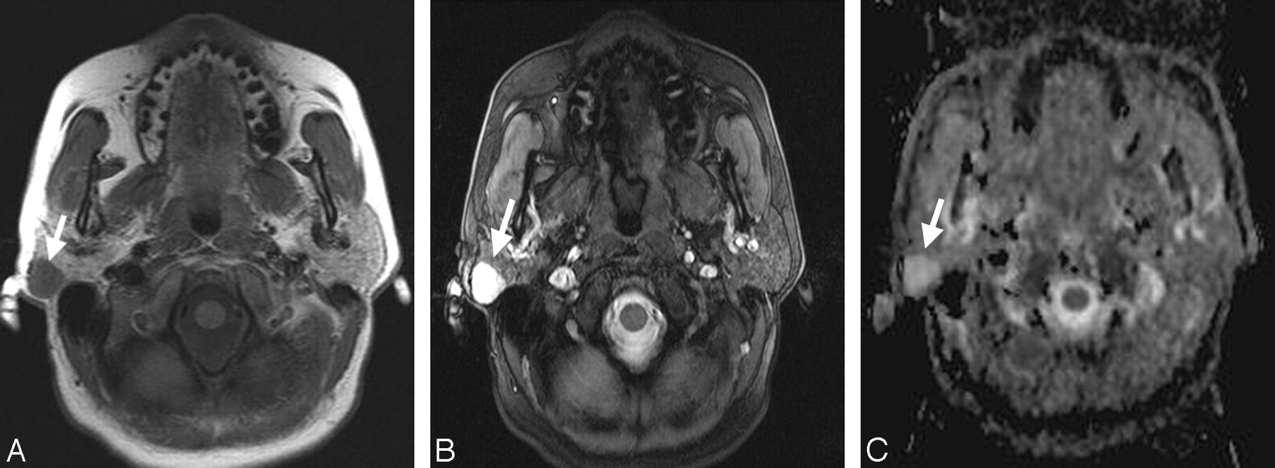
- Fig 2.
A, Transverse T1-weighted spin-echo MR image (TR, 500 ms; TE, 14 ms) from a 69-year-old male patient with a histologically proved pleomorphic adenoma within the right parotid gland (arrow). The mass shows muscle-isointense signal intensity. B, The corresponding T2-weighted 3D true FISP (TR, 700 ms; TE, 2.29 ms) image has a bright lesion within the right parotid gland (arrow). C, The corresponding ADC map shows an easily detectable bright signal intensity of the lesion (mean ADC value: 2.14 × 10⁻³ mm2/s; arrow).
- Fig 3.
A, Transverse T1-weighted spin-echo MR image (TR, 500 ms; TE, 14 ms) from a 49-year-old female patient with a histologically proved Warthin tumor in the left parotid gland (arrow). B, The T2-weighted image (3D true FISP; TR, 700 ms; TE, 2.29 ms) also shows a bright signal intensity of the lesion (arrow) with a lesser extent but appearance similar to that of the pleomorphic adenoma in Fig 2B. C, On the basis of ADC maps, the difference between the pleomorphic adenoma in Fig 2C and the Warthin tumor shown (arrow) becomes obvious (mean ADC value: 0.85 × 10⁻³ mm2/s).


Tables
Diagnosis No. of Lesions (n = 136) Pleomorphic adenoma 43 Warthin tumor 32 Myoepithelial adenoma 6 Lipoma 3 Basal cell adenoma 1 Cystadenoma 1 Inverted ductal papilloma 1 Mucoepidermoid carcinoma 16 Salivary duct carcinoma 11 Acinic cell carcinoma 10 Basal cell adenocarcinoma 9 Adenoid cystic carcinoma 1 Epithelial-myoepithelial carcinoma 1 Carcinoma ex pleomorphic adenoma 1 - Table 2:
Mean ADC values of all histologically proved entities of parotid gland tumors (n = 136)*
Diagnosis Mean ADC values ± SD (95% CI) (× 10−3 mm2/s) Pleomorphic adenoma (n = 43) 2.09 ± 0.16 (2.03–2.14) Warthin tumor (n = 32) 0.89 ± 0.16 (0.82–0.96) Myoepithelial adenoma (n = 6) 1.86 ± 0.18 (1.83–1.91) Lipoma (n = 3) 0.62 ± 0.21 (0.54–0.76) Basal cell adenoma (n = 1) 1.23 Cystadenoma (n = 1) 2.29 Inverted ductal papilloma (n = 1) 1.99 Mucoepidermoid carcinoma (n = 16) 1.05 ± 0.03 (0.97–1.14) Salivary duct carcinoma (n = 11) 1.10 ± 0.09 (0.89–1.31) Acinic cell carcinoma (n = 10) 0.79 ± 0.33 (0.68–0.88) Basal cell adenocarcinoma (n = 9) 0.96 ± 0.29 (0.71–1.12) Adenoid cystic carcinoma (n = 1) 0.87 Epithelial-myoepithelial carcinoma (n = 1) 0.92 Carcinoma ex pleomorphic adenoma (n = 1) 1.14 Note:—CI indicates confidence interval; ADC, apparent diffusion coefficient.
* In case of a single tumor, SD and 95% CI were not calculated.
- Table 3:
Level of significance of calculated mean ADC values comparing all observed entities (n > 1)
Warthin Tumors Myoepithelial Adenomas Lipomas Mucoepidermoid Carcinomas Salivary Duct Carcinomas Acinic Cell Carcinomas Basal Cell Adenocarcinomas Pleomorphic adenomas <.001 .054* <.001 <.001 <.001 <.001 <.001 Warthin tumors <.001 .013 .094* .037 .396* .604* Myoepithelial adenomas .009 .133* .014 .032 .082* Lipomas .024 .022 .48* .222* Mucoepidermoid carcinomas .430* .246* .569* Salivary duct carcinoma .195* .446* Acinic cell carcinomas .616* * Non-statistically significant differences.


