Advertisement

Main menu

  • Home
  • Content
    • Current Issue
    • Accepted Manuscripts
    • Article Preview
    • Past Issue Archive
    • Video Articles
    • AJNR Case Collection
    • Case of the Week Archive
    • Case of the Month Archive
    • Classic Case Archive
  • Special Collections
    • AJNR Awards
    • Low-Field MRI
    • Alzheimer Disease
    • ASNR Foundation Special Collection
    • Photon-Counting CT
    • View All
  • Multimedia
    • AJNR Podcasts
    • AJNR SCANtastic
    • Trainee Corner
    • MRI Safety Corner
    • Imaging Protocols
  • For Authors
    • Submit a Manuscript
    • Submit a Video Article
    • Submit an eLetter to the Editor/Response
    • Manuscript Submission Guidelines
    • Statistical Tips
    • Fast Publishing of Accepted Manuscripts
    • Graphical Abstract Preparation
    • Imaging Protocol Submission
    • Author Policies
  • About Us
    • About AJNR
    • Editorial Board
    • Editorial Board Alumni
  • More
    • Become a Reviewer/Academy of Reviewers
    • Subscribers
    • Permissions
    • Alerts
    • Feedback
    • Advertisers
    • ASNR Home

User menu

  • Alerts
  • Log in

Search

  • Advanced search
American Journal of Neuroradiology

American Journal of Neuroradiology

ASHNR American Society of Functional Neuroradiology ASHNR American Society of Pediatric Neuroradiology ASSR
  • Alerts
  • Log in

Advanced Search

  • Home
  • Content
    • Current Issue
    • Accepted Manuscripts
    • Article Preview
    • Past Issue Archive
    • Video Articles
    • AJNR Case Collection
    • Case of the Week Archive
    • Case of the Month Archive
    • Classic Case Archive
  • Special Collections
    • AJNR Awards
    • Low-Field MRI
    • Alzheimer Disease
    • ASNR Foundation Special Collection
    • Photon-Counting CT
    • View All
  • Multimedia
    • AJNR Podcasts
    • AJNR SCANtastic
    • Trainee Corner
    • MRI Safety Corner
    • Imaging Protocols
  • For Authors
    • Submit a Manuscript
    • Submit a Video Article
    • Submit an eLetter to the Editor/Response
    • Manuscript Submission Guidelines
    • Statistical Tips
    • Fast Publishing of Accepted Manuscripts
    • Graphical Abstract Preparation
    • Imaging Protocol Submission
    • Author Policies
  • About Us
    • About AJNR
    • Editorial Board
    • Editorial Board Alumni
  • More
    • Become a Reviewer/Academy of Reviewers
    • Subscribers
    • Permissions
    • Alerts
    • Feedback
    • Advertisers
    • ASNR Home
  • Follow AJNR on Twitter
  • Visit AJNR on Facebook
  • Follow AJNR on Instagram
  • Join AJNR on LinkedIn
  • RSS Feeds

AJNR Awards, New Junior Editors, and more. Read the latest AJNR updates

OtherBRAIN

Reversible Encephalopathy after Cardiac Transplantation: Histologic Evidence of Endothelial Activation, T-Cell Specific Trafficking, and Vascular Endothelial Growth Factor Expression

C. Horbinski, W.S. Bartynski, E. Carson-Walter, R.L. Hamilton, H.P. Tan and S. Cheng
American Journal of Neuroradiology March 2009, 30 (3) 588-590; DOI: https://doi.org/10.3174/ajnr.A1311
C. Horbinski
  • Find this author on Google Scholar
  • Find this author on PubMed
  • Search for this author on this site
W.S. Bartynski
  • Find this author on Google Scholar
  • Find this author on PubMed
  • Search for this author on this site
E. Carson-Walter
  • Find this author on Google Scholar
  • Find this author on PubMed
  • Search for this author on this site
R.L. Hamilton
  • Find this author on Google Scholar
  • Find this author on PubMed
  • Search for this author on this site
H.P. Tan
  • Find this author on Google Scholar
  • Find this author on PubMed
  • Search for this author on this site
S. Cheng
  • Find this author on Google Scholar
  • Find this author on PubMed
  • Search for this author on this site
  • Article
  • Figures & Data
  • Info & Metrics
  • Responses
  • References
  • PDF
Loading

Abstract

SUMMARY: Reversible encephalopathy after transplantation is well recognized. The condition is commonly thought to be related to immune suppression, and a characteristic brain imaging pattern is typically recognized with vasogenic edema in the parietal and occipital regions, typically termed posterior reversible encephalopathy syndrome (PRES). We report the case of a patient with reversible encephalopathy after cardiac transplantation with brain biopsy evidence of endothelial activation, selective intravascular/perivascular T-cell trafficking, and VEGF expression in astrocytes, neurons, and the endothelium.

Reversible encephalopathy after transplantation is well recognized. A characteristic brain-imaging pattern is typically seen with vasogenic edema in the parietal and occipital regions, typically termed posterior reversible encephalopathy syndrome (PRES). We report the case of a patient with reversible encephalopathy after cardiac transplantation with brain biopsy evidence of endothelial activation, selective intravascular/perivascular T-cell trafficking, and endothelial/cellular vascular endothelial growth factor (VEGF) expression.

Case Report

The patient is a 57-year-old man status postorthotopic heart transplantation for ischemic cardiomyopathy. Routine cardiac biopsy results were normal until 5 months after the transplantation when mild rejection was noted (grade I). One month later, he experienced altered mentation concerning for tacrolimus toxicity, rejection, or infection. Empiric antibiotics and antiviral agents were initiated, but results of work-up for bacterial and virus infections, including lumbar puncture and blood cultures, were negative, and serum tacrolimus levels were within normal limits (13.2 ng/mL; laboratory upper limit of normal, 16 ng/mL). The patient clinically improved and was discharged to home.

One week later, he again presented with confusion. Results of repeated blood cultures and lumbar puncture were negative. On day 5, he experienced a grand mal seizure. At that time, white blood cell count was 3.4 × 10³ μL−1 (77% polymorphonuclear leukocytes, 8% bands, 10% lymphocytes), platelet count was 190 × 103 μL−1, and serum glucose level was 80 mg/dL. Emergent outside CT suggested a “subacute stroke,” and he was transferred to the University Hospital for further management.

On transfer, the patient was lethargic and febrile (102.1°F) but with normal blood pressure readings (range, 114–138/41–66 mm Hg). Brain MR imaging examination demonstrated bifrontal signal intensity abnormality (Fig 1A) consistent with vasogenic edema without restricted diffusion or infarction. Concern remained high for underlying infection; antibiotics were reinstated.

Fig 1.
  • Download figure
  • Open in new tab
  • Download powerpoint
Fig 1.

MR imaging studies of reversible encephalopathy. A, Axial MR fluid-attenuated inversion recovery image obtained at toxicity demonstrates vasogenic edema in the frontal lobes bilaterally (arrows). Minor small-vessel disease changes were also present (arrowhead). Diffusion sequence demonstrated T2 shinethrough, but true restricted diffusion was not present. B, Axial MR fluid-attenuated inversion recovery image obtained 1 month after right frontal lobe biopsy demonstrates near-complete resolution of the edema in the left frontal lobe (large arrow). Postbiopsy changes are present in the right frontal lobe (small arrows) with resolution of the previously present vasogenic edema deep to the biopsy changes. Residual small-vessel disease changes are again noted (arrowhead).

After 6 days, some clinical improvement was noted, but seizure activity persisted and a second MR imaging examination continued to demonstrate bifrontal edema. A positive stool toxin for Clostridium difficile was identified, but results of comprehensive infection work-up (blood/urine/CSF) remained otherwise negative. Concern at this point included undefined brain infection or possible posttransplant lymphoproliferative disease, and it was elected to perform a biopsy of the frontal lobe lesion on the right.

Histologic examination demonstrated evidence of endothelial activation, diffuse vasogenic edema, reactive astrocytes, and reactive microglia (Fig 2A and B). Results of leukocyte common antigen stain demonstrated T-cell (CD3 antigen positive) specific trafficking with intravascular, transmural, and perivascular T-lymphocytes (Fig 3). T-cell trafficking was nonselective with both helper (CD4) and cytotoxic (CD8) subtypes represented. Results of specific antibody staining VEGF and in situ hybridization for VEGF messenger RNA were also positive (Fig 4).

Fig 2.
  • Download figure
  • Open in new tab
  • Download powerpoint
Fig 2.

Early microscopic pathologic features of reversible encephalopathy, hematoxylin-eosin stain. A, Hematoxylin- eosin–stained sections showed many small blood vessels with reactive endothelial cells (black arrow) in the white matter demonstrating cellular and nuclear enlargement as well as mild interstitial edema (white arrow). Some vessels appeared nonreactive and are visible as a comparison (arrowhead). No vasculitis, demyelination, or axonal swelling was seen. Some scattered erythrocytes are present from the biopsy procedure, but no hemosiderin-laden macrophages were seen. B, These reactive blood vessels were also present in the gray matter, seen adjacent to a neuron (arrow), along with reactive astrocytes (arrowheads).

Fig 3.
  • Download figure
  • Open in new tab
  • Download powerpoint
Fig 3.

Early microscopic pathologic feature of reversible encephalopathy on immunohistochemical examination. A, CD45 (leukocyte common antigen) shows scattered lymphocytes in the intravascular (arrowhead) and perivascular (arrow) areas. The vessels identified by the arrows are shown in higher magnification in Figs B and C. B, Higher magnification of arrowhead in Fig 3A. Lymphocytes (T-cells) are regularly noted within the intravascular space (arrows) surrounded by endothelium (arrowhead). Other leukocytes (polymorphonuclear leukocytes, monocytes, macrophages) were not identified, suggesting selective T-lymphocyte adherence. C, Higher magnification of arrowhead in Fig 3A. Lymphocytes (T-cells) have traversed the endothelium (end point of the lymphocyte trafficking process) and are in the perivascular space (arrow). D, These lymphocytes were CD3-positive T-cells (arrow). No CD20-positive B-cells were present, and CD68 immunostaining identified only a modest number of activated microglia with no macrophages (not shown). Results of immunohistochemical and in situ hybridization studies were negative for toxoplasmosis, herpes simplex virus 1 and 2, Epstein-Barr virus, and JC virus (not shown). Results of tissue cultures were also negative.

Fig 4.
  • Download figure
  • Open in new tab
  • Download powerpoint
Fig 4.

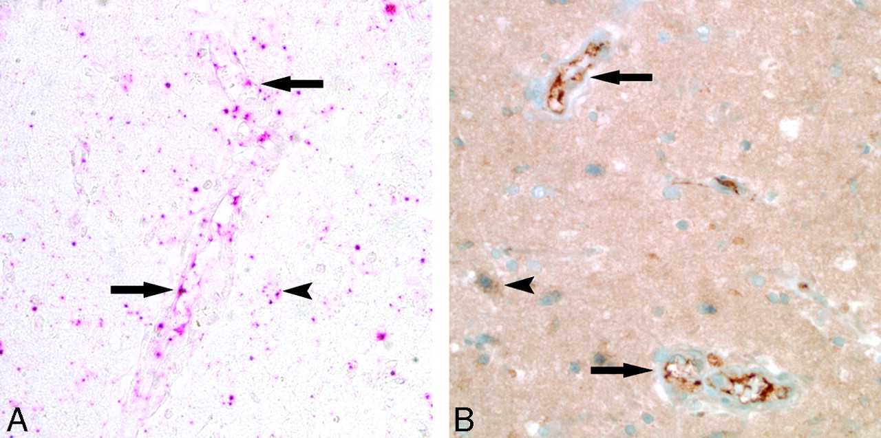
VEGF expression in reversible encephalopathy. A, In situ hybridization with a probe specific to VEGF messenger RNA showed robust transcription (red pigment) in the endothelium (arrows), astrocytes (arrowhead), and neurons. Results of biopsy of control brain tissue were negative (not shown). B, Immunohistochemical study for VEGF protein expression showed strong staining (deep brown pigment) in the brain tissue, including the endothelium (arrows), astrocytes (arrowhead), and neurons.

Trafficking leukocytes were not B-cells (CD20), and no polymorphonuclear cells, monocytes, or macrophages were identified on hematoxylin-eosin sections. Focal lymphocyte accumulation was not present.

Tacrolimus was empirically switched to cyclosporine, the patient's mental status continued to improve, and he was discharged to home. Follow-up MR imaging examination 1 month after discharge (Fig 1B) demonstrated near-complete reversal of the bifrontal vasogenic edema.

Discussion

Because most cases of reversible encephalopathy are diagnosed solely by clinical and radiologic findings, reports describing histologic findings in the acute phase of toxicity are not common. Of those, some are in the postmortem setting where the pathologic process was severe; in such cases, diffuse white matter edema with thrombotic microangiopathy, ischemia, and perivascular lymphocytic infiltration were described.1,2 Reports of biopsy being performed on material in the premortem setting have generally shown vasogenic white matter edema, perivascular lymphocytes, reactive astrocytosis,3–6 and perivascular macrophages.7,8 Endothelial immunoreactivity to tumor necrosis factor-alpha has been demonstrated.4 Some white matter pallor has been attributed to demyelination,9 but this feature more likely represents focally pronounced edema.4 The case of our patient suggests that endothelial activation (with subsequent edema) and perivascular trafficking of T-cells play important roles in the pathogenesis of reversible encephalopathy.

The classic form of reversible encephalopathy is PRES, typically demonstrating vasogenic edema in the parieto-occipital region, frontal lobes, temporo-occipital junction, and cerebellum,10 but other reversible states or presentations are recognized, including isolated brain stem or basal ganglion edema and reversible lesions in the splenium of the corpus callosum.11,12 PRES is recognized after transplantation but is also seen in other complex systemic processes including preeclampsia/eclampsia, infection/sepsis/shock, autoimmune disease, and postcancer chemotherapy. Although the mechanism is as yet unproven, the associated conditions have immune-related antigenic challenge as a common characteristic.13

The current case has elements of classic posttransplant neurotoxicity (altered mentation, reversible brain edema, recent mild rejection) lacking only the typical PRES edema distribution. Indeed, this unusual pattern was the main reason biopsy was performed. Histologic demonstration of T-cell, but not B-cell, trafficking (intravascular, transmural, perivascular T-cells) suggests selective T-cell activation, whereas VEGF (previously vascular permeability factor) expression, likewise, suggests tissue hypoxemia and is likely responsible for the vasogenic edema.

No evidence of local brain infection to account for the T-cell response was identified (histologic examination, immunohistochemical examination, tissue culture). Results of systemic testing for bacterial and virus infection were negative.

A mechanism for the developing vasogenic edema has been proposed on the basis of T-cell activation: endothelial cell activation or injury, T-cell trafficking, plus-or-minus superimposed vasoconstriction with resultant hypoperfusion or hypoxemia, VEGF expression, and increased endothelial permeability.13 Endothelial activation (histologically endothelial cell and nucleus enlargement) and selective T-cell trafficking could certainly lead to restricted brain blood flow with reduced perfusion and hypoxemia. In this case, recent low-grade rejection (reaction to transplant endothelium) or the presence of colon flora imbalance with C difficile might have been the foreign protein antigen source that led to this pathologic sequence of events. The findings in this case suggest that reversible encephalopathy after transplantation may be because of an aberrant T-cell mediated immune response.

References

  1. ↵
    Bartynski WS, Zeigler Z, Spearman MP, et al. Etiology of cortical and white matter lesions in cyclosporin-A and FK-506 neurotoxicity. AJNR Am J Neuroradiol 2001;22:1901–14
    Abstract/FREE Full Text
  2. ↵
    Kilinc M, Benli S, Can U, et al. FK 506-induced fulminant leukoencephalopathy after kidney transplantation: case report. Transplant Proc 2002;34:1182–84
    CrossRefPubMed
  3. ↵
    Lanzino G, Cloft H, Hemstreet MK, et al. Reversible posterior leukoencephalopathy following organ transplantation. Description of two cases. Clin Neurol Neurosurg 1997;99:222–26
    CrossRefPubMed
  4. ↵
    Lavigne CM, Shrier DA, Ketkar M, et al. Tacrolimus leukoencephalopathy: a neuropathologic confirmation. Neurology 2004;63:1132–33
    FREE Full Text
  5. Schiff D, Lopes MB. Neuropathological correlates of reversible posterior leukoencephalopathy. Neurocrit Care 2005;2:303–05
    CrossRefPubMed
  6. ↵
    Thyagarajan GK, Cobanoglu A, Johnston W. FK506-induced fulminant leukoencephalopathy after single-lung transplantation. Ann Thorac Surg 1997;64:1461–64
    CrossRefPubMed
  7. ↵
    Misawa A, Takeuchi Y, Hibi S, et al. FK506-induced intractable leukoencephalopathy following allogeneic bone marrow transplantation. Bone Marrow Transplant 2000;25:331–34
    CrossRefPubMed
  8. ↵
    Schuuring J, Wesseling P, Verrips A. Severe tacrolimus leukoencephalopathy after liver transplantation. AJNR Am J Neuroradiol 2003;24:2085–88
    Abstract/FREE Full Text
  9. ↵
    Small SL, Fukui MB, Bramblett GT, et al. Immunosuppression-induced leukoencephalopathy from tacrolimus (FK506). Ann Neurol 1996;40:575–80
    CrossRefPubMed
  10. ↵
    Bartynski WS. Posterior reversible encephalopathy syndrome, part 1: fundamental imaging and clinical features. AJNR Am J Neuroradiol 2008;29:1036–42
    Abstract/FREE Full Text
  11. ↵
    Ahn KJ, You WJ, Jeong SL, et al. Atypical manifestations of reversible posterior leukoencephalopathy syndrome: findings on diffusion imaging and ADC mapping. Neuroradiology 2004;46:978–83
    CrossRefPubMed
  12. ↵
    Takanashi J, Barkovich AJ, Shiihara T, et al. Widening spectrum of a reversible splenial lesion with transiently reduced diffusion. AJNR Am J Neuroradiol 2006;27:836–38
    Abstract/FREE Full Text
  13. ↵
    Bartynski WS. Posterior reversible encephalopathy syndrome, part 2: controversies surrounding pathophysiology of vasogenic edema. AJNR Am J Neuroradiol 2008;29:1043–49
    Abstract/FREE Full Text
  • Received July 23, 2008.
  • Accepted after revision August 5, 2008.
  • Copyright © American Society of Neuroradiology
View Abstract
PreviousNext
Back to top

In this issue

American Journal of Neuroradiology: 30 (3)
American Journal of Neuroradiology
Vol. 30, Issue 3
March 2009
  • Table of Contents
  • Index by author
Advertisement
Print
Download PDF
Email Article

Thank you for your interest in spreading the word on American Journal of Neuroradiology.

NOTE: We only request your email address so that the person you are recommending the page to knows that you wanted them to see it, and that it is not junk mail. We do not capture any email address.

Enter multiple addresses on separate lines or separate them with commas.
Reversible Encephalopathy after Cardiac Transplantation: Histologic Evidence of Endothelial Activation, T-Cell Specific Trafficking, and Vascular Endothelial Growth Factor Expression
(Your Name) has sent you a message from American Journal of Neuroradiology
(Your Name) thought you would like to see the American Journal of Neuroradiology web site.
CAPTCHA
This question is for testing whether or not you are a human visitor and to prevent automated spam submissions.
Cite this article
C. Horbinski, W.S. Bartynski, E. Carson-Walter, R.L. Hamilton, H.P. Tan, S. Cheng
Reversible Encephalopathy after Cardiac Transplantation: Histologic Evidence of Endothelial Activation, T-Cell Specific Trafficking, and Vascular Endothelial Growth Factor Expression
American Journal of Neuroradiology Mar 2009, 30 (3) 588-590; DOI: 10.3174/ajnr.A1311

Citation Manager Formats

  • BibTeX
  • Bookends
  • EasyBib
  • EndNote (tagged)
  • EndNote 8 (xml)
  • Medlars
  • Mendeley
  • Papers
  • RefWorks Tagged
  • Ref Manager
  • RIS
  • Zotero
0 Responses
Respond to this article
Share
Bookmark this article
Reversible Encephalopathy after Cardiac Transplantation: Histologic Evidence of Endothelial Activation, T-Cell Specific Trafficking, and Vascular Endothelial Growth Factor Expression
C. Horbinski, W.S. Bartynski, E. Carson-Walter, R.L. Hamilton, H.P. Tan, S. Cheng
American Journal of Neuroradiology Mar 2009, 30 (3) 588-590; DOI: 10.3174/ajnr.A1311
del.icio.us logo Twitter logo Facebook logo Mendeley logo
  • Tweet Widget
  • Facebook Like
  • Google Plus One
Purchase

Jump to section

  • Article
    • Abstract
    • Case Report
    • Discussion
    • References
  • Figures & Data
  • Info & Metrics
  • Responses
  • References
  • PDF

Related Articles

  • No related articles found.
  • PubMed
  • Google Scholar

Cited By...

  • Clinical outcomes and risk factors for posterior reversible encephalopathy syndrome in systemic lupus erythematosus: a multicentric case-control study
  • The posterior reversible encephalopathy syndrome in HIV infection
  • Could Increased Vascular Endothelial Growth Factor Induced by Interleukin 17 Be the Cause of Posterior Reversible Encephalopathy Syndrome in Systemic Lupus Erythematosus?
  • Dr. Varaprasad and Dr. Agrawal reply
  • Reversible Cerebral Vasoconstriction Syndrome: Treatment with Combined Intra-Arterial Verapamil Infusion and Intracranial Angioplasty
  • Type of Edema in Posterior Reversible Encephalopathy Syndrome Depends on Serum Albumin Levels: An MR Imaging Study in 28 Patients
  • Influenza A Encephalopathy, Cerebral Vasculopathy, and Posterior Reversible Encephalopathy Syndrome: Combined Occurrence in a 3-Year-Old Child
  • Hemorrhage in Posterior Reversible Encephalopathy Syndrome: Imaging and Clinical Features
  • Crossref (50)
  • Google Scholar

This article has been cited by the following articles in journals that are participating in Crossref Cited-by Linking.

  • Posterior reversible encephalopathy syndrome: clinical and radiological manifestations, pathophysiology, and outstanding questions
    Jennifer E Fugate, Alejandro A Rabinstein
    The Lancet Neurology 2015 14 9
  • Hemorrhage in Posterior Reversible Encephalopathy Syndrome: Imaging and Clinical Features
    H.M. Hefzy, W.S. Bartynski, J.F. Boardman, D. Lacomis
    American Journal of Neuroradiology 2009 30 7
  • Posterior reversible encephalopathy syndrome: The endothelial hypotheses
    A. Marra, M. Vargas, P. Striano, L. Del Guercio, P. Buonanno, G. Servillo
    Medical Hypotheses 2014 82 5
  • Posterior reversible encephalopathy syndrome: do predisposing risk factors make a difference in MRI appearance?
    Christina Mueller-Mang, Thomas Mang, Agnes Pirker, Katharina Klein, Christine Prchla, Daniela Prayer
    Neuroradiology 2009 51 6
  • Blood Pressure Fluctuations in Posterior Reversible Encephalopathy Syndrome
    Alejandro A. Rabinstein, Jay Mandrekar, Ryan Merrell, Osman S. Kozak, Olayemi Durosaro, Jennifer E. Fugate
    Journal of Stroke and Cerebrovascular Diseases 2012 21 4
  • A multi-disciplinary model of risk factors for fatal outcome in posterior reversible encephalopathy syndrome
    Lea M. Alhilali, Arich R. Reynolds, Saeed Fakhran
    Journal of the Neurological Sciences 2014 347 1-2
  • Posterior Reversible Encephalopathy Syndrome Associated with Hemorrhage
    Rosalyn M. Aranas, Shyam Prabhakaran, Vivien H. Lee
    Neurocritical Care 2009 10 3
  • Imaging of Atypical and Complicated Posterior Reversible Encephalopathy Syndrome
    Amin F. Saad, Ruchir Chaudhari, Max Wintermark
    Frontiers in Neurology 2019 10
  • Reversible Cerebral Vasoconstriction Syndrome: Treatment with Combined Intra-Arterial Verapamil Infusion and Intracranial Angioplasty
    H. Farid, J.K. Tatum, C. Wong, V.V. Halbach, S.W. Hetts
    American Journal of Neuroradiology 2011 32 10
  • Immune system activation in the pathogenesis of posterior reversible encephalopathy syndrome
    Zheng Chen, Gui-Quan Shen, Alexander Lerner, Bo Gao
    Brain Research Bulletin 2017 131

More in this TOC Section

  • Evaluating the Effects of White Matter Multiple Sclerosis Lesions on the Volume Estimation of 6 Brain Tissue Segmentation Methods
  • Quiet PROPELLER MRI Techniques Match the Quality of Conventional PROPELLER Brain Imaging Techniques
  • Predictors of Reperfusion in Patients with Acute Ischemic Stroke
Show more BRAIN

Similar Articles

Advertisement

Indexed Content

  • Current Issue
  • Accepted Manuscripts
  • Article Preview
  • Past Issues
  • Editorials
  • Editor's Choice
  • Fellows' Journal Club
  • Letters to the Editor
  • Video Articles

Cases

  • Case Collection
  • Archive - Case of the Week
  • Archive - Case of the Month
  • Archive - Classic Case

More from AJNR

  • Trainee Corner
  • Imaging Protocols
  • MRI Safety Corner

Multimedia

  • AJNR Podcasts
  • AJNR Scantastics

Resources

  • Turnaround Time
  • Submit a Manuscript
  • Submit a Video Article
  • Submit an eLetter to the Editor/Response
  • Manuscript Submission Guidelines
  • Statistical Tips
  • Fast Publishing of Accepted Manuscripts
  • Graphical Abstract Preparation
  • Imaging Protocol Submission
  • Evidence-Based Medicine Level Guide
  • Publishing Checklists
  • Author Policies
  • Become a Reviewer/Academy of Reviewers
  • News and Updates

About Us

  • About AJNR
  • Editorial Board
  • Editorial Board Alumni
  • Alerts
  • Permissions
  • Not an AJNR Subscriber? Join Now
  • Advertise with Us
  • Librarian Resources
  • Feedback
  • Terms and Conditions
  • AJNR Editorial Board Alumni

American Society of Neuroradiology

  • Not an ASNR Member? Join Now

© 2025 by the American Society of Neuroradiology All rights, including for text and data mining, AI training, and similar technologies, are reserved.
Print ISSN: 0195-6108 Online ISSN: 1936-959X

Powered by HighWire