
Article Figures & Data
Figures
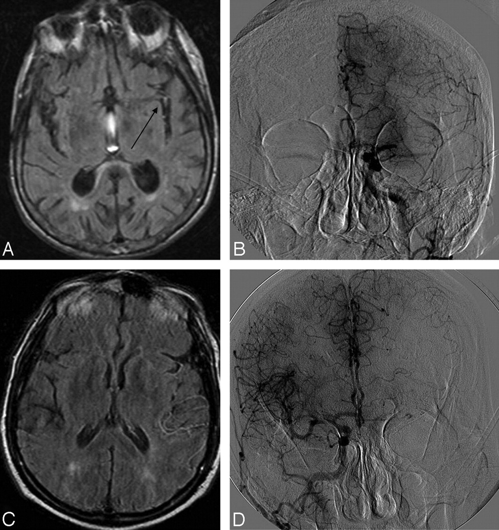
- Fig 1.
Two cases of retrograde leptomeningeal collateral flow in areas corresponding to FVH. A and B, Case 1: FLAIR demonstrates FVH (arrow) in the Sylvian fissure with an angiogram (B) showing grade 3 collaterals from the ipsilateral ACA. C and D, Case 2: FLAIR (C) demonstrates temporoparietal FVH with ACA-MCA leptomeningeal collaterals on the angiogram (D).
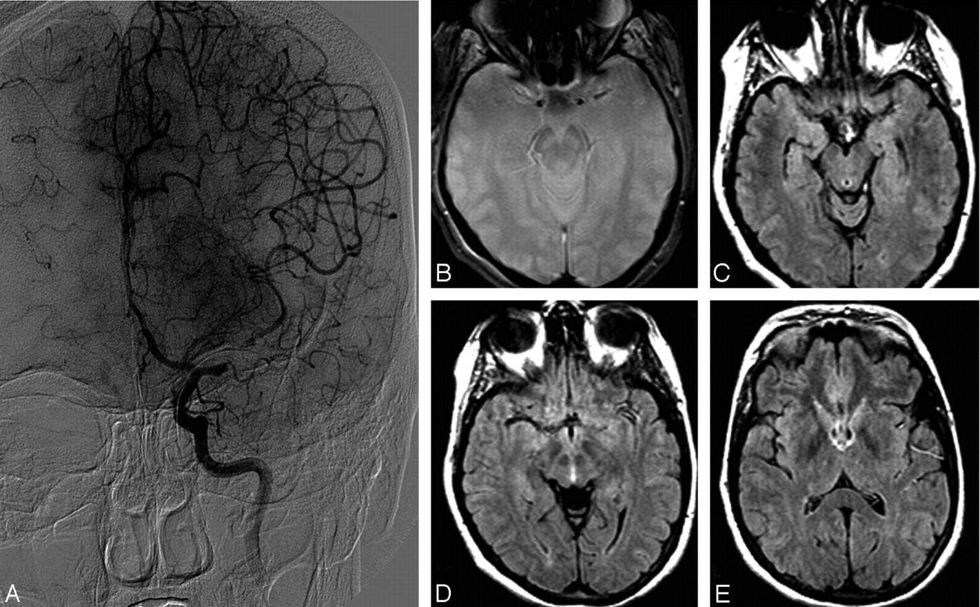
- Fig 2.
A case showing grade 4 collaterals on angiography (A) and FVH (C−E) distal to thrombus, demonstrated as an area of blooming artifact (B).


Tables
Features Data No. of patients 74 Age (yr) Mean, 63.4 Sex 36 Women (48%) Median time from MRI to angiography (hr) 2.9 (IQR, 1.1–4.7) FVH present 53/74 (72%) FVH in arterial territory of ischemia 53/53 (100%) Location of FVH Sylvian fissure 50/53 (94%) Temporal lobe 47/53 (89%) Frontal lobe 28/53 (53%) Parietal lobe 18/53 (34%) Discontinuity or gap in FVH 26/53 (49%) FVH not noted on angiography 21 (28%) Angiographic findings in FVH-negative cases MCA M1 occlusion 1 MCA M1 stenosis with anterograde flow 2 MCA M2 occlusion 2 Distal (M3 or greater) occlusion 8 ICA occlusion without MCA occlusion 2 Vertebrobasilar occlusion 3 Posterior cerebral occlusion 2 Anterior choroidal occlusion 1 Note:—MRI indicates MR imaging; FVH, fluid-attenuated inversion recovery vascular hyperintensities; MCA, middle cerebral artery; ICA, internal carotid artery.
- Table 2:
The ASITN/SIR collateral-flow grading system to determine angiographic collateral grade on pretreatment angiography
ASTIN/SIR Collateral Grade Definition 0 No collaterals visible to the ischemic site 1 Slow collaterals to the periphery of the ischemic site with persistence of some of the defect 2 Rapid collaterals to the periphery of ischemic site with persistence of some of the defect and to only a portion of the ischemic territory 3 Collaterals with slow but complete angiographic blood flow of the ischemic bed by the late venous phase 4 Complete and rapid collateral blood flow to the vascular bed in the entire ischemic territory by retrograde perfusion Note:—ASTIN/SIR indicates American Society of Interventional and Therapeutic Neuroradiology/Society of Interventional Radiology.7
In this issue


Jump to section
Related Articles
Cited By...
- Hyperintense vessel sign in vertebrobasilar dolichoectasia
- FLAIR Vascular Hyperintensities as a Surrogate of Collaterals in Acute Stroke: DWI Matters
- Association between fluid-attenuated inversion recovery vascular hyperintensity and outcome varies with different lesion patterns in patients with intravenous thrombolysis
- Ivy Sign in Moyamoya Disease: A Comparative Study of the FLAIR Vascular Hyperintensity Sign Against Contrast-Enhanced MRI
- The Association between FLAIR Vascular Hyperintensity and Stroke Outcome Varies with Time from Onset
- Hyperintense Vessels, Collateralization, and Functional Outcome in Patients With Stroke Receiving Endovascular Treatment
- FLAIR vascular hyperintensities predict early ischemic recurrence in TIA
- Do Fluid-Attenuated Inversion Recovery Vascular Hyperintensities Represent Good Collaterals before Reperfusion Therapy?
- Acute Ischemic Stroke Therapy Overview
- Fluid-Attenuated Inversion Recovery Vascular Hyperintensity Topography, Novel Imaging Marker for Revascularization in Middle Cerebral Artery Occlusion
- Fluid-Attenuated Inversion Recovery Vascular Hyperintensities-Diffusion-Weighted Imaging Mismatch Identifies Acute Stroke Patients Most Likely to Benefit From Recanalization
- Susceptibility Vessel Sign on MRI Predicts Favorable Clinical Outcome in Patients with Anterior Circulation Acute Stroke Treated with Mechanical Thrombectomy
- Hyperintense Vessels on T2-PROPELLER-FLAIR in Patients with Acute MCA Stroke: Prediction of Arterial Stenosis and Perfusion Abnormality
- Do FLAIR Vascular Hyperintensities beyond the DWI Lesion Represent the Ischemic Penumbra?
- Correlation of clot imaging with endovascular recanalization in internal carotid artery terminus occlusion
- FLAIR vascular hyperintensity resolution in a TIA patient: Clinical-radiologic correlation
- Elevated Cerebral Blood Volume Contributes to Increased FLAIR Signal in the Cerebral Sulci of Propofol-Sedated Children
- Hyperintense Basilar Artery on FLAIR MR Imaging: Diagnostic Accuracy and Clinical Impact in Patients with Acute Brain Stem Stroke
- Clinical Significance of Fluid-Attenuated Inversion Recovery Vascular Hyperintensities in Transient Ischemic Attack
- Guidelines for the Early Management of Patients With Acute Ischemic Stroke: A Guideline for Healthcare Professionals From the American Heart Association/American Stroke Association
- Hyperintense Vessels on Acute Stroke Fluid-Attenuated Inversion Recovery Imaging: Associations With Clinical and Other MRI Findings
- Systematic Review of Methods for Assessing Leptomeningeal Collateral Flow
- Fluid-Attenuated Inversion Recovery Images and Stroke Outcome After Thrombolysis
- Fluid-Attenuated Inversion Recovery Vascular Hyperintensities: An Important Imaging Marker for Cerebrovascular Disease
- Decrease in Leptomeningeal Ivy Sign on Fluid-Attenuated Inversion Recovery Images after Cerebral Revascularization in Patients with Moyamoya Disease