
Article Figures & Data
Figures
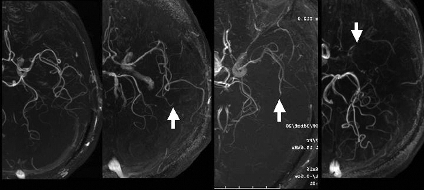
- Fig 1.
Degree of visualization of the ipsilateral MCA on brain MRA was graded as follows: all M3 branches of the left MCA could be visualized to the cortical surface (grade A), one M3 branch could not be visualized to the cortical surface (grade B, arrow), one M2 branch could not be visualized along its course (grade C, arrow), and the M1 could not be visualized along its course (grade D, arrow).
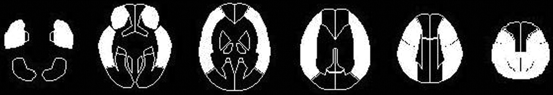
- Fig 2.
Diagrams showing the ROIs of a 3D stereotaxic ROI template. The white ROIs (precentral, central, parietal, angular, and temporal segments) indicate territories perfused by the bilateral MCAs.
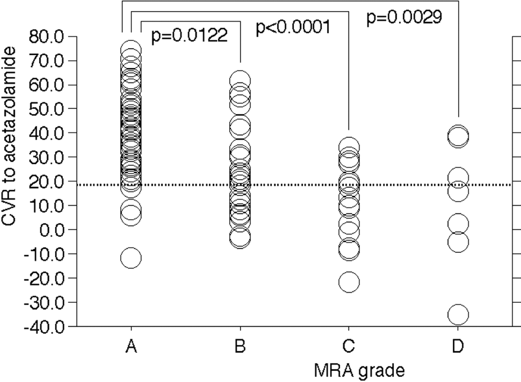
- Fig 3.
Comparison of CVR to acetazolamide among the 4 MRA grades in cerebral hemispheres with lesions.
- Fig 4.
A 70-year-old man with symptomatic left ICA stenosis (90%) exhibiting MRA grade C (Fig 1). Single-photon emission CT scan shows reduction of resting cerebral blood flow (upper images) and poor acetazolamide-induced increases in perfusion (lower images) in the left MCA territory.


Tables
Concordance rates in interobserver and intraobserver agreements of MRA grading in 108 cerebral hemispheres with lesions
First Review by First Observer Second Review by First Observer First Review by Second Observer MRA Grade Number of Hemispheres MRA Grades Number of Hemispheres Concordance Rate in Intraobserver Agreement MRA Grade Number of Hemispheres Concordance Rate in Interobserver Agreement A 59 A 59 1.00 A 59 1.00 B 0 B 0 C 0 C 0 D 0 D 0 B 26 A 0 0.85 A 0 0.85 B 22 B 22 C 4 C 4 D 0 D 0 C 16 A 0 0.94 A 0 0.88 B 1 B 1 C 15 C 14 D 0 D 1 D 7 A 0 1.00 A 0 0.71 B 0 B 0 C 0 C 2 D 7 D 5 Note:—MRA indicates MR angiography.
In this issue


Jump to section
Related Articles
Cited By...
- Estimating Flow Direction of Circle of Willis Using Dynamic Arterial Spin-Labeling MR Angiography
- Predicting Impaired Cerebrovascular Reactivity and Hyperperfusion Syndrome with BeamSAT MRI in Carotid Artery Stenosis
- Fractional Flow on TOF-MRA as a Measure of Stroke Risk in Children with Intracranial Arterial Stenosis
- The Association between FLAIR Vascular Hyperintensity and Stroke Outcome Varies with Time from Onset