
Article Figures & Data
Figures
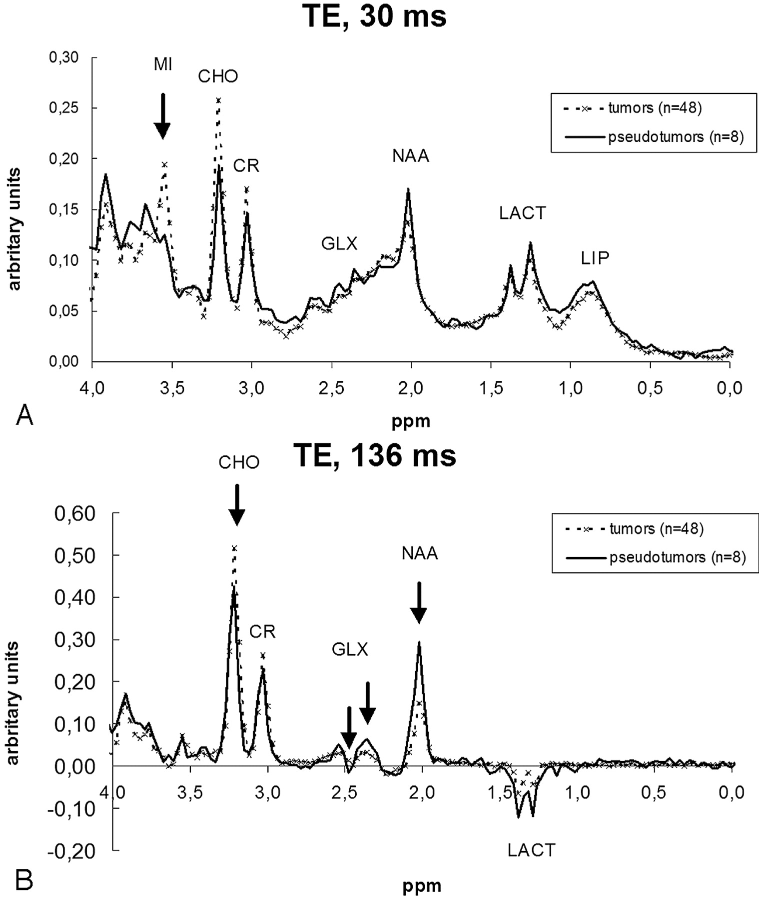
- Fig 1.
Average spectra of tumors and pseudotumors calculated with the cases included in the training-set. Arrows depict the points that showed significant differences between the 2 groups in the statistical analysis. A, Short TE (TE, 30 ms) spectra showed significant differences at 3.55 ppm (MI). Although differences in the mean spectra can also be seen at 2.02 ppm (NAA) and 3.22 ppm (CHO), no statistical significance was reached. B, Long TE (TE, 136 ms) spectra showed significant differences at 2.02 ppm (NAA), 2.36 and 2.48 ppm (GLX), and 3.22 ppm (CHO).
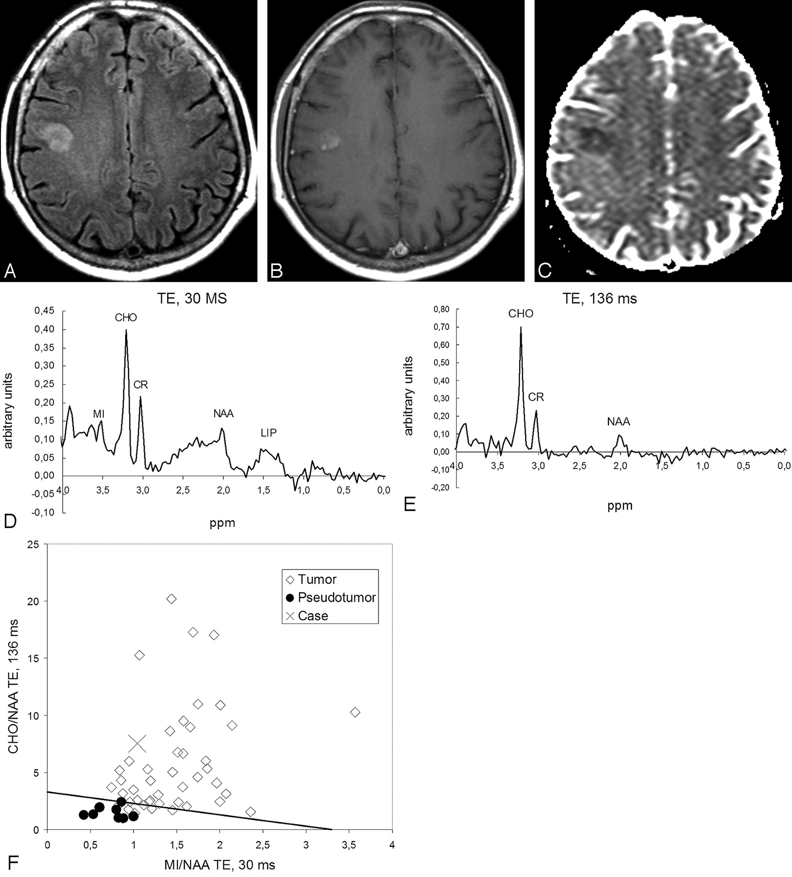
- Fig 2.
Case 81. Anaplastic oligoastrocytoma. A, FLAIR-weighted images show a hyperintense subcortical lesion on the right frontal lobe. B, The lesion shows slight enhancement on T1-weighted images obtained after contrast administration. C, Decrease in the apparent diffusion coefficient (ADC) is seen on the ADC map. The possibility of a cortical infarct was considered. D, 1H-MR spectroscopy at short TE shows slight increase of the mIns/NAA ratio, high CHO, and low NAA. E, 1H-MR spectroscopy at long TE shows high CHO and low NAA levels. F, Graph constructed with the mIns/NAA ratio at short TE (x axis) and CHO/NAA at long TE (y axis) of the universe (latent space) of cases of the training-set projects the case (cross) in the tumoral area. The straight line in the graph depicts the cutting point between tumor and pseudotumor on the basis of the combined ratio value (formula, x + y = 3.33). Values obtained for the classifiers were mIns/NAA ratio at short TE, 1.04 (>0.90, suggesting tumor); CHO/NAA ratio at long TE, 7.52 (>1.90, suggesting tumor), and combined ratio, 8.56 (>3.3, suggesting tumor). The lesion was an anaplastic oligoastrocytoma on the pathologic assessment.
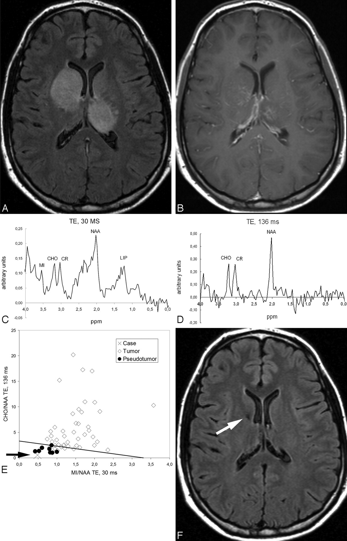
- Fig 3.
Case 68. Unspecific benign pseudotumoral mass. A, FLAIR-weighted images show enlargement and hyperintensity of the head of the right caudate nucleus and left thalamus. B, T1-weighted images after contrast administration show slight discontinuous contrast enhancement. C, 1H-MR spectroscopy at short TE. Ratios between CHO, Cr, and NAA are within the normal range. D, 1H-MR spectroscopy at long TE shows no significant anomalies in the ratios between CHO, Cr, and NAA. E, Graph constructed with the mIns/NAA ratio at short TE (x axis) and CHO/NAA ratio at long TE (y axis) of the universe (latent space) of cases of the training-set projects the case (cross) in the nontumoral area. The straight line in the graph depicts the cutting point between tumor and pseudotumor on the basis of the combined ratio value (formula, x + y = 3.33). The arrow highlights the position of the case. Values obtained for the classifiers were mIns/NAA ratio at short TE, 0.48 (<0.90, suggesting nontumor); CHO/NAA ratio at long TE, 0.55 (<1.90, suggesting nontumor); and combined ratio, 1.03 (<3.3, suggesting nontumor). The pathologic assessment of a stereotactic biopsy was “inflammatory changes.” F, Follow-up with axial FLAIR images obtained in 8 months shows complete resolution of previous abnormalities in the images. Note a small hypointense area on the head of the caudate nucleus (arrow) corresponding to the biopsy area.


Tables
Case No. Follow-up (Months/Evolution of Lesion) Histologic Examination Final Diagnosis 2 77 m/Focal atrophy No Acute arterial infarct 13 26 m/Lesion regression. Detection of new lesions on follow-up Yes, brain tissue without abnormalities Multiple sclerosis 17 14 m/Lesion regression No Acute disseminated encephalomyelitis 28 30 m/Lesion regression Yes, brain tissue without abnormalities Multiple sclerosis 33 2 m/Lesion regression No Unspecific 34 75 m/Lesion regression. Detection of new lesions on follow-up Yes, inflammatory, gliosis Multiple sclerosis 42 35 m/Focal atrophy Yes, inflammatory, gliosis Unspecific 52 30 m/Lesion regression. Resolution of contrast enhancement Yes, inflammatory, gliosis Unspecific 60 8 m/Lesion regression. Resolution of contrast enhancement No Multiple sclerosis 62 27 m/Lesion regression Yes, inflammatory Unspecific 64 20 m/Lesion regression. Resolution of contrast enhancement No Unspecific 68 8 m/Lesion resolution Yes, inflammatory Unspecific 72 6 m/Lesion regression. Focal atrophy No Acute arterial infarct 78 11 m/Lesion regression No Acute arterial infarct 80 4 m/Lesion regression No Venous infarct 82 8 m/Lesion regression No Multiple sclerosis Diagnosis Number of Cases Tumors Training-Set Test-Set Low-grade astrocytoma (WHO grade II) 19 4 Oligodendroglioma (WHO grade II) 4 3 Oligoastrocytoma (WHO grade II) 3 1 Anaplastic astrocytoma (WHO grade III) 16 11 Anaplastic oligoastrocytoma (WHO grade III) 6 1 Pseudotumors Training-Set Test-Set Acute infarct 1 3 Multiple sclerosis 3 2 Acute disseminated encephalomyelitis 1 0 No specific diagnosis 3 3 Total 56 28 - Table 3:
Confidence rating of 5 neuroradiologists in the discrimination between tumor and pseudotumor in 28 test-cases, before and after having available spectroscopic information
Step 1: Evaluation with MR Imaging Alone Step 2: Evaluation with MR Imaging and MR Spectroscopy 0* 1* 2* 3* 4* TOTAL 0* 3 2 3 1 0 9 1* 4 4 4 13 0 25 2* 0 2 0 10 1 13 3* 0 1 8 11 27 47 4* 0 0 0 1 45 46 TOTAL 7 9 15 36 73 140 Note:—The numbers in the table represent the number of times that each combination of scores was given by participating neuroradiologists.
* Modified scores: 0, quite certainly the wrong diagnosis; 1, probably the wrong diagnosis; 2, equivocal; 3, probably the right diagnosis; and 4, quite certainly the right diagnosis.
In this issue


Jump to section
Related Articles
Cited By...
- Diagnostic Accuracy of MR Spectroscopic Imaging and 18F-FET PET for Identifying Glioma: A Biopsy-Controlled Hybrid PET/MRI Study
- Spatial Relationship of Glioma Volume Derived from 18F-FET PET and Volumetric MR Spectroscopy Imaging: A Hybrid PET/MRI Study
- Utility of Proton MR Spectroscopy for Differentiating Typical and Atypical Primary Central Nervous System Lymphomas from Tumefactive Demyelinating Lesions
- MR Imaging of Neoplastic Central Nervous System Lesions: Review and Recommendations for Current Practice
- Proton MR Spectroscopy Provides Relevant Prognostic Information in High-Grade Astrocytomas
- Intracranial dural arteriovenous fistula presenting as an enhancing lesion of the medulla