
Article Figures & Data
Figures
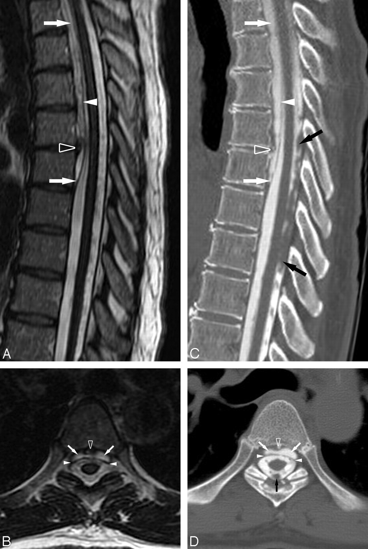
- Fig 1.
A, Sagittal T2-weighted (TR, 3300 msec; TE, 100.8 msec) image of the thoracic spine defines the caudal aspect of the epidural fluid collection at T6–T7. A prominent central disk extrusion is present at T5–T6. B, Axial T2-weighted (TR, 2600 msec; TE, 103.1 msec) image through the T5–T6 disk extrusion further delineates the ventral epidural fluid collection displacing the dura posteriorly. Note the prominent T2 hypointensity on the cord surface secondary to superficial siderosis. C, D, Postmyelography CT images in similar planes and at analogous levels to the MR images demonstrate that the ventral epidural fluid collection is opacified by intrathecal contrast to a degree similar to CSF within the thecal sac, confirming the presence of an active CSF leak. The prominent T5–T6 disk extrusion is partially calcified. Ventral epidural fluid collection (white arrows), dura (white arrowheads), T5–T6 disk extrusion (open arrowhead), and subarachnoid clot (black arrows) are shown.
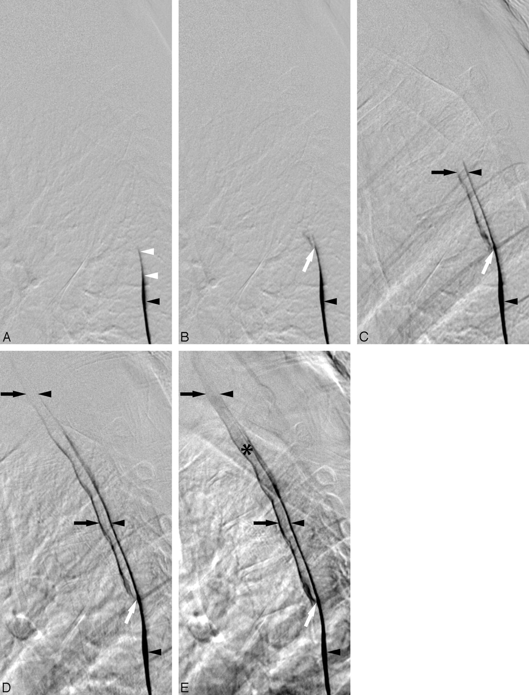
- Fig 2.
Lateral views of the thoracic spine acquired during digital subtraction myelography with the patient in the prone position. For temporal reference, the initial image is designated as time of (A) 0.0 second, and the subsequent images are at (B) 0.4 second, (C) 1.4 seconds, (D) 3.6 seconds, and (E) 11.2 seconds. Contrast is seen to progressively extend cephalad dependently within the thecal sac (black arrowheads). At T6–T7, the contrast slightly deviates dorsally because of mass effect on the dura by the caudal aspect of the epidural fluid collection (white arrowheads). Contrast focally extravasates through the ventral dural tear at T5–T6 (white arrow) into the epidural collection (black arrows). Contrast extends cephalad within both the thecal sac and the epidural collection. Ultimately, the epidural fluid collection becomes more dense (*) because of its smaller volume relative to the subarachnoid space.


In this issue


Jump to section
Related Articles
Cited By...
- Azygos Vein Stenosis in Frontotemporal Dementia Sagging Brain Syndrome
- Lateral Spinal CSF Leaks in Patients with Spontaneous Intracranial Hypotension: Radiologic-Anatomic Study of Different Variants
- Optic Nerve Sheath MR Imaging Measurements in Patients with Orthostatic Headaches and Normal Findings on Conventional Imaging Predict the Presence of an Underlying CSF-Venous Fistula
- Mechanism of chronic iatrogenic CSF leak following dural puncture-ventral dural leak: case report
- Conebeam CT as an Adjunct to Digital Subtraction Myelography for Detection of CSF-Venous Fistulas
- Modified Dynamic CT Myelography for Type 1 and 2 CSF Leaks: A Procedural Approach
- Surgical Ligation of Spinal CSF-Venous Fistulas after Transvenous Embolization in Patients with Spontaneous Intracranial Hypotension
- Diskogenic Dural Defect Is the Reason for the Ventral Location of the Epidural Spinal Fluid Collection Seen in Superficial Siderosis
- Diagnostic Yield of Lateral Decubitus Digital Subtraction Myelogram Stratified by Brain MRI Findings
- Spinal CSF-Venous Fistulas in Morbidly and Super Obese Patients with Spontaneous Intracranial Hypotension
- Safety of Consecutive Bilateral Decubitus Digital Subtraction Myelography in Patients with Spontaneous Intracranial Hypotension and Occult CSF Leak
- Spine MRI in Spontaneous Intracranial Hypotension for CSF Leak Detection: Nonsuperiority of Intrathecal Gadolinium to Heavily T2-Weighted Fat-Saturated Sequences
- Renal Excretion of Contrast on CT Myelography: A Specific Marker of CSF Leak
- Spontaneous Intracranial Hypotension: A Systematic Imaging Approach for CSF Leak Localization and Management Based on MRI and Digital Subtraction Myelography
- Spinal meningeal diverticula, spontaneous intracranial hypotension, and superficial siderosis
- A classification system of spontaneous spinal CSF leaks
- The "Hyperdense Paraspinal Vein" Sign: A Marker of CSF-Venous Fistula
- False localizing sign of cervico-thoracic CSF leak in spontaneous intracranial hypotension
- Beyond superficial siderosis: Introducing "duropathies"
- When Should I Do Dynamic CT Myelography? Predicting Fast Spinal CSF Leaks in Patients with Spontaneous Intracranial Hypotension
- Superficial siderosis associated with abundant {tau} and {alpha}-synuclein accumulation
- Neuroimaging in Superficial Siderosis: An In-Depth Look