Advertisement

Main menu

  • Home
  • Content
    • Current Issue
    • Accepted Manuscripts
    • Article Preview
    • Past Issue Archive
    • Video Articles
    • AJNR Case Collection
    • Case of the Week Archive
    • Case of the Month Archive
    • Classic Case Archive
  • Special Collections
    • Advancing NeuroMRI with High-Relaxivity Contrast Agents
    • AJNR Awards
    • Low-Field MRI
    • Alzheimer Disease
    • ASNR Foundation Special Collection
    • Photon-Counting CT
    • View All
  • Multimedia
    • AJNR Podcasts
    • AJNR SCANtastic
    • Trainee Corner
    • MRI Safety Corner
    • Imaging Protocols
  • For Authors
    • Submit a Manuscript
    • Submit a Video Article
    • Submit an eLetter to the Editor/Response
    • Manuscript Submission Guidelines
    • Statistical Tips
    • Fast Publishing of Accepted Manuscripts
    • Graphical Abstract Preparation
    • Imaging Protocol Submission
    • Author Policies
  • About Us
    • About AJNR
    • Editorial Board
    • Editorial Board Alumni
  • More
    • Become a Reviewer/Academy of Reviewers
    • Subscribers
    • Permissions
    • Alerts
    • Feedback
    • Advertisers
    • ASNR Home

User menu

  • Alerts
  • Log in

Search

  • Advanced search
American Journal of Neuroradiology

American Journal of Neuroradiology

ASHNR American Society of Functional Neuroradiology ASHNR American Society of Pediatric Neuroradiology ASSR
  • Alerts
  • Log in

Advanced Search

  • Home
  • Content
    • Current Issue
    • Accepted Manuscripts
    • Article Preview
    • Past Issue Archive
    • Video Articles
    • AJNR Case Collection
    • Case of the Week Archive
    • Case of the Month Archive
    • Classic Case Archive
  • Special Collections
    • Advancing NeuroMRI with High-Relaxivity Contrast Agents
    • AJNR Awards
    • Low-Field MRI
    • Alzheimer Disease
    • ASNR Foundation Special Collection
    • Photon-Counting CT
    • View All
  • Multimedia
    • AJNR Podcasts
    • AJNR SCANtastic
    • Trainee Corner
    • MRI Safety Corner
    • Imaging Protocols
  • For Authors
    • Submit a Manuscript
    • Submit a Video Article
    • Submit an eLetter to the Editor/Response
    • Manuscript Submission Guidelines
    • Statistical Tips
    • Fast Publishing of Accepted Manuscripts
    • Graphical Abstract Preparation
    • Imaging Protocol Submission
    • Author Policies
  • About Us
    • About AJNR
    • Editorial Board
    • Editorial Board Alumni
  • More
    • Become a Reviewer/Academy of Reviewers
    • Subscribers
    • Permissions
    • Alerts
    • Feedback
    • Advertisers
    • ASNR Home
  • Follow AJNR on Twitter
  • Visit AJNR on Facebook
  • Follow AJNR on Instagram
  • Join AJNR on LinkedIn
  • RSS Feeds

AJNR Awards, New Junior Editors, and more. Read the latest AJNR updates


Improved Turnaround Times | Median time to first decision: 12 days

Research ArticleBrain
Open Access

The Effect of Exercise on the Cerebral Vasculature of Healthy Aged Subjects as Visualized by MR Angiography

E. Bullitt, F.N. Rahman, J.K. Smith, E. Kim, D. Zeng, L.M. Katz and B.L. Marks
American Journal of Neuroradiology November 2009, 30 (10) 1857-1863; DOI: https://doi.org/10.3174/ajnr.A1695
E. Bullitt
  • Find this author on Google Scholar
  • Find this author on PubMed
  • Search for this author on this site
F.N. Rahman
  • Find this author on Google Scholar
  • Find this author on PubMed
  • Search for this author on this site
J.K. Smith
  • Find this author on Google Scholar
  • Find this author on PubMed
  • Search for this author on this site
E. Kim
  • Find this author on Google Scholar
  • Find this author on PubMed
  • Search for this author on this site
D. Zeng
  • Find this author on Google Scholar
  • Find this author on PubMed
  • Search for this author on this site
L.M. Katz
  • Find this author on Google Scholar
  • Find this author on PubMed
  • Search for this author on this site
B.L. Marks
  • Find this author on Google Scholar
  • Find this author on PubMed
  • Search for this author on this site
  • Article
  • Figures & Data
  • Info & Metrics
  • Responses
  • References
  • PDF
Loading

Article Figures & Data

Figures

  • Fig 1.
    • Download figure
    • Open in new tab
    • Download powerpoint
    Fig 1.

    A, Axial section of an MRA image. B, Anteroposterior view of a 3D vessel model generated from MRA. Cyan indicates the left middle cerebral circulation; red, the anterior cerebral distribution; blue, the right middle cerebral circulation; gold, the posterior circulation.

  • Fig 2.
    • Download figure
    • Open in new tab
    • Download powerpoint
    Fig 2.

    Illustration of abnormal tortuosity. A, Drawing of a healthy intracerebral vessel. The vessel is gently curved. B, Drawing of abnormal tortuosity by SOAM. There are irregular sharp high-frequency low-amplitude curves superimposed on the basic vessel shape. C, Drawing of abnormal tortuosity by ICM. The vessel has elongated and possesses marked C- or S-shaped curves. D, 3D rendering of abnormal SOAM and abnormal ICM values in the same vessel segmented from MRA in 2 different subjects. Left: Healthy vessel. Right: Vessel in a patient with cancer. The vessel possesses abnormal tortuosity by both SOAM and ICM.

  • Fig 3.
    • Download figure
    • Open in new tab
    • Download powerpoint
    Fig 3.

    Scatterplots giving vessel-attribute measures in high-activity (light gray) and low-activity (dark gray) subjects. The 3 horizontal lines along each set of scatterplots represent the mean and the mean ± 1 SD. Stars indicate a significant difference (P < .05) between subjects of high and low activity as determined by at least 1 of the statistical measures used. A, Vessel number. B, Average vessel radius (AVR). C, Tortuosity measured by SOAM. D, Tortuosity measured by ICM. H indicates high; L, low; W.H., whole-head circulation; Ant, anterior cerebral circulation; Post, posterior circulation; Left, left middle cerebral circulation; Right, right middle cerebral circulation.
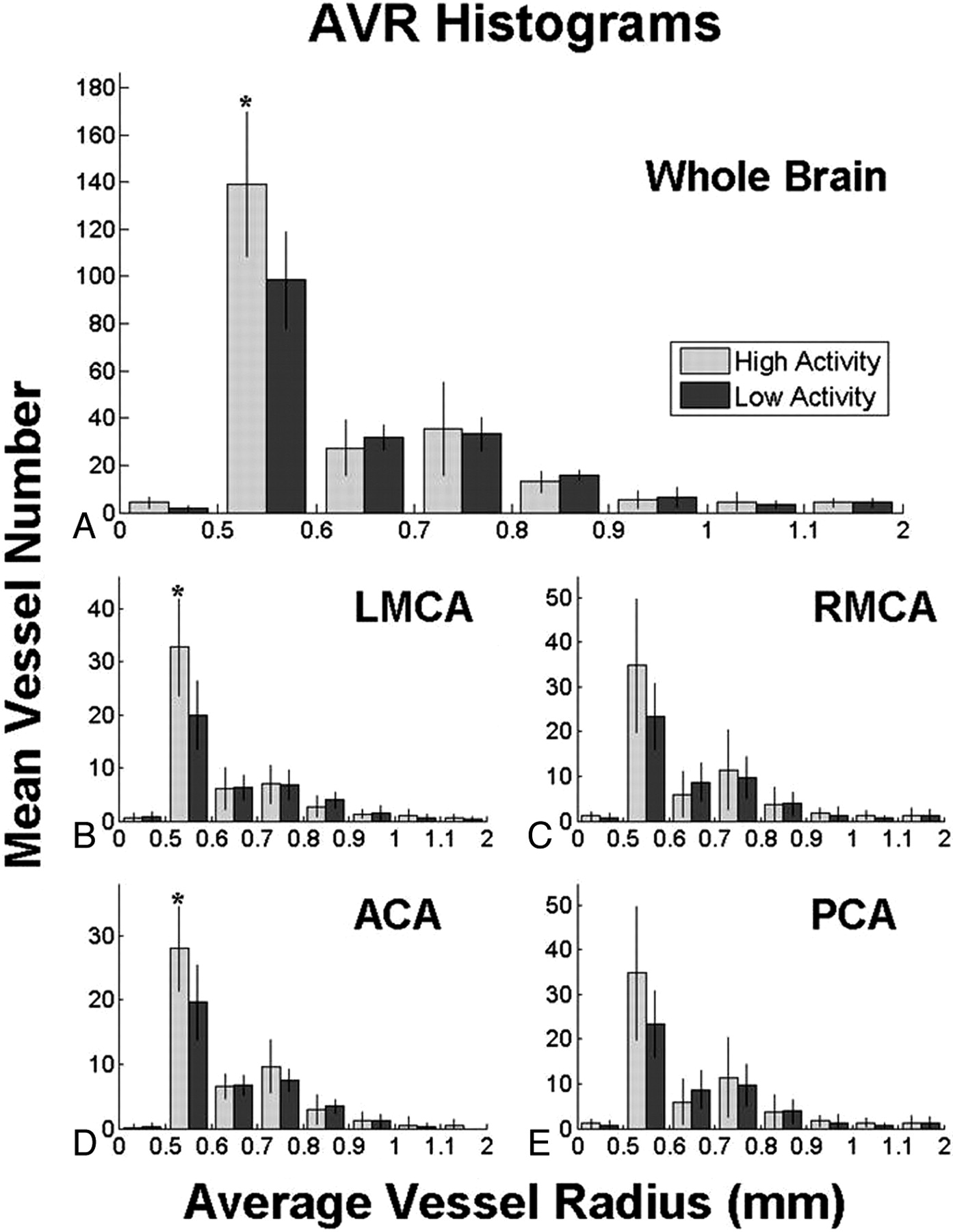
  • Fig 4.
    • Download figure
    • Open in new tab
    • Download powerpoint
    Fig 4.

    Comparison of high-activity (light gray) and low-activity (dark gray) groups by the number of vessels of graded radius. Colored bars represent the mean value and vertical lines, the SD. Stars denote statistically significant differences (P < .05) between activity groups. Note that the smallest and largest bins have ranges of >0.1 mm to encompass all vessels. PCA indicates posterior circulation.

PreviousNext
Back to top

In this issue

American Journal of Neuroradiology: 30 (10)
American Journal of Neuroradiology
Vol. 30, Issue 10
1 Nov 2009
  • Table of Contents
  • Index by author
Advertisement
Print
Download PDF
Email Article

Thank you for your interest in spreading the word on American Journal of Neuroradiology.

NOTE: We only request your email address so that the person you are recommending the page to knows that you wanted them to see it, and that it is not junk mail. We do not capture any email address.

Enter multiple addresses on separate lines or separate them with commas.
The Effect of Exercise on the Cerebral Vasculature of Healthy Aged Subjects as Visualized by MR Angiography
(Your Name) has sent you a message from American Journal of Neuroradiology
(Your Name) thought you would like to see the American Journal of Neuroradiology web site.
CAPTCHA
This question is for testing whether or not you are a human visitor and to prevent automated spam submissions.
Cite this article
E. Bullitt, F.N. Rahman, J.K. Smith, E. Kim, D. Zeng, L.M. Katz, B.L. Marks
The Effect of Exercise on the Cerebral Vasculature of Healthy Aged Subjects as Visualized by MR Angiography
American Journal of Neuroradiology Nov 2009, 30 (10) 1857-1863; DOI: 10.3174/ajnr.A1695

Citation Manager Formats

  • BibTeX
  • Bookends
  • EasyBib
  • EndNote (tagged)
  • EndNote 8 (xml)
  • Medlars
  • Mendeley
  • Papers
  • RefWorks Tagged
  • Ref Manager
  • RIS
  • Zotero
0 Responses
Respond to this article
Share
Bookmark this article
The Effect of Exercise on the Cerebral Vasculature of Healthy Aged Subjects as Visualized by MR Angiography
E. Bullitt, F.N. Rahman, J.K. Smith, E. Kim, D. Zeng, L.M. Katz, B.L. Marks
American Journal of Neuroradiology Nov 2009, 30 (10) 1857-1863; DOI: 10.3174/ajnr.A1695
del.icio.us logo Twitter logo Facebook logo Mendeley logo
  • Tweet Widget
  • Facebook Like
  • Google Plus One
Purchase

Jump to section

  • Article
    • Abstract
    • Materials and Methods
    • Results
    • Discussion
    • Conclusions
    • Footnotes
    • References
  • Figures & Data
  • Info & Metrics
  • Responses
  • References
  • PDF

Related Articles

  • No related articles found.
  • PubMed
  • Google Scholar

Cited By...

  • Determinants of cerebral blood flow and arterial transit time in healthy older adults
  • Crossref
  • Google Scholar

This article has not yet been cited by articles in journals that are participating in Crossref Cited-by Linking.

More in this TOC Section

  • Usefulness of Quantitative Susceptibility Mapping for the Diagnosis of Parkinson Disease
  • White Matter Alterations in the Brains of Patients with Active, Remitted, and Cured Cushing Syndrome: A DTI Study
  • Qualitative and Quantitative Analysis of MR Imaging Findings in Patients with Middle Cerebral Artery Stroke Implanted with Mesenchymal Stem Cells
Show more Brain

Similar Articles

Advertisement

Indexed Content

  • Current Issue
  • Accepted Manuscripts
  • Article Preview
  • Past Issues
  • Editorials
  • Editor's Choice
  • Fellows' Journal Club
  • Letters to the Editor
  • Video Articles

Cases

  • Case Collection
  • Archive - Case of the Week
  • Archive - Case of the Month
  • Archive - Classic Case

More from AJNR

  • Trainee Corner
  • Imaging Protocols
  • MRI Safety Corner
  • Book Reviews

Multimedia

  • AJNR Podcasts
  • AJNR Scantastics

Resources

  • Turnaround Time
  • Submit a Manuscript
  • Submit a Video Article
  • Submit an eLetter to the Editor/Response
  • Manuscript Submission Guidelines
  • Statistical Tips
  • Fast Publishing of Accepted Manuscripts
  • Graphical Abstract Preparation
  • Imaging Protocol Submission
  • Evidence-Based Medicine Level Guide
  • Publishing Checklists
  • Author Policies
  • Become a Reviewer/Academy of Reviewers
  • News and Updates

About Us

  • About AJNR
  • Editorial Board
  • Editorial Board Alumni
  • Alerts
  • Permissions
  • Not an AJNR Subscriber? Join Now
  • Advertise with Us
  • Librarian Resources
  • Feedback
  • Terms and Conditions
  • AJNR Editorial Board Alumni

American Society of Neuroradiology

  • Not an ASNR Member? Join Now

© 2025 by the American Society of Neuroradiology All rights, including for text and data mining, AI training, and similar technologies, are reserved.
Print ISSN: 0195-6108 Online ISSN: 1936-959X

Powered by HighWire