Advertisement

Main menu

  • Home
  • Content
    • Current Issue
    • Accepted Manuscripts
    • Article Preview
    • Past Issue Archive
    • Video Articles
    • AJNR Case Collection
    • Case of the Week Archive
    • Case of the Month Archive
    • Classic Case Archive
  • Special Collections
    • AJNR Awards
    • Low-Field MRI
    • Alzheimer Disease
    • ASNR Foundation Special Collection
    • Photon-Counting CT
    • View All
  • Multimedia
    • AJNR Podcasts
    • AJNR SCANtastic
    • Trainee Corner
    • MRI Safety Corner
    • Imaging Protocols
  • For Authors
    • Submit a Manuscript
    • Submit a Video Article
    • Submit an eLetter to the Editor/Response
    • Manuscript Submission Guidelines
    • Statistical Tips
    • Fast Publishing of Accepted Manuscripts
    • Graphical Abstract Preparation
    • Imaging Protocol Submission
    • Author Policies
  • About Us
    • About AJNR
    • Editorial Board
    • Editorial Board Alumni
  • More
    • Become a Reviewer/Academy of Reviewers
    • Subscribers
    • Permissions
    • Alerts
    • Feedback
    • Advertisers
    • ASNR Home

User menu

  • Alerts
  • Log in

Search

  • Advanced search
American Journal of Neuroradiology

American Journal of Neuroradiology

ASHNR American Society of Functional Neuroradiology ASHNR American Society of Pediatric Neuroradiology ASSR
  • Alerts
  • Log in

Advanced Search

  • Home
  • Content
    • Current Issue
    • Accepted Manuscripts
    • Article Preview
    • Past Issue Archive
    • Video Articles
    • AJNR Case Collection
    • Case of the Week Archive
    • Case of the Month Archive
    • Classic Case Archive
  • Special Collections
    • AJNR Awards
    • Low-Field MRI
    • Alzheimer Disease
    • ASNR Foundation Special Collection
    • Photon-Counting CT
    • View All
  • Multimedia
    • AJNR Podcasts
    • AJNR SCANtastic
    • Trainee Corner
    • MRI Safety Corner
    • Imaging Protocols
  • For Authors
    • Submit a Manuscript
    • Submit a Video Article
    • Submit an eLetter to the Editor/Response
    • Manuscript Submission Guidelines
    • Statistical Tips
    • Fast Publishing of Accepted Manuscripts
    • Graphical Abstract Preparation
    • Imaging Protocol Submission
    • Author Policies
  • About Us
    • About AJNR
    • Editorial Board
    • Editorial Board Alumni
  • More
    • Become a Reviewer/Academy of Reviewers
    • Subscribers
    • Permissions
    • Alerts
    • Feedback
    • Advertisers
    • ASNR Home
  • Follow AJNR on Twitter
  • Visit AJNR on Facebook
  • Follow AJNR on Instagram
  • Join AJNR on LinkedIn
  • RSS Feeds

AJNR Awards, New Junior Editors, and more. Read the latest AJNR updates

Case ReportSpine

Spinal Epidural Lubricant Grease Collection Mimicking Traumatic Spinal Epidural Hematoma

A.U. Kalnins, B. Geryk, W. Olivero and T.A. Kim
American Journal of Neuroradiology November 2009, 30 (10) 1847-1849; DOI: https://doi.org/10.3174/ajnr.A1670
A.U. Kalnins
  • Find this author on Google Scholar
  • Find this author on PubMed
  • Search for this author on this site
B. Geryk
  • Find this author on Google Scholar
  • Find this author on PubMed
  • Search for this author on this site
W. Olivero
  • Find this author on Google Scholar
  • Find this author on PubMed
  • Search for this author on this site
T.A. Kim
  • Find this author on Google Scholar
  • Find this author on PubMed
  • Search for this author on this site
  • Article
  • Figures & Data
  • Info & Metrics
  • Responses
  • References
  • PDF
Loading

Abstract

SUMMARY: We report an unusual case of a patient with an epidural fluid collection with signal characteristics mimicking an epidural hematoma. The patient presented with myelopathy caused by thoracic spinal cord compression after a traumatic injury to the chest and back. The injury was caused by high-pressure injection of industrial-grade lubricant grease. This case demonstrates that cord compression can be caused by exogenous material in the setting of trauma that can mimic an acute epidural hematoma.

The evolution of the MR imaging signal intensity characteristics of a spinal epidural hematoma is thought to follow the MR imaging characteristics of a hemorrhage in the brain.1,2 This case demonstrates an unusual occurrence in which exogenous fluid mimicked the clinical and radiologic manifestations of early subacute traumatic spinal epidural hemorrhage.

Case Report

The patient was a 31-year-old man who sustained a traumatic injury to the right neck and right chest wall while trying to repair a pneumatic device on a hydraulic injection instrument of a bulldozer. The patient was struck in the neck with a “metal plug,” resulting in a puncture of the right anterior neck and right upper chest wall. The patient was airlifted to a trauma center, and on arrival, he was noted to be hemodynamically stable and was ambulatory. The initial neurologic examination by the trauma service in the emergency department was reported to be normal. The patient was taken to the operating room for local exploration of the right neck wound, and a right thoracostomy was completed for drainage of the fluid, presumed to be a hemopneumothorax. A neck CT angiographic examination was done, which showed general soft tissue injury involving the chest wall and posterior paraspinous muscles, a small residual left pneumothorax, and hypoattenuated epidural fluid collection.

On day 2 of admission, a Foley catheter was inserted because of the patient's inability to void. During morning rounds on day 4 of admission, the patient exhibited new neurologic deficits with bilateral leg numbness, tingling, and motor deficits, with the right worse than the left. Neurosurgical consultation was obtained, along with spine CT and MR imaging.

MR imaging of the thoracic spine revealed a posterior epidural collection, which was interpreted as an epidural hematoma, extending from the T3 to the T6 level with some residual collection extending into the lower thoracic and upper lumbar spinal canal (Fig 1).

Fig 1.
  • Download figure
  • Open in new tab
  • Download powerpoint
Fig 1.

Sagittal T1 and T2 turbo-spin-echo sequences of the posterior spinal canal. A, Sagittal T1 spin-echo sequence revealing mixed signal intensity in the posterior spinal canal most marked from approximately the T1 level to the T7 level, with minimal findings extending into the lower thoracic and upper lumbar levels. There is an area of high signal intensity extending from the T3 to the T5 level (arrows). B, Sagittal T2 turbo-spin-echo sequence revealing mixed signal intensity in the posterior spinal canal most marked from approximately the T1 level to the T7 level, with minimal findings extending into the lower thoracic and upper lumbar levels. There is an area of low signal intensity extending from the T3 to the T5 level (arrows).

The patient was emergently taken to the operating room, and a decompressive laminectomy was performed from the T2 to the T6 level. There was a large collection of what was presumed to be pus and possibly old blood within the right paraspinous muscles at approximately T2 through T4. Once the ligamentum flavum was opened up, immediately more apparent dark epidural fluid was encountered, which was thought to represent a collection of pus and old blood. This fluid was evacuated and sent for culture. Initially thought to be pus, the viscous fluid was shown to represent industrial grease with some inflammatory cells. The same thick viscous fluid was evacuated from the epidural space and from within the muscles of the right upper back.

Additional investigation revealed that the patient had sustained a high-pressure injection of lubricant grease from the hydraulic device of a bulldozer, with pressures exceeding 3000 psi. The lubricant grease injection was postulated to have passed through the apex of the lung, potentially the mediastinal space, and into the posterior thorax, ending in the thoracic epidural space.

The lubricant grease was identified as Conoco Phillips Dynalife GC-LB, an automotive lithium complex grease.3 This lubricant grease was placed along with normal saline in a glass jar and imaged with the same scanner with the same T1 and T2 acquisition parameters. The acquired images demonstrate similar MR imaging signal intensity characteristics of the epidural fluid collection: hyperintensity on T1-weighted imaging and hypointensity on T2-weighted imaging, with normal expected hypointensity and hyperintensity of the saline, respectively (Fig 2). These signal intensity characteristics mimic those of an early subacute epidural hemorrhage (>3 days).1

Fig 2.
  • Download figure
  • Open in new tab
  • Download powerpoint
Fig 2.

T1- and T2-weighted images of the saline and lubricant grease. A, T1-weighted image reveals hypointensity of the saline and hyperintensity of the lubricant grease. B, T2-weighted image reveals hyperintensity of the saline and hypointensity of the lubricant grease

Discussion

Traumatic spinal epidural hematomas (TSEHs) are relatively uncommon, occurring in less than 2% of all spinal injuries, but their sequelae, including cord compression, can be serious, which makes early diagnosis essential.4 Imaging plays a crucial role in this diagnosis, and MR imaging is the best tool for evaluation of a potential TSEH.

MR imaging of an early subacute spinal epidural hematoma displays a heterogeneously high signal intensity on T1-weighted imaging and a heterogeneously low signal intensity on T2-weighted imaging.1,2 In the case of our patient, the presumed acute spinal epidural hematoma had similar T1 and T2 signal intensity characteristics.

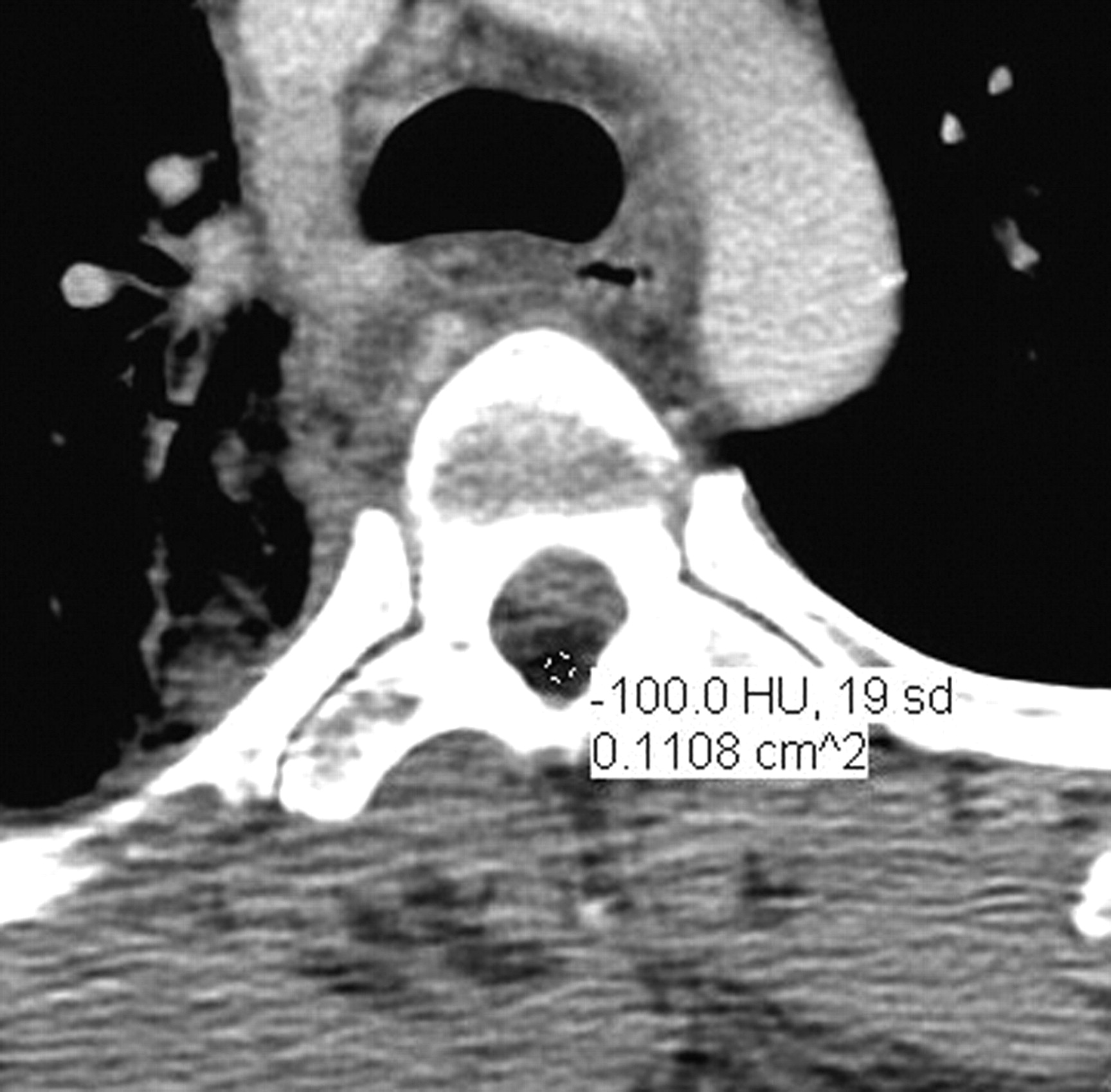
CT imaging of a spinal epidural hematoma displays a high-attenuation collection within the spinal canal.1 From a retrospective standpoint, the intraspinal epidural collection reported on CT angiography of the neck in this patient revealed a Hounsfield unit (HU) attenuation of −100 HU, in the range of fat, which is typically −90 HU, rather than fluid, which typically has Hounsfield values of approximately 0 HU (Fig 3).5

Fig 3.
  • Download figure
  • Open in new tab
  • Download powerpoint
Fig 3.

Axial CT revealing evidence of an epidural fluid collection in the thoracic spine displacing the cord anteriorly and to the left. The fluid collection has a attenuation of −100 HU.

We are not aware of any previous case reports in which cord compression was caused by an exogenous fluid that entered the epidural space through a blast injury to the thorax. The delayed neurologic symptoms are consistent with previously reported cases of TSEH in which cord compression does not manifest itself until some time (7 days to 2.5 months) after the initial injury.6,7 In a review of high-pressure injection injuries of a foreign material into the body, the cause of delayed symptoms is described as arising from the combination of the additional volume and evolving inflammatory response because of the presence of a foreign substance.8 These factors may help account for the delayed onset of symptoms in our patient.

In conclusion, we report an unusual presentation of a traumatic spinal epidural fluid collection, caused by a high-pressure injection injury of lubricant grease to the upper thorax and into the thoracic spinal canal. The spinal epidural fluid collection mimicked the appearance of an early subacute spinal epidural hematoma on spinal MR imaging. Again, the case of our patient illustrates the importance of detailed clinical information, including a thorough understanding of the mechanism of trauma at the time of interpretation of spinal imaging studies, since cord compression can rarely be caused by nonhemorrhagic fluid collection.

Acknowledgment

We thank Gail Mann, radiology technologist, for her assistance.

Footnotes

  • Previously presented at: Annual Meeting of the American Society of Neuroradiology and Neuroradiology Education and Research Foundation Symposium, May 31–June 5, 2008; New Orleans, La.

References

  1. 1.↵
    1. Harris JH.,
    2. Mirvis SE.
    The Radiology of Acute Cervical Spine Trauma. Baltimore: Williams & Wilkins; 1996:161
  2. 2.↵
    1. Bradley WG Jr.
    . MR appearance of hemorrhage in the brain. Radiology 1993:189:15–26
    CrossRefPubMed
  3. 3.↵
    ConocoPhillips. Home >Brands and Products >Conoco >Greases. 2009 02/08 [cited 2009 March 26]; Available at: http://www.conocophillipslubricants.com/documents/conoco/greases/FOB%20Dynalife%20GCLB%20TDS%20Web.pdf. Accessed September 23, 2009
  4. 4.↵
    1. Foo D,
    2. Rossier AB
    . Post-traumatic spinal epidural hematoma. Neurosurgery 1982:11:25–32
    PubMed
  5. 5.↵
    1. Huda W
    . Review of Radiologic Physics. Baltimore: Lippincott Williams & Wilkins; 2002:122
  6. 6.↵
    1. Newey ML,
    2. Cain CM
    . Delayed focal neurological deficit secondary to a cervico-thoracic spinal cord epidural haematoma. Eur Spine J 1999;8:332–33
    CrossRefPubMed
  7. 7.↵
    1. Cuenca PJ,
    2. Tulley EB,
    3. Devita D,
    4. et al
    . Delayed traumatic spinal epidural hematoma with spontaneous resolution of symptoms. J Emerg Med 2004;27:37–41
    CrossRefPubMed
  8. 8.↵
    1. Hogan CJ,
    2. Ruland RT
    . High-pressure injection injuries to the upper extremity: a review of the literature. J Orthop Trauma 2006:20:503–11
    CrossRefPubMed
  • Received March 31, 2009.
  • Accepted after revision April 7, 2009.
  • Copyright © American Society of Neuroradiology
View Abstract
PreviousNext
Back to top

In this issue

American Journal of Neuroradiology: 30 (10)
American Journal of Neuroradiology
Vol. 30, Issue 10
1 Nov 2009
  • Table of Contents
  • Index by author
Advertisement
Print
Download PDF
Email Article

Thank you for your interest in spreading the word on American Journal of Neuroradiology.

NOTE: We only request your email address so that the person you are recommending the page to knows that you wanted them to see it, and that it is not junk mail. We do not capture any email address.

Enter multiple addresses on separate lines or separate them with commas.
Spinal Epidural Lubricant Grease Collection Mimicking Traumatic Spinal Epidural Hematoma
(Your Name) has sent you a message from American Journal of Neuroradiology
(Your Name) thought you would like to see the American Journal of Neuroradiology web site.
CAPTCHA
This question is for testing whether or not you are a human visitor and to prevent automated spam submissions.
Cite this article
A.U. Kalnins, B. Geryk, W. Olivero, T.A. Kim
Spinal Epidural Lubricant Grease Collection Mimicking Traumatic Spinal Epidural Hematoma
American Journal of Neuroradiology Nov 2009, 30 (10) 1847-1849; DOI: 10.3174/ajnr.A1670

Citation Manager Formats

  • BibTeX
  • Bookends
  • EasyBib
  • EndNote (tagged)
  • EndNote 8 (xml)
  • Medlars
  • Mendeley
  • Papers
  • RefWorks Tagged
  • Ref Manager
  • RIS
  • Zotero
0 Responses
Respond to this article
Share
Bookmark this article
Spinal Epidural Lubricant Grease Collection Mimicking Traumatic Spinal Epidural Hematoma
A.U. Kalnins, B. Geryk, W. Olivero, T.A. Kim
American Journal of Neuroradiology Nov 2009, 30 (10) 1847-1849; DOI: 10.3174/ajnr.A1670
del.icio.us logo Twitter logo Facebook logo Mendeley logo
  • Tweet Widget
  • Facebook Like
  • Google Plus One
Purchase

Jump to section

  • Article
    • Abstract
    • Case Report
    • Discussion
    • Acknowledgment
    • Footnotes
    • References
  • Figures & Data
  • Info & Metrics
  • Responses
  • References
  • PDF

Related Articles

  • No related articles found.
  • PubMed
  • Google Scholar

Cited By...

  • No citing articles found.
  • Crossref (2)
  • Google Scholar

This article has been cited by the following articles in journals that are participating in Crossref Cited-by Linking.

  • Diagnostic uncertainty following orbital injury from a grease gun
    Muhammed Jawad, Krishna Yamini, Ziyaad Sultan, Sarah Coupland, James Hsuan, Austin McCormick
    Orbit 2023 42 3
  • Rare Diseases and Syndromes of the Spinal Cord
    Ibrahim M. Eltorai
    2016

More in this TOC Section

  • Bern Score Validity for SIH
  • MP2RAGE 7T in MS Lesions of the Cervical Spine
  • Deep Learning for STIR Spine MRI Quality
Show more Spine

Similar Articles

Advertisement

Indexed Content

  • Current Issue
  • Accepted Manuscripts
  • Article Preview
  • Past Issues
  • Editorials
  • Editor's Choice
  • Fellows' Journal Club
  • Letters to the Editor
  • Video Articles

Cases

  • Case Collection
  • Archive - Case of the Week
  • Archive - Case of the Month
  • Archive - Classic Case

More from AJNR

  • Trainee Corner
  • Imaging Protocols
  • MRI Safety Corner
  • Book Reviews

Multimedia

  • AJNR Podcasts
  • AJNR Scantastics

Resources

  • Turnaround Time
  • Submit a Manuscript
  • Submit a Video Article
  • Submit an eLetter to the Editor/Response
  • Manuscript Submission Guidelines
  • Statistical Tips
  • Fast Publishing of Accepted Manuscripts
  • Graphical Abstract Preparation
  • Imaging Protocol Submission
  • Evidence-Based Medicine Level Guide
  • Publishing Checklists
  • Author Policies
  • Become a Reviewer/Academy of Reviewers
  • News and Updates

About Us

  • About AJNR
  • Editorial Board
  • Editorial Board Alumni
  • Alerts
  • Permissions
  • Not an AJNR Subscriber? Join Now
  • Advertise with Us
  • Librarian Resources
  • Feedback
  • Terms and Conditions
  • AJNR Editorial Board Alumni

American Society of Neuroradiology

  • Not an ASNR Member? Join Now

© 2025 by the American Society of Neuroradiology All rights, including for text and data mining, AI training, and similar technologies, are reserved.
Print ISSN: 0195-6108 Online ISSN: 1936-959X

Powered by HighWire