Advertisement

Main menu

  • Home
  • Content
    • Current Issue
    • Accepted Manuscripts
    • Article Preview
    • Past Issue Archive
    • Video Articles
    • AJNR Case Collection
    • Case of the Week Archive
    • Case of the Month Archive
    • Classic Case Archive
  • Special Collections
    • AJNR Awards
    • Low-Field MRI
    • Alzheimer Disease
    • ASNR Foundation Special Collection
    • Photon-Counting CT
    • View All
  • Multimedia
    • AJNR Podcasts
    • AJNR SCANtastic
    • Trainee Corner
    • MRI Safety Corner
    • Imaging Protocols
  • For Authors
    • Submit a Manuscript
    • Submit a Video Article
    • Submit an eLetter to the Editor/Response
    • Manuscript Submission Guidelines
    • Statistical Tips
    • Fast Publishing of Accepted Manuscripts
    • Graphical Abstract Preparation
    • Imaging Protocol Submission
    • Author Policies
  • About Us
    • About AJNR
    • Editorial Board
    • Editorial Board Alumni
  • More
    • Become a Reviewer/Academy of Reviewers
    • Subscribers
    • Permissions
    • Alerts
    • Feedback
    • Advertisers
    • ASNR Home

User menu

  • Alerts
  • Log in

Search

  • Advanced search
American Journal of Neuroradiology

American Journal of Neuroradiology

ASHNR American Society of Functional Neuroradiology ASHNR American Society of Pediatric Neuroradiology ASSR
  • Alerts
  • Log in

Advanced Search

  • Home
  • Content
    • Current Issue
    • Accepted Manuscripts
    • Article Preview
    • Past Issue Archive
    • Video Articles
    • AJNR Case Collection
    • Case of the Week Archive
    • Case of the Month Archive
    • Classic Case Archive
  • Special Collections
    • AJNR Awards
    • Low-Field MRI
    • Alzheimer Disease
    • ASNR Foundation Special Collection
    • Photon-Counting CT
    • View All
  • Multimedia
    • AJNR Podcasts
    • AJNR SCANtastic
    • Trainee Corner
    • MRI Safety Corner
    • Imaging Protocols
  • For Authors
    • Submit a Manuscript
    • Submit a Video Article
    • Submit an eLetter to the Editor/Response
    • Manuscript Submission Guidelines
    • Statistical Tips
    • Fast Publishing of Accepted Manuscripts
    • Graphical Abstract Preparation
    • Imaging Protocol Submission
    • Author Policies
  • About Us
    • About AJNR
    • Editorial Board
    • Editorial Board Alumni
  • More
    • Become a Reviewer/Academy of Reviewers
    • Subscribers
    • Permissions
    • Alerts
    • Feedback
    • Advertisers
    • ASNR Home
  • Follow AJNR on Twitter
  • Visit AJNR on Facebook
  • Follow AJNR on Instagram
  • Join AJNR on LinkedIn
  • RSS Feeds

AJNR Awards, New Junior Editors, and more. Read the latest AJNR updates

Review ArticleReview Articles
Open Access

[18F] Fluorodeoxyglucose–Positron-Emission Tomography and MR Imaging Coregistration for Presurgical Evaluation of Medically Refractory Epilepsy

K.K. Lee and N. Salamon
American Journal of Neuroradiology November 2009, 30 (10) 1811-1816; DOI: https://doi.org/10.3174/ajnr.A1637
K.K. Lee
  • Find this author on Google Scholar
  • Find this author on PubMed
  • Search for this author on this site
N. Salamon
  • Find this author on Google Scholar
  • Find this author on PubMed
  • Search for this author on this site
  • Article
  • Figures & Data
  • Info & Metrics
  • Responses
  • References
  • PDF
Loading

Article Figures & Data

Figures

  • Fig 1.
    • Download figure
    • Open in new tab
    • Download powerpoint
    Fig 1.

    MTS in a 31-year-old woman. A, FDG-PET image was read as having normal findings. B, However, the FLAIR image shows some question of increased T2 signal intensity in the right hippocampus. C, FDG-PET/MR imaging coregistration demonstrates subtle hypometabolism of the right mesial temporal structures. Because neurocoginitive testing also demonstrated impaired function of right mesial temporal structures, right anteromedial temporal lobectomy and hippocampectomy were performed. FDG-PET/MR imaging coregistration facilitated localization of the metabolically abnormal area that was subtle when read by FDG-PET alone.

  • Fig 2.
    • Download figure
    • Open in new tab
    • Download powerpoint
    Fig 2.

    FCD type II in a 5-year-old girl. A, T2-weighted MR image demonstrates hyperintensity in the left frontal white matter suggestive of FCD. B, With FDG-PET/MR imaging coregistration, a focal area of hypometabolism is seen correlating to the lesion. Pathologic findings of the surgical resection specimen were consistent with FCD type II.
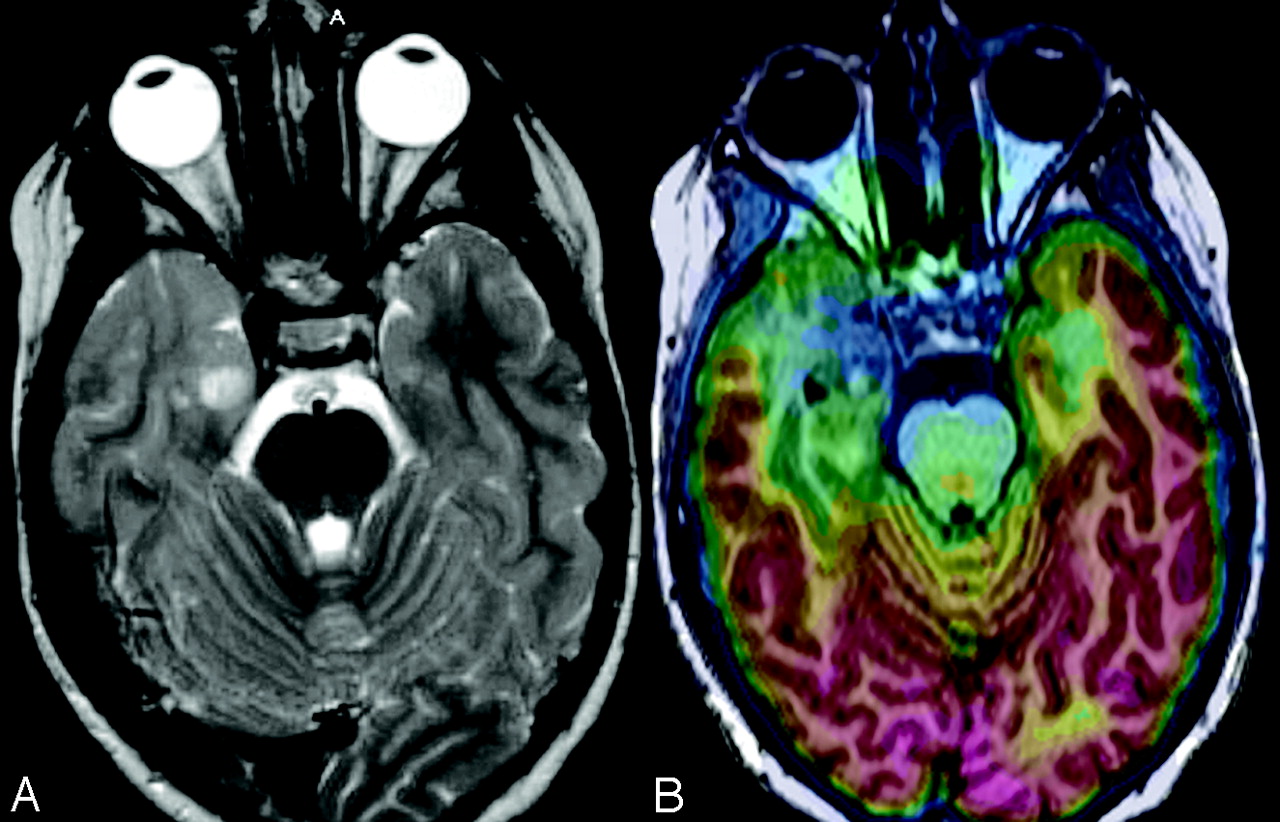
  • Fig 3.
    • Download figure
    • Open in new tab
    • Download powerpoint
    Fig 3.

    FCD type I in a 3-year-old boy. A, T2-weighted MR image shows questionable hyperintensity and atrophy in the left temporal pole. B, FDG-PET/MR imaging coregistration shows that this area corresponds to an area of mild hypometabolism. After a left lateral temporal lobe resection, pathologic findings were consistent with FCD type I.

  • Fig 4.
    • Download figure
    • Open in new tab
    • Download powerpoint
    Fig 4.

    Tumor with surrounding FCD in a 7-year-old girl. A, T2-weighted MR image shows a lesion in the right mesiotemporal lobe with perilesional blurring of the gray and white matter in the right anterior temporal pole. B, FDG-PET/MR imaging coregistration demonstrates relative hypometabolism in right temporal lobe localized at the lesion and the surrounding abnormal gray and white matter. After a right anterior temporal lobectomy, pathologic findings of the surgical specimen showed a neoplasm with features of oligoastrocytoma with surrounding FCD type I.

  • Fig 5.
    • Download figure
    • Open in new tab
    • Download powerpoint
    Fig 5.

    Tumor with surrounding FCD in an 18-year-old woman. A, FLAIR sequence shows hyperintensity of the left amygdala and temporal lobe. B, FDG-PET/MR imaging coregistration demonstrates an area of hypometabolism extending beyond the lesion seen with MR imaging. After surgical resection, ganglioglioma was seen on pathology of the lesion (white arrow). Pathology of the area of hypometabolism surrounding the lesion (yellow arrow) showed histology consistent with FCD type I.

  • Fig 6.
    • Download figure
    • Open in new tab
    • Download powerpoint
    Fig 6.

    TSC in a 5-year-old girl. A, T2 axial image shows numerous hyperintense tubers on both temporal lobes. B, FDG-PET/MR imaging coregistration demonstrates an area of hypometabolism in the right temporal pole that is significantly larger than the tuber size. EEG demonstrated the presence of right temporal interictal discharges suggesting an epileptogenic zone in this area. Right temporal lobectomy was performed with good postsurgical seizure control.

PreviousNext
Back to top

In this issue

American Journal of Neuroradiology: 30 (10)
American Journal of Neuroradiology
Vol. 30, Issue 10
1 Nov 2009
  • Table of Contents
  • Index by author
Advertisement
Print
Download PDF
Email Article

Thank you for your interest in spreading the word on American Journal of Neuroradiology.

NOTE: We only request your email address so that the person you are recommending the page to knows that you wanted them to see it, and that it is not junk mail. We do not capture any email address.

Enter multiple addresses on separate lines or separate them with commas.
[18F] Fluorodeoxyglucose–Positron-Emission Tomography and MR Imaging Coregistration for Presurgical Evaluation of Medically Refractory Epilepsy
(Your Name) has sent you a message from American Journal of Neuroradiology
(Your Name) thought you would like to see the American Journal of Neuroradiology web site.
CAPTCHA
This question is for testing whether or not you are a human visitor and to prevent automated spam submissions.
Cite this article
K.K. Lee, N. Salamon
[18F] Fluorodeoxyglucose–Positron-Emission Tomography and MR Imaging Coregistration for Presurgical Evaluation of Medically Refractory Epilepsy
American Journal of Neuroradiology Nov 2009, 30 (10) 1811-1816; DOI: 10.3174/ajnr.A1637

Citation Manager Formats

  • BibTeX
  • Bookends
  • EasyBib
  • EndNote (tagged)
  • EndNote 8 (xml)
  • Medlars
  • Mendeley
  • Papers
  • RefWorks Tagged
  • Ref Manager
  • RIS
  • Zotero
0 Responses
Respond to this article
Share
Bookmark this article
[18F] Fluorodeoxyglucose–Positron-Emission Tomography and MR Imaging Coregistration for Presurgical Evaluation of Medically Refractory Epilepsy
K.K. Lee, N. Salamon
American Journal of Neuroradiology Nov 2009, 30 (10) 1811-1816; DOI: 10.3174/ajnr.A1637
del.icio.us logo Twitter logo Facebook logo Mendeley logo
  • Tweet Widget
  • Facebook Like
  • Google Plus One
Purchase

Jump to section

  • Article
    • Abstract
    • Presurgical Evaluation and FDG-PET/MR Imaging Coregistration for Medically Refractory Epilepsy at UCLA
    • MTS
    • FCD
    • Epileptogenic Tumors
    • TSC
    • Discussion and Future Directions
    • Conclusions
    • References
  • Figures & Data
  • Info & Metrics
  • Responses
  • References
  • PDF

Related Articles

  • No related articles found.
  • PubMed
  • Google Scholar

Cited By...

  • Clinical Value of Hybrid TOF-PET/MR Imaging-Based Multiparametric Imaging in Localizing Seizure Focus in Patients with MRI-Negative Temporal Lobe Epilepsy
  • Crossref (44)
  • Google Scholar

This article has been cited by the following articles in journals that are participating in Crossref Cited-by Linking.

  • PET/MR Imaging: Technical Aspects and Potential Clinical Applications
    Drew A. Torigian, Habib Zaidi, Thomas C. Kwee, Babak Saboury, Jayaram K. Udupa, Zang-Hee Cho, Abass Alavi
    Radiology 2013 267 1
  • Imaging in the surgical treatment of epilepsy
    John S. Duncan
    Nature Reviews Neurology 2010 6 10
  • Current status and future role of brain PET/MRI in clinical and research settings
    P. Werner, H. Barthel, A. Drzezga, O. Sabri
    European Journal of Nuclear Medicine and Molecular Imaging 2015 42 3
  • Clinical PET/MRI: 2018 Update
    Stephen M. Broski, Ajit H. Goenka, Bradley J. Kemp, Geoffrey B. Johnson
    American Journal of Roentgenology 2018 211 2
  • Validation of FDG-PET/MRI coregistration in nonlesional refractory childhood epilepsy
    Sebastià Rubí, Xavier Setoain, Antonio Donaire, Núria Bargalló, Francesc Sanmartí, Mar Carreño, Jordi Rumià, Anna Calvo, Javier Aparicio, Jaume Campistol, Francesca Pons
    Epilepsia 2011 52 12
  • PET/MRI and PET/MRI/SISCOM coregistration in the presurgical evaluation of refractory focal epilepsy
    S. Fernández, A. Donaire, E. Serès, X. Setoain, N. Bargalló, C. Falcón, F. Sanmartí, I. Maestro, J. Rumià, L. Pintor, T. Boget, J. Aparicio, M. Carreño
    Epilepsy Research 2015 111
  • Neurologic Applications of PET/MR Imaging
    Michelle M. Miller-Thomas, Tammie L.S. Benzinger
    Magnetic Resonance Imaging Clinics of North America 2017 25 2
  • FDG‐PET and magnetoencephalography in presurgical workup of children with localization‐related nonlesional epilepsy
    Elysa Widjaja, Amer Shammas, Reza Vali, Hiroshi Otsubo, Ayako Ochi, O. Carter Snead, Cristina Go, Martin Charron
    Epilepsia 2013 54 4
  • Clinical Value of Hybrid TOF-PET/MR Imaging–Based Multiparametric Imaging in Localizing Seizure Focus in Patients with MRI-Negative Temporal Lobe Epilepsy
    K. Shang, J. Wang, X. Fan, B. Cui, J. Ma, H. Yang, Y. Zhou, G. Zhao, J. Lu
    American Journal of Neuroradiology 2018 39 10
  • From PET/CT to PET/MRI: Advances in Instrumentation and Clinical Applications
    Zhenhua Hu, Weidong Yang, Haixiao Liu, Kun Wang, Chengpeng Bao, Tianming Song, Jing Wang, Jie Tian
    Molecular Pharmaceutics 2014 11 11

More in this TOC Section

  • An Atlas of Neonatal Neurovascular Imaging Anatomy as Depicted with Microvascular Imaging: The Intracranial Arteries
  • An Atlas of Neonatal Neurovascular Imaging Anatomy as Depicted with Microvascular Imaging: The Intracranial Veins
  • Clinical Translation of Hyperpolarized 13C Metabolic Probes for Glioma Imaging
Show more Review articles

Similar Articles

Advertisement

Indexed Content

  • Current Issue
  • Accepted Manuscripts
  • Article Preview
  • Past Issues
  • Editorials
  • Editor's Choice
  • Fellows' Journal Club
  • Letters to the Editor
  • Video Articles

Cases

  • Case Collection
  • Archive - Case of the Week
  • Archive - Case of the Month
  • Archive - Classic Case

More from AJNR

  • Trainee Corner
  • Imaging Protocols
  • MRI Safety Corner
  • Book Reviews

Multimedia

  • AJNR Podcasts
  • AJNR Scantastics

Resources

  • Turnaround Time
  • Submit a Manuscript
  • Submit a Video Article
  • Submit an eLetter to the Editor/Response
  • Manuscript Submission Guidelines
  • Statistical Tips
  • Fast Publishing of Accepted Manuscripts
  • Graphical Abstract Preparation
  • Imaging Protocol Submission
  • Evidence-Based Medicine Level Guide
  • Publishing Checklists
  • Author Policies
  • Become a Reviewer/Academy of Reviewers
  • News and Updates

About Us

  • About AJNR
  • Editorial Board
  • Editorial Board Alumni
  • Alerts
  • Permissions
  • Not an AJNR Subscriber? Join Now
  • Advertise with Us
  • Librarian Resources
  • Feedback
  • Terms and Conditions
  • AJNR Editorial Board Alumni

American Society of Neuroradiology

  • Not an ASNR Member? Join Now

© 2025 by the American Society of Neuroradiology All rights, including for text and data mining, AI training, and similar technologies, are reserved.
Print ISSN: 0195-6108 Online ISSN: 1936-959X

Powered by HighWire