
Article Figures & Data
Figures
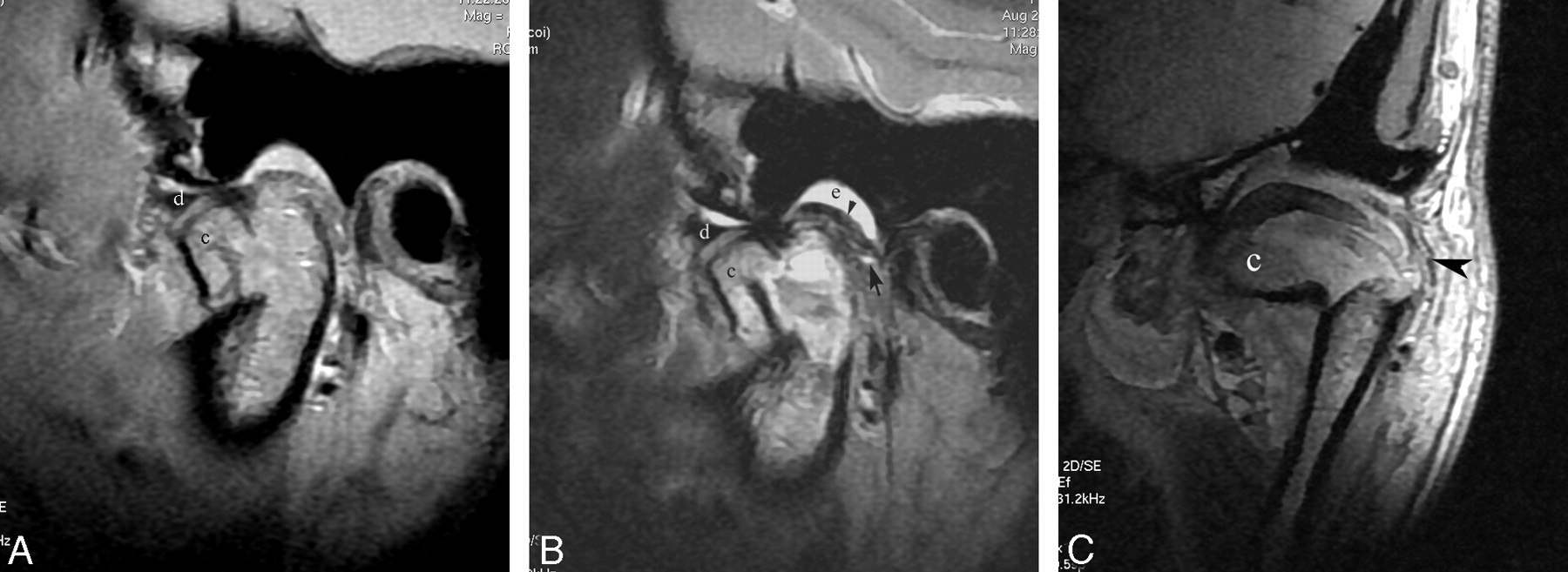
- Fig 1.
Patient with dislocated mandibular condylar fracture. Sagittal proton attenuation-weighted image (A) and T2-weighted image (B) demonstrate that the fractured condyle (c) is displaced in the anteroinferior direction and out of the glenoid fossa. The TMJ disk (d) is also displaced in the same direction as the fractured fragment. On T2-weighted image (B), the joint effusion (e) is only identified in the upper joint space, and the dotted high signal intensity is found in the retrodiskal tissue (black arrow). The faint inferoposterior attachment and well-defined superior posterior attachment (black arrowhead) of the disk are visible. Coronal proton attenuation-weighted image (C) shows the fractured fragment (c) and well-defined joint capsule (black arrowhead).
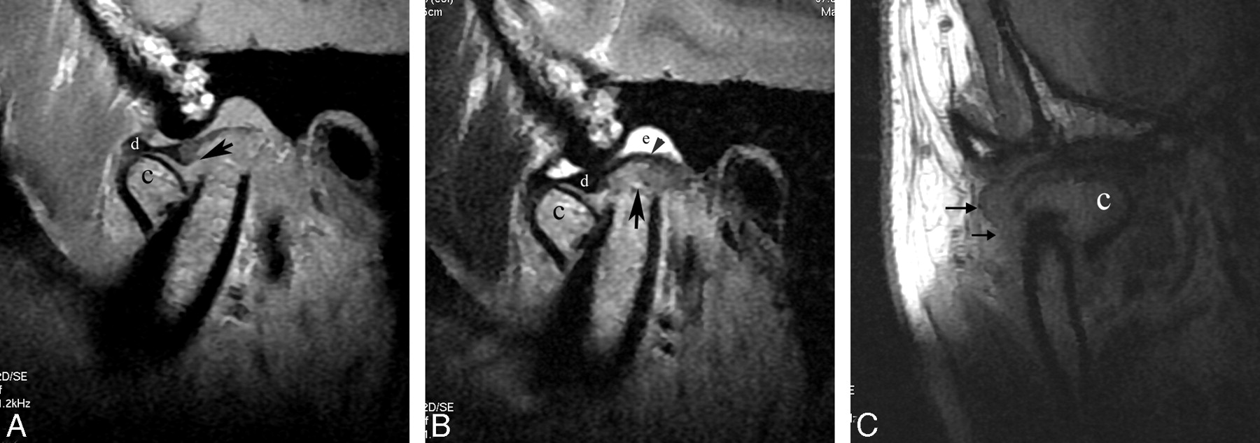
- Fig 2.
Patient with dislocated mandibular condyle fracture. Sagittal proton attenuation-weighted image (A) and T2-weighted image (B) demonstrate that the fractured condyle (c) is anteroinferiorly displaced. The TMJ disk (d) is located superior to the fractured fragment but is anteroinferiorly displaced relative to the remaining mandibular ramus. The tear of the inferoposterior attachment of the disk (black arrow on A) is shown. T2-weighted image (B) demonstrates the dotted high signal intensity of retrodiskal tissue (black arrow), joint effusion of the upper joint compartment (e), and well-defined superior posterior attachment (black arrowhead) of the disk. Coronal proton attenuation-weighted image (C) reveals the tear of the joint capsule (black arrows) and fractured fragment (c) located on the medial side of the mandibular ramus.
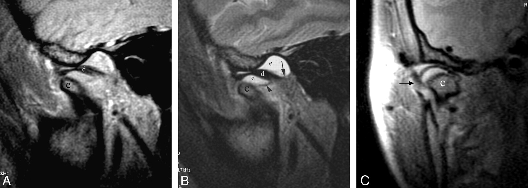
- Fig 3.
Patient with a dislocated mandibular condylar fracture. Sagittal proton attenuation-weighted image (A) and T2-weighted image (B) demonstrate that both the fractured condyle (c) and the TMJ disk (d) are displaced in the same direction (anterior and inferior direction). The joint effusion (e) with well-defined superior (black arrow) and inferior (black arrowhead) posterior attachments is identified in both the upper and lower compartments of the TMJ on T2-weighted image (B). Coronal proton attenuation-weighted image (C) shows the ill-defined joint capsule (black arrow) and fractured condyle (c) located in the medial side of the mandibular ramus.
- Fig 4.
Patient with nondislocated mandibular condyle fracture. Sagittal proton attenuation-weighted image (A) demonstrates interrupted cortical bone (white arrowhead) of the condyle (c) and avulsion of the TMJ disk (white arrow). However, the location of the TMJ disk is normal. Coronal proton attenuation-weighted image (B) shows a condylar head fracture (c). The outline of the lateral side of the joint capsule is completed (white arrow).


Tables
Different soft tissue changes of TMJ between group 1 and group 2 on MR images
Abnormal MR Imaging Findings Group 1 (%) Group 2 (%) Total (%) P Value Joint effusion 95 (88%) 6 (60%) 101 (85.6%) > .05 Disk displacement 105 (97.2%) 3 (30%) 108 (91.5%) < .01 Disk deformity 10 (9.3%) 1 (10%) 11 (9.3%) > .05 Disk perforation 9 (8.3%) 2 (20%) 10 (8.5%) > .05 Abnormal superoposterior attachment of disk 42 (38.9%) 3 (30%) 45 (38.1%) > .05 Abnormal inferoposterior attachment of disk 96 (88.9%) 7 (70%) 103 (87.3%) > .05 Abnormal signal intensity of retrodiskal tissue 98 (88.3%) 5 (50%) 103 (87.3%) < .05 Abnormal joint capsule 94 (87%) 7 (70%) 101 (85.6%) > .05
In this issue


Jump to section
Related Articles
Cited By...
- No citing articles found.