
Article Figures & Data
Figures
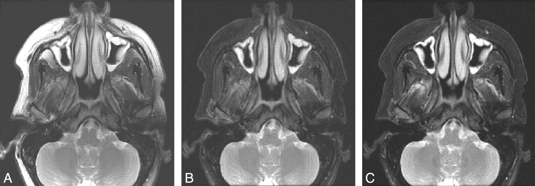
- Fig 1.
T2-weighted images obtained with a conventional FSE acquisition with CHESS (A), the 2PD technique (B), and the fTED technique (C). The images obtained with use of the 2 Dixon techniques provide uniform fat suppression throughout the FOV, whereas the fat suppression achieved by the conventional CHESS FSE technique is unsatisfactory in the anterior facial region.
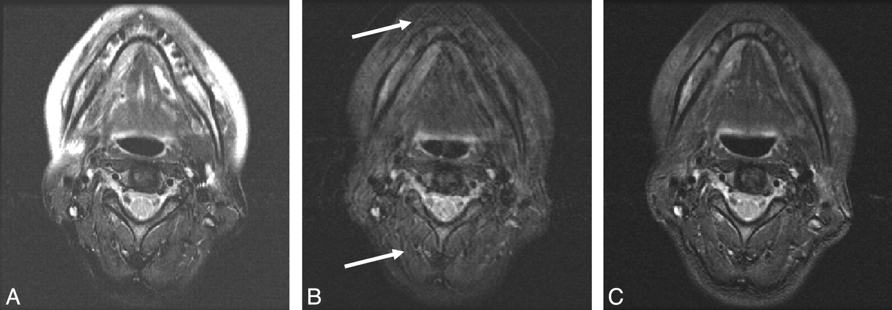
- Fig 2.
T2-weighted images obtained with a conventional FSE acquisition with CHESS (A), the 2PD technique (B), and the fTED technique (C). The images obtained with use of the 2 Dixon techniques again provide subjectively better and more uniform fat suppression than the images obtained with the conventional CHESS FSE technique. However, motion artifacts are noted to be present in the images obtained with 2PD (arrows). In comparison, the fTED images show uniform fat suppression and best overall image quality.
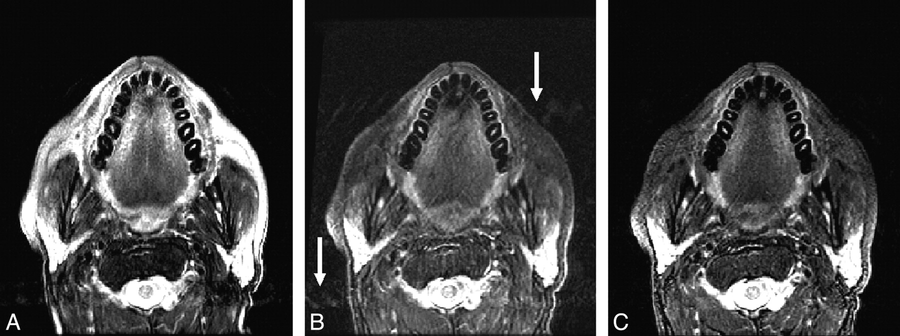
- Fig 3.
T2-weighted images of another patient obtained with a conventional FSE acquisition with CHESS (A), the 2PD technique (B), and the fTED technique (C). As in Fig 2, fat suppression in the 2 Dixon images (B and C) was more uniform than that of the image by CHESS (A). In contrast, motion artifacts (at the levels by the arrows) were most severe in the image by the 2PD technique.


In this issue


Jump to section
Related Articles
Cited By...
- Development and implementation of optimized endogenous contrast sequences for delineation in adaptive radiotherapy on a 1.5T MR-Linear-accelerator (MR-Linac): A prospective R-IDEAL Stage 0-2a quantitative/qualitative evaluation of in vivo site-specific quality-assurance using a 3D T2 fat-suppressed platform for head and neck cancer
- Optimal Fat Suppression in Head and Neck MRI: Comparison of Multipoint Dixon with 2 Different Fat-Suppression Techniques, Spectral Presaturation and Inversion Recovery, and STIR