
Article Figures & Data
Figures
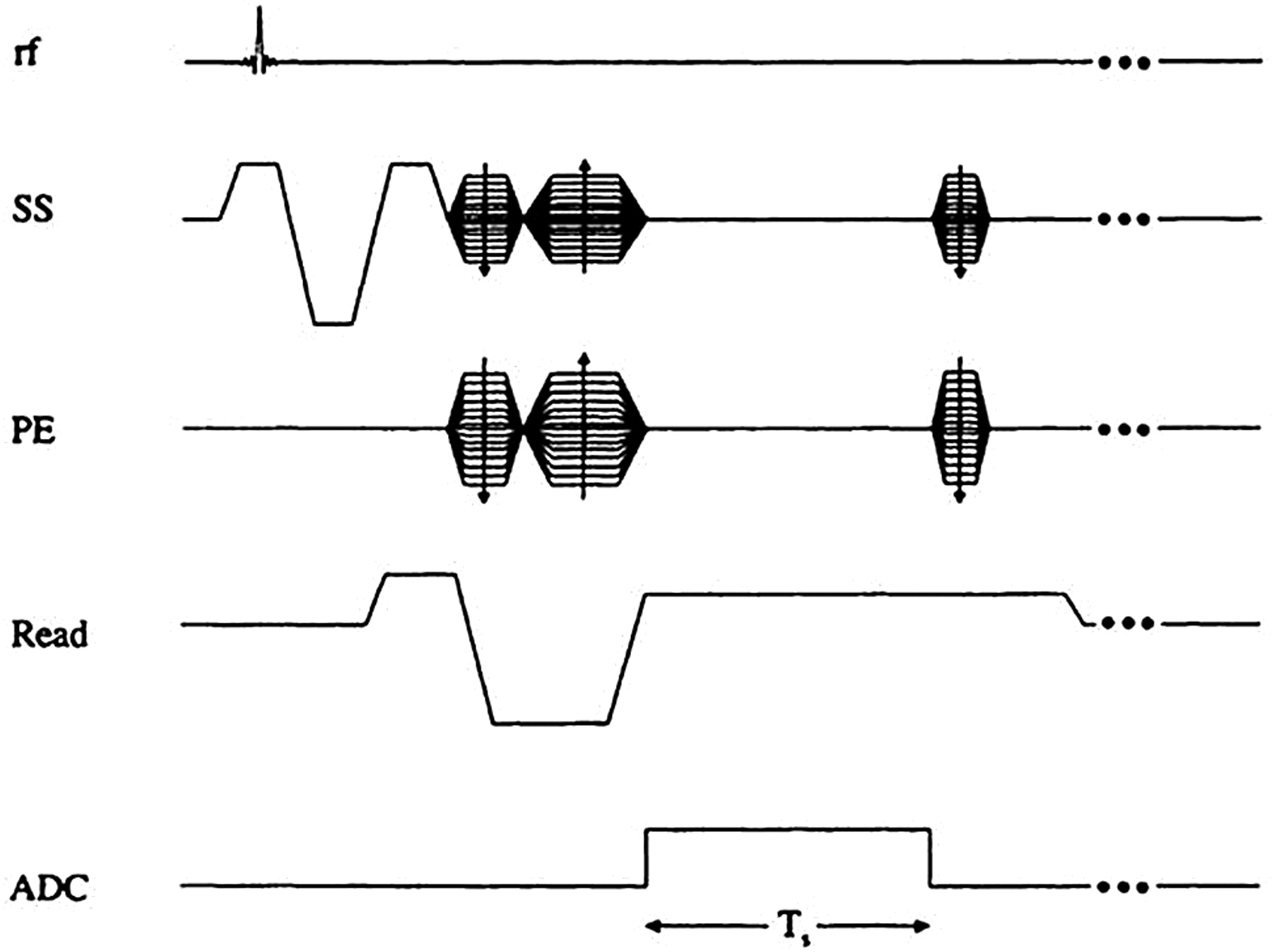
- Fig 1.
Gradient-echo sequence design. Ts refers to the sampling time interval. The gradient pulses are designed to give first-order flow compensation. SS indicates the slice-select gradient; PE, the phase-encoding gradient; ADC, analog-to-digital conversion.
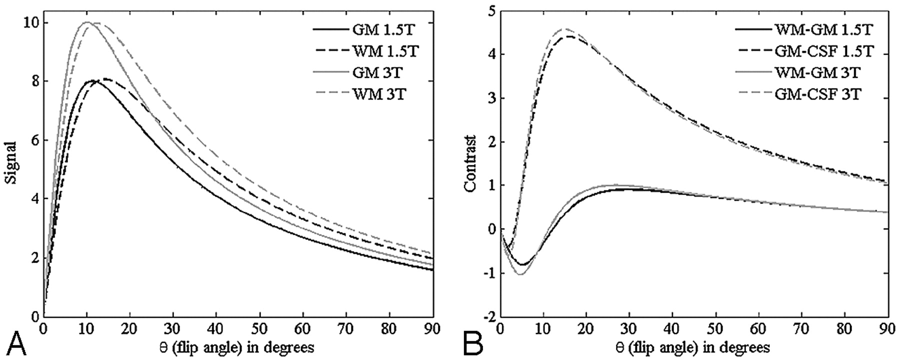
- Fig 2.
A, Plots showing the signal-intensity behavior as a function of flip angle for gray matter (GM) and white matter (WM) at field strengths 1.5T and 3T. B, Plots showing the corresponding contrast between GM-WM and GM-CSF. These curves are calculated taking into consideration the higher signal intensity available at 3T and assuming that bandwidth is correspondingly increased at 3T to ensure equivalent geometric distortion. Tissue parameters are given in Table 1. Note that the GM/WM contrast is highest around 20° at 3T and there is a minimum or reversal of contrast around 5° (where GM is now brighter than WM; ie, it is a high-contrast spin echo-weighted image).
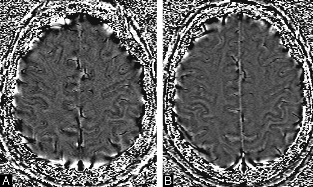
- Fig 3.
A, Raw phase image. B, HP-filtered phase image with a central filter size of 32 × 32; C, Filtered-phase image with a central filter size of 64 × 64.
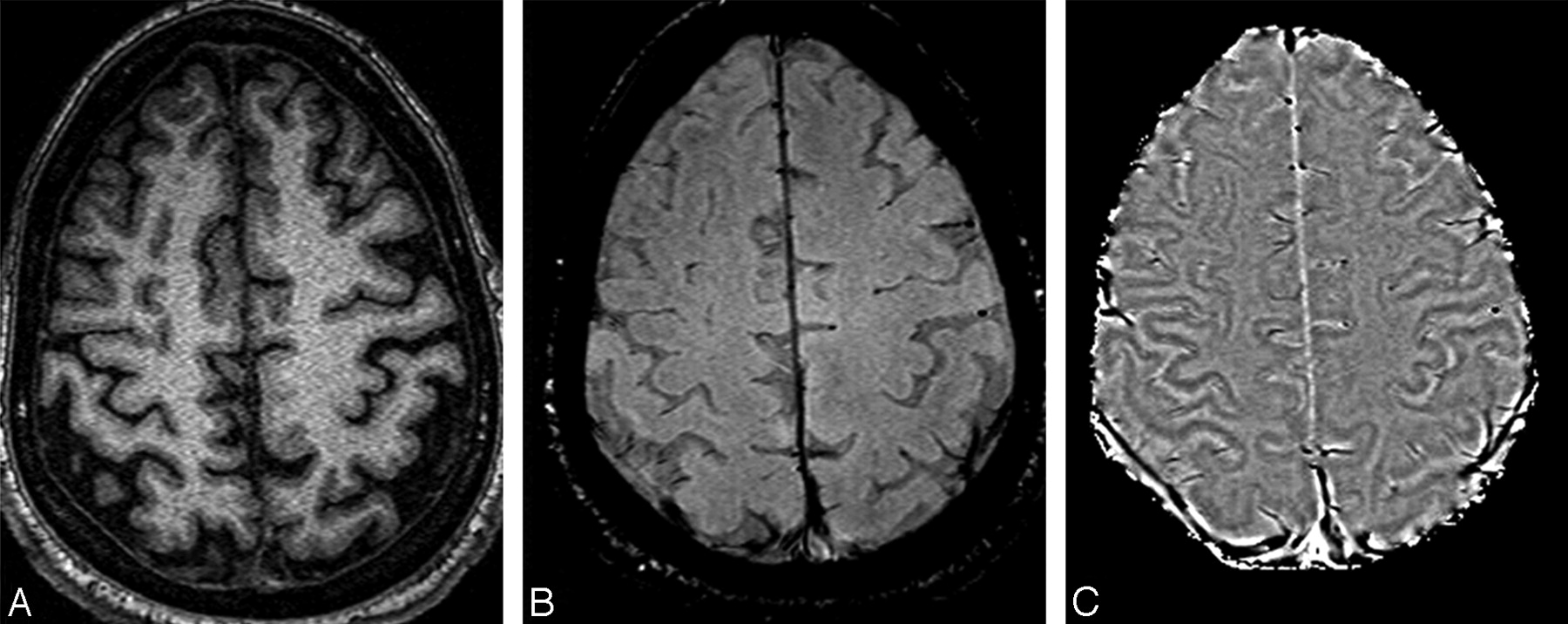
- Fig 4.
Short-echo T1-weighted image (A), compared with the SWI long-echo gradient-echo processed magnitude (B) and HP-filtered phase data (C).
- Fig 5.
A, HP-filtered phase image in the midbrain acquired at 4T with TE = 15 ms. B, India ink-stained cadaver brain section showing a strong correspondence to variations in signal intensity as seen with SWI HP-filtered phase.
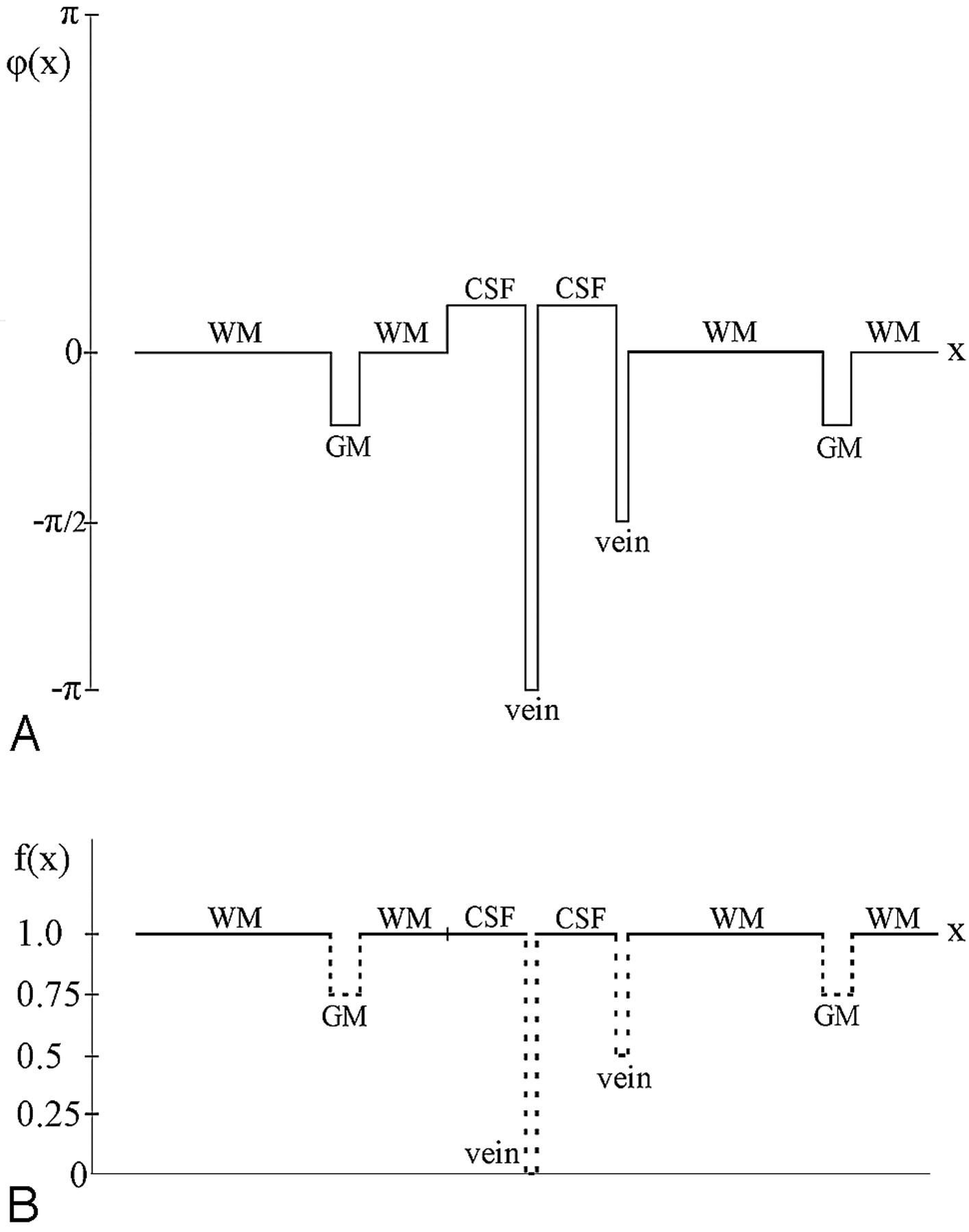
- Fig 6.
Pictorial depiction of the phase-masking process. A, Phase profile in a filtered-phase image. B, Corresponding profile of the mask created from A. Once the mask is raised to the fourth power, the vein that has a phase of −π / 2 and a mask value of 0.5 will become 1-/16 and, therefore, this vein will be almost as well suppressed as the vein with a phase of −π and f(x) = 0. GM indicates gray matter; WM, white matter.
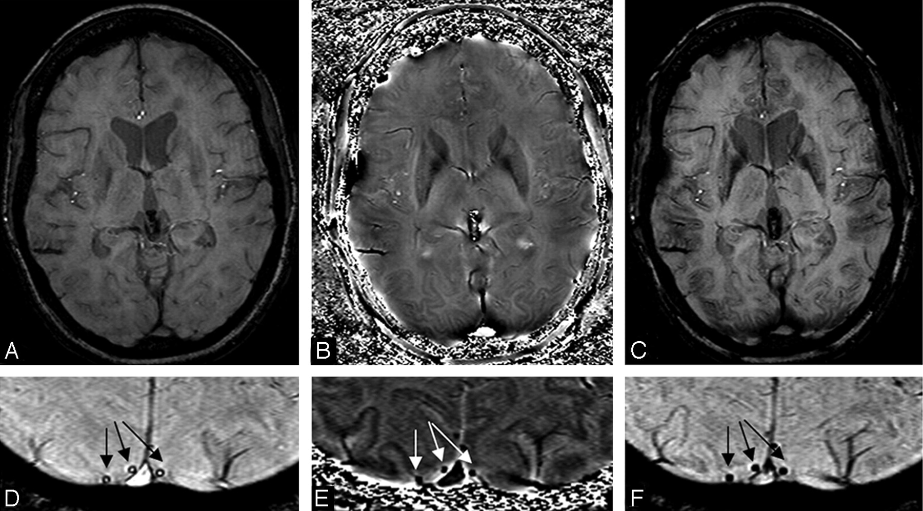
- Fig 7.
A, Unprocessed original SWI magnitude image. B, HP-filtered phase image. C, Processed SWI magnitude image (ie, after phase-mask multiplication with m = 4). D, Unprocessed original SWI magnitude image showing the dark hypointense ring around vein cross-sections (arrows). E, HP-filtered phase image showing the veins have low signal-intensity (arrows). F, Processed SWI magnitude image showing that the veins now appear with uniform low signal intensity (arrows).
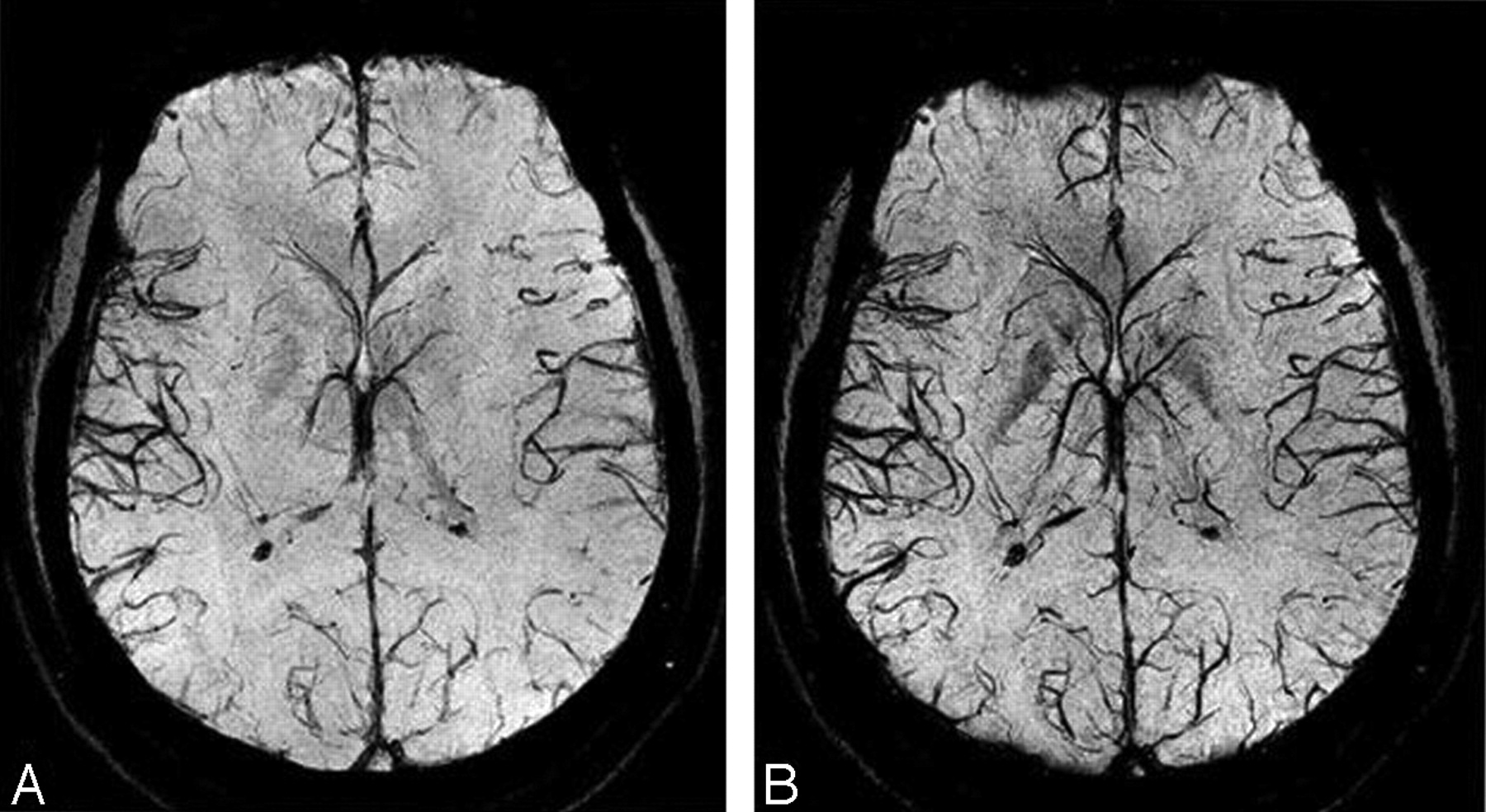
- Fig 8.
mIP data over the original magnitude images (A) and over the processed SWI images (B). The mIP is carried out over 7 sections (representing a 14-mm slab thickness). The images were collected at 3T. There is a dropout of signal intensity in the frontal part of the brain, but otherwise the vessel visibility is much improved in B. In the future, this frontal dropout should not be a problem when the air/tissue geometries are corrected (Fig 9).
- Fig 9.
Results of the improved processing methodology. A, Original SWI phase image. B, Simulated phase due to air/tissue geometry at 40 ms. C, Result of subtracting A from B through the complex division. D, Result of HP filtering of A. E, Result of HP filtering of C. F, Corresponding unprocessed magnitude SWI image. G, Processed SWI magnitude image by using a phase mask from D. H, Processed SWI magnitude by using a phase mask from E. I, Result of subtracting G from H. Corresponding magnitude and phase images have been adjusted to the same, but appropriate, contrast levels. The size of the central filter in the HP process is 64 × 64.
- Fig 10.
A and B, Phase images, keeping the product BoTE constant. A phase image acquired at 1.5T (A) and the same subject at 3T (B) show the same overall contrast but with a better SNR. The gray/white matter contrast in these images comes from the increased MR-visible iron content in the gray matter, giving it an appearance similar to a T1-weighted scan.
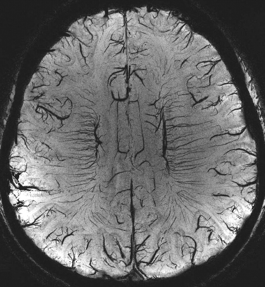
- Fig 11.
A sample mIP from 7T data, with a resolution of 215 × 215 × 1000 μ, TE = 16 ms, TR = 45 ms, FA = 25°, and mIP over 8 sections. Image courtesy of Ge Y and Barnes S.
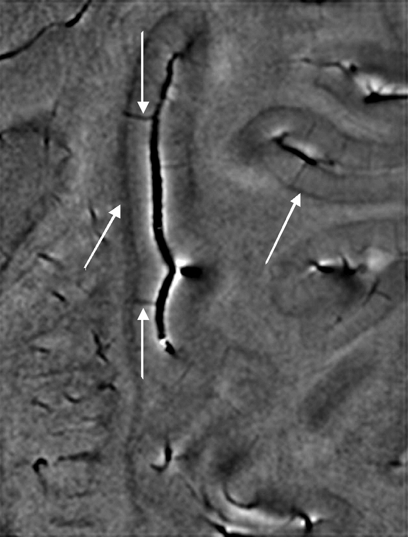
- Fig 12.
A zoomed image from the same dataset shown in Fig 11 reveals a dark band between the gray matter and white matter, which we assume represents the white matter arcuate fibers (diagonal arrows). The small vessels joining the pial veins are the venules (vertical arrows). Image courtesy of Ge Y and Barnes S.


Tables
GM WM CSF Spin density % 80 65 100 1.5T 1000 ms 650 ms 4000 ms 3T 1283 ms 838 ms 4000 ms Note:—GM indicates gray matter; WM, white matter; CSF, cerebrospinal fluid. CSF is taken to have 100% water content.
- Table 2:
Range of experimental parameters advised for SWI data collection at different field strengths*
FS FA (degrees) TR (ms) TE (ms) BW/pixel (Hz/pixel) 1.5T 18–25 (20) 50–60 (60) 40 70–100 3T 12–17 (12) 25–35 (30) 20 80–100 4T 8–15 (12) 25–35 (28) 15 80–120 7T 11–17 (14) 25–35 (30) 10–16 100–150 Note:—FS indicates field strength; FA, flip angle; BW, bandwidth.
* The values given in parentheses for FA and TR are the ones generally used by the authors. TEs given here are optimized for maximum signal cancellation from venous vasculature. Slightly shorter or longer TEs can be used for appropriate applications. However, note that for implementing shorter TEs, higher BW would be required, which would adversely affect the SNR of the image. One way to avoid this is to reduce the TR appropriately and cover more sections of the brain, which can recover some of the lost SNR.
In this issue


Jump to section
- Article
- Abstract
- Gradient-Echo Imaging
- Magnetic Susceptibility
- Creating Susceptibility-Weighted Filtered-Phase Images
- Marrying Magnitude and Phase Images to Create Magnitude SWI Data
- Advanced Phase Processing
- Recommended Imaging Parameters at Different Field Strengths
- Interpreting SWI Data
- Future Directions
- Acknowledgments
- Footnotes
- References
- Figures & Data
- Info & Metrics
- Responses
- References