Advertisement

Main menu

  • Home
  • Content
    • Current Issue
    • Accepted Manuscripts
    • Article Preview
    • Past Issue Archive
    • Video Articles
    • AJNR Case Collection
    • Case of the Week Archive
    • Case of the Month Archive
    • Classic Case Archive
  • Special Collections
    • AJNR Awards
    • Low-Field MRI
    • Alzheimer Disease
    • ASNR Foundation Special Collection
    • Photon-Counting CT
    • View All
  • Multimedia
    • AJNR Podcasts
    • AJNR SCANtastic
    • Trainee Corner
    • MRI Safety Corner
    • Imaging Protocols
  • For Authors
    • Submit a Manuscript
    • Submit a Video Article
    • Submit an eLetter to the Editor/Response
    • Manuscript Submission Guidelines
    • Statistical Tips
    • Fast Publishing of Accepted Manuscripts
    • Graphical Abstract Preparation
    • Imaging Protocol Submission
    • Author Policies
  • About Us
    • About AJNR
    • Editorial Board
    • Editorial Board Alumni
  • More
    • Become a Reviewer/Academy of Reviewers
    • Subscribers
    • Permissions
    • Alerts
    • Feedback
    • Advertisers
    • ASNR Home

User menu

  • Alerts
  • Log in

Search

  • Advanced search
American Journal of Neuroradiology

American Journal of Neuroradiology

ASHNR American Society of Functional Neuroradiology ASHNR American Society of Pediatric Neuroradiology ASSR
  • Alerts
  • Log in

Advanced Search

  • Home
  • Content
    • Current Issue
    • Accepted Manuscripts
    • Article Preview
    • Past Issue Archive
    • Video Articles
    • AJNR Case Collection
    • Case of the Week Archive
    • Case of the Month Archive
    • Classic Case Archive
  • Special Collections
    • AJNR Awards
    • Low-Field MRI
    • Alzheimer Disease
    • ASNR Foundation Special Collection
    • Photon-Counting CT
    • View All
  • Multimedia
    • AJNR Podcasts
    • AJNR SCANtastic
    • Trainee Corner
    • MRI Safety Corner
    • Imaging Protocols
  • For Authors
    • Submit a Manuscript
    • Submit a Video Article
    • Submit an eLetter to the Editor/Response
    • Manuscript Submission Guidelines
    • Statistical Tips
    • Fast Publishing of Accepted Manuscripts
    • Graphical Abstract Preparation
    • Imaging Protocol Submission
    • Author Policies
  • About Us
    • About AJNR
    • Editorial Board
    • Editorial Board Alumni
  • More
    • Become a Reviewer/Academy of Reviewers
    • Subscribers
    • Permissions
    • Alerts
    • Feedback
    • Advertisers
    • ASNR Home
  • Follow AJNR on Twitter
  • Visit AJNR on Facebook
  • Follow AJNR on Instagram
  • Join AJNR on LinkedIn
  • RSS Feeds

AJNR Awards, New Junior Editors, and more. Read the latest AJNR updates

Research ArticleBRAIN

Predictors of Hemorrhage Following Intra-Arterial Thrombolysis for Acute Ischemic Stroke: The Role of Pial Collateral Formation

G.A. Christoforidis, C. Karakasis, Y. Mohammad, L.P. Caragine, M. Yang and A.P. Slivka
American Journal of Neuroradiology January 2009, 30 (1) 165-170; DOI: https://doi.org/10.3174/ajnr.A1276
G.A. Christoforidis
  • Find this author on Google Scholar
  • Find this author on PubMed
  • Search for this author on this site
C. Karakasis
  • Find this author on Google Scholar
  • Find this author on PubMed
  • Search for this author on this site
Y. Mohammad
  • Find this author on Google Scholar
  • Find this author on PubMed
  • Search for this author on this site
L.P. Caragine
  • Find this author on Google Scholar
  • Find this author on PubMed
  • Search for this author on this site
M. Yang
  • Find this author on Google Scholar
  • Find this author on PubMed
  • Search for this author on this site
A.P. Slivka
  • Find this author on Google Scholar
  • Find this author on PubMed
  • Search for this author on this site
  • Article
  • Figures & Data
  • Info & Metrics
  • Responses
  • References
  • PDF
Loading

Article Figures & Data

Figures

  • Tables
  • Fig 1.
    • Download figure
    • Open in new tab
    • Download powerpoint
    Fig 1.

    This diagram depicts the scoring of the anatomic extent of pial collateral formation from the ACA territory to the MCA territory during occlusion of the M1 segment. Scoring corresponds to angiographically visible retrograde opacification of the MCA segments on the delayed venous phase.9 Each distinctly textured segment depicts the furthest extent of retrograde opacification identified on cerebral angiography for each pial collateral score.

  • Fig 2.
    • Download figure
    • Open in new tab
    • Download powerpoint
    Fig 2.

    Bar graph displays the rates of any hemorrhage (black) and significant hemorrhage (hemorrhage volume >25 mL, gray) based on the pial collateral score.
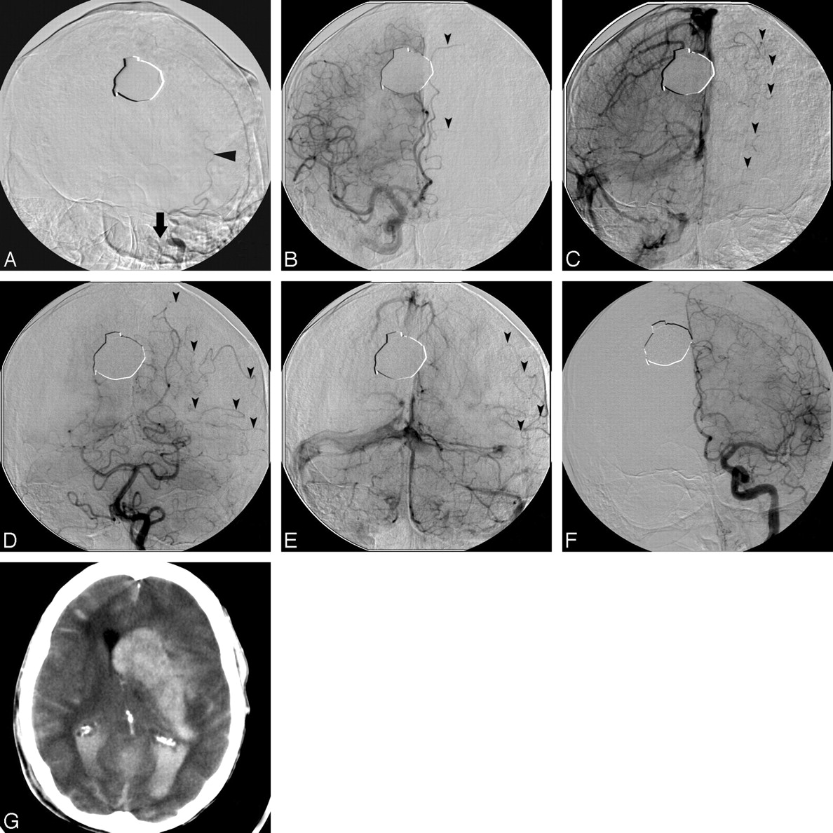
  • Fig 3.
    • Download figure
    • Open in new tab
    • Download powerpoint
    Fig 3.

    Large hemorrhagic conversion following reperfusion in a patient with distal left internal carotid occlusion with poor pial collateral formation. A, Left internal carotid arteriogram demonstrates a distal carotid internal occlusion (arrow) with refluxed contrast filling of the occipital artery (arrowhead, A). B–E, Arteriograms of the right internal carotid (B and C) and left vertebral arteries (D and E) demonstrate no circle of Willis collaterals on either the late arterial phase (arrowheads, B and D) or the late venous phase (arrowheads, C and E). Poor pial collateral formation reconstitutes, at best, the M4 branches of the MCA (arrowheads, D and E). F and G, Twenty-four hours following reperfusion (F), the patient experienced a large hemorrhage (G) and subsequently died.
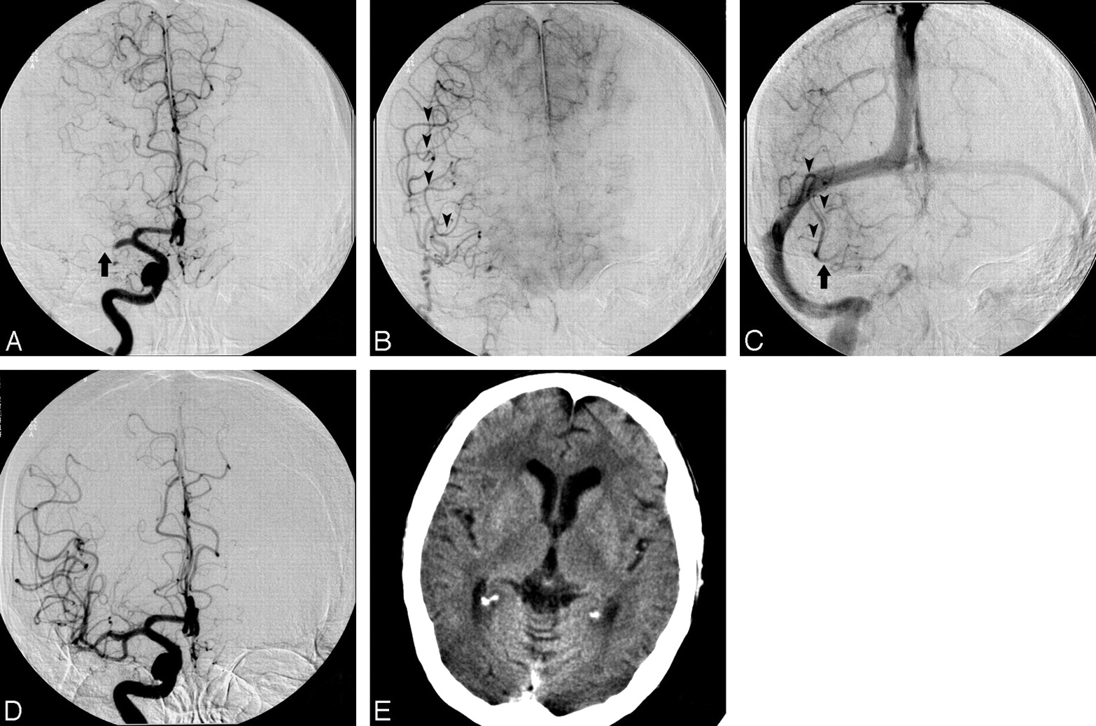
  • Fig 4.
    • Download figure
    • Open in new tab
    • Download powerpoint
    Fig 4.

    Arterial (A), parenchymal (B), and late venous (C) phase arteriograms in a patient with right MCA occlusion (A, arrow) demonstrate good pial collateral formation (B and C, arrowheads) with reconstitution of the entire MCA territory, including the M1 segment (arrow, C). D and E, Following reperfusion (D), the patient had no hemorrhage or infarction (E).

Tables

  • Figures
    • View popup
    Table 1:

    Logistic regression analysis for any hemorrhage

    PredictorOR (95% CI)Estimate (SE)P > χ2*
    Poor pial collateral score3.03 (1.09–8.71)1.11 (0.52).0342
    Platelets <200K†2.95 (1.06–8.57)1.08 (0.53).0403
    Diabetes4.82 (1.49–16.9)0.787 (0.306).0100
    Time to treat >180 min12.0 (1.77–253)2.47 (1.17).0333
    Intercept−0.81 (0.52).1138
    • Note:—OR indicates odds ratio.

    • * Variables tested with P > .05 were rejected using backward selection (whole-model test, P < .0001; r2 = 0.202).

    • † 200K = 200,000/μL.

    • View popup
    Table 2:

    Logistic regression analysis for significant hemorrhage (volume >25 mL)

    Predictive factorOR (95% CI)Estimate (SE)P > χ2*
    Poor pial collateral score13.1 (2.84–96.0)2.57 (0.860).0028
    Platelets <200K8.14 (1.73–60.3)2.10 (0.868).0157
    Intercept0.190 (0.520).715
    • Note:—OR indicates odds ratio; CI, confidence interval.

    • * Variables tested with P > .05 were rejected using backward selection (whole-model test, P < .0001; r2 = 0.202).

    • View popup
    Table 3:

    Hemorrhage rates versus pial collateral formation and platelet level*

    ParameterRate of Any HemorrhageP Value (Pearson)Rate of Significant HemorrhageP Value (Pearson)
    Total26/104 (25.0%)10/104 (9.61%)
    Good collaterals13/72 (18.1%).01422/72 (2.78%).0004
    Poor collaterals13/32 (40.6%)8/32 (25.0%)
    Platelets <200K16/42 (38.1%).01118/42 (19.0%).0072
    Platelets >200K10/62 (16.1%)2/62 (3.23 %)
    • * Shows significantly higher rates of any hemorrhage and significant hemorrhage (volume > 25 mL) among patients with poor pial collaterals and platelet levels <200,000/μL.

PreviousNext
Back to top

In this issue

American Journal of Neuroradiology: 30 (1)
American Journal of Neuroradiology
Vol. 30, Issue 1
January 2009
  • Table of Contents
  • Index by author
Advertisement
Print
Download PDF
Email Article

Thank you for your interest in spreading the word on American Journal of Neuroradiology.

NOTE: We only request your email address so that the person you are recommending the page to knows that you wanted them to see it, and that it is not junk mail. We do not capture any email address.

Enter multiple addresses on separate lines or separate them with commas.
Predictors of Hemorrhage Following Intra-Arterial Thrombolysis for Acute Ischemic Stroke: The Role of Pial Collateral Formation
(Your Name) has sent you a message from American Journal of Neuroradiology
(Your Name) thought you would like to see the American Journal of Neuroradiology web site.
CAPTCHA
This question is for testing whether or not you are a human visitor and to prevent automated spam submissions.
Cite this article
G.A. Christoforidis, C. Karakasis, Y. Mohammad, L.P. Caragine, M. Yang, A.P. Slivka
Predictors of Hemorrhage Following Intra-Arterial Thrombolysis for Acute Ischemic Stroke: The Role of Pial Collateral Formation
American Journal of Neuroradiology Jan 2009, 30 (1) 165-170; DOI: 10.3174/ajnr.A1276

Citation Manager Formats

  • BibTeX
  • Bookends
  • EasyBib
  • EndNote (tagged)
  • EndNote 8 (xml)
  • Medlars
  • Mendeley
  • Papers
  • RefWorks Tagged
  • Ref Manager
  • RIS
  • Zotero
0 Responses
Respond to this article
Share
Bookmark this article
Predictors of Hemorrhage Following Intra-Arterial Thrombolysis for Acute Ischemic Stroke: The Role of Pial Collateral Formation
G.A. Christoforidis, C. Karakasis, Y. Mohammad, L.P. Caragine, M. Yang, A.P. Slivka
American Journal of Neuroradiology Jan 2009, 30 (1) 165-170; DOI: 10.3174/ajnr.A1276
del.icio.us logo Twitter logo Facebook logo Mendeley logo
  • Tweet Widget
  • Facebook Like
  • Google Plus One
Purchase

Jump to section

  • Article
    • Abstract
    • Materials and Methods
    • Results
    • Discussion
    • Conclusions
    • Acknowledgments
    • Footnotes
    • References
  • Figures & Data
  • Info & Metrics
  • Responses
  • References
  • PDF

Related Articles

  • No related articles found.
  • PubMed
  • Google Scholar

Cited By...

  • Systematic review protocol to assess artificial intelligence diagnostic accuracy performance in detecting acute ischaemic stroke and large-vessel occlusions on CT and MR medical imaging
  • Multi-ancestry genetic study in 5,876 patients identifies an association between excitotoxic genes and early outcomes after acute ischemic stroke
  • The impact of general anesthesia, baseline ASPECTS, time to treatment, and IV tPA on intracranial hemorrhage after neurothrombectomy: pooled analysis of the SWIFT PRIME, SWIFT, and STAR trials
  • Guidelines for evaluation and management of cerebral collateral circulation in ischaemic stroke 2017
  • Collateral status affects the onset-to-reperfusion time window for good outcome
  • Role of Genetic Variation in Collateral Circulation in the Evolution of Acute Stroke: A Multimodal Magnetic Resonance Imaging Study
  • Comparison of four different collateral scores in acute ischemic stroke by CT angiography
  • Fluid-Attenuated Inversion Recovery Vascular Hyperintensity Topography, Novel Imaging Marker for Revascularization in Middle Cerebral Artery Occlusion
  • Comparing Vessel Imaging: Noncontrast Computed Tomography/Computed Tomographic Angiography Should Be the New Minimum Standard in Acute Disabling Stroke
  • Determinants of Intracranial Hemorrhage Occurrence and Outcome after Neurothrombectomy Therapy: Insights from the Solitaire FR With Intention For Thrombectomy Randomized Trial
  • Poor Collateral Circulation Assessed by Multiphase Computed Tomographic Angiography Predicts Malignant Middle Cerebral Artery Evolution After Reperfusion Therapies
  • Impact of Time-to-Reperfusion on Outcome in Patients with Poor Collaterals
  • Congenic Fine-Mapping Identifies a Major Causal Locus for Variation in the Native Collateral Circulation and Ischemic Injury in Brain and Lower Extremity
  • Diabetes Mellitus, Admission Glucose, and Outcomes After Stroke Thrombolysis: A Registry and Systematic Review
  • Factors associated with rapid neurological improvement 24 h following intra-arterial thrombolytic treatment for acute ischemic stroke
  • Reperfusion Rates Following Intra-Arterial Thrombolysis for Acute Ischemic Stroke: The Influence of the Method for Alteplase Delivery
  • Endovascular Therapy of 623 Patients With Anterior Circulation Stroke
  • Systematic Review of Methods for Assessing Leptomeningeal Collateral Flow
  • Extending the Time Window for Endovascular Procedures According to Collateral Pial Circulation
  • Patient Outcomes With Endovascular Embolectomy Therapy for Acute Ischemic Stroke: A Study of the National Inpatient Sample: 2006 to 2008
  • Predictors of Subarachnoid Hemorrhage in Acute Ischemic Stroke With Endovascular Therapy
  • Genetic Architecture Underlying Variation in Extent and Remodeling of the Collateral Circulation
  • Higher Rates of Mortality but Not Morbidity Follow Intracranial Mechanical Thrombectomy in the Elderly
  • Impact of Baseline Tissue Status (Diffusion-Weighted Imaging Lesion) Versus Perfusion Status (Severity of Hypoperfusion) on Hemorrhagic Transformation
  • Occurrence and Predictors of Futile Recanalization following Endovascular Treatment among Patients with Acute Ischemic Stroke: A Multicenter Study
  • Crossref (105)
  • Google Scholar

This article has been cited by the following articles in journals that are participating in Crossref Cited-by Linking.

  • Hemorrhagic Transformation after Ischemic Stroke in Animals and Humans
    Glen C Jickling, DaZhi Liu, Boryana Stamova, Bradley P Ander, Xinhua Zhan, Aigang Lu, Frank R Sharp
    Journal of Cerebral Blood Flow & Metabolism 2014 34 2
  • Vascular remodeling after ischemic stroke: Mechanisms and therapeutic potentials
    Jialing Liu, Yongting Wang, Yosuke Akamatsu, Chih Cheng Lee, R. Anne Stetler, Michael T. Lawton, Guo-Yuan Yang
    Progress in Neurobiology 2014 115
  • Wide Genetic Variation in the Native Pial Collateral Circulation is a Major Determinant of Variation in Severity of Stroke
    Hua Zhang, Pranay Prabhakar, Robert Sealock, James E Faber
    Journal of Cerebral Blood Flow & Metabolism 2010 30 5
  • Diabetes Mellitus, Admission Glucose, and Outcomes After Stroke Thrombolysis
    Jean-Philippe Desilles, Elena Meseguer, Julien Labreuche, Bertrand Lapergue, Gaia Sirimarco, Jaime Gonzalez-Valcarcel, Philippa Lavallée, Lucie Cabrejo, Celine Guidoux, Isabelle Klein, Pierre Amarenco, Mikael Mazighi
    Stroke 2013 44 7
  • Systematic Review of Methods for Assessing Leptomeningeal Collateral Flow
    F. McVerry, D.S. Liebeskind, K.W. Muir
    American Journal of Neuroradiology 2012 33 3
  • Occurrence and Predictors of Futile Recanalization following Endovascular Treatment among Patients with Acute Ischemic Stroke: A Multicenter Study
    H.M. Hussein, A.L. Georgiadis, G. Vazquez, J.T. Miley, M.Z. Memon, Y.M. Mohammad, G.A. Christoforidis, N. Tariq, A.I. Qureshi
    American Journal of Neuroradiology 2010 31 3
  • Endovascular Therapy of 623 Patients With Anterior Circulation Stroke
    Aekaterini Galimanis, Simon Jung, Marie-Luise Mono, Urs Fischer, Oliver Findling, Anja Weck, Niklaus Meier, Gian Marco De Marchis, Caspar Brekenfeld, Marwan El-Koussy, Heinrich P. Mattle, Marcel Arnold, Gerhard Schroth, Jan Gralla
    Stroke 2012 43 4
  • Guidelines for evaluation and management of cerebral collateral circulation in ischaemic stroke 2017
    Liping Liu, Jing Ding, Xinyi Leng, Yuehua Pu, Li-An Huang, Anding Xu, Ka Sing Lawrence Wong, Xin Wang, Yongjun Wang
    Stroke and Vascular Neurology 2018 3 3
  • Extending the Time Window for Endovascular Procedures According to Collateral Pial Circulation
    Marc Ribo, Alan Flores, Marta Rubiera, Jorge Pagola, Joao Sargento-Freitas, David Rodriguez-Luna, Pilar Coscojuela, Olga Maisterra, Socorro Piñeiro, Francisco J. Romero, Jose Alvarez-Sabin, Carlos A. Molina
    Stroke 2011 42 12
  • Genetic Architecture Underlying Variation in Extent and Remodeling of the Collateral Circulation
    Shiliang Wang, Hua Zhang, Xuming Dai, Robert Sealock, James E. Faber
    Circulation Research 2010 107 4

More in this TOC Section

  • Multimodal CT Provides Improved Performance for Lacunar Infarct Detection
  • Optimal MRI Sequence for Identifying Occlusion Location in Acute Stroke: Which Value of Time-Resolved Contrast-Enhanced MRA?
  • Evaluating the Effects of White Matter Multiple Sclerosis Lesions on the Volume Estimation of 6 Brain Tissue Segmentation Methods
Show more BRAIN

Similar Articles

Advertisement

Indexed Content

  • Current Issue
  • Accepted Manuscripts
  • Article Preview
  • Past Issues
  • Editorials
  • Editor's Choice
  • Fellows' Journal Club
  • Letters to the Editor
  • Video Articles

Cases

  • Case Collection
  • Archive - Case of the Week
  • Archive - Case of the Month
  • Archive - Classic Case

More from AJNR

  • Trainee Corner
  • Imaging Protocols
  • MRI Safety Corner

Multimedia

  • AJNR Podcasts
  • AJNR Scantastics

Resources

  • Turnaround Time
  • Submit a Manuscript
  • Submit a Video Article
  • Submit an eLetter to the Editor/Response
  • Manuscript Submission Guidelines
  • Statistical Tips
  • Fast Publishing of Accepted Manuscripts
  • Graphical Abstract Preparation
  • Imaging Protocol Submission
  • Evidence-Based Medicine Level Guide
  • Publishing Checklists
  • Author Policies
  • Become a Reviewer/Academy of Reviewers
  • News and Updates

About Us

  • About AJNR
  • Editorial Board
  • Editorial Board Alumni
  • Alerts
  • Permissions
  • Not an AJNR Subscriber? Join Now
  • Advertise with Us
  • Librarian Resources
  • Feedback
  • Terms and Conditions
  • AJNR Editorial Board Alumni

American Society of Neuroradiology

  • Not an ASNR Member? Join Now

© 2025 by the American Society of Neuroradiology All rights, including for text and data mining, AI training, and similar technologies, are reserved.
Print ISSN: 0195-6108 Online ISSN: 1936-959X

Powered by HighWire