
Article Figures & Data
Figures
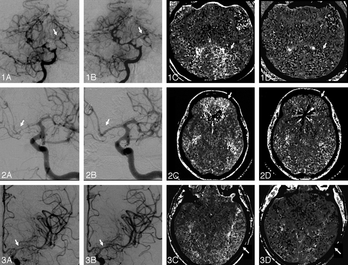
- Fig 1.
Panels 1, 2, and 3 illustrate cases 1, 2, and 3, respectively. Panels A–D show pre- and posttreatment angiograms and MTT maps, respectively. Panel 1: A, Severe vasospasm of the left PCA, which had a marked response to nicardipine infusion in the basilar artery (1B) resulting in 28% reduction in MTT (1C, -D) and 62% increase in CBF (not shown) at the left PCA territory. Panel 2: A, Severe vasospasm of the left ACA, which had a marked response to PTA of the A1 segment and nicardipine infusion (2B), resulting in 68% reduction in MTT (2C, -D) and 89% increase in CBF (not shown) at the left ACA territory. Panel 3: A, Moderate vasospasm of the left MCA, which had only a mild angiographic response to nicardipine infusion (3B). C and D, Despite the suboptimal angiographic result, there was a 37% reduction in MTT and 69% increase in CBF (not shown) at the left MCA territory. This likely reflects the nicardipine effect in the microcirculation.

Tables
Patient data summary
No. Post-SAH Day Age (yr) Sex HH Grade Fisher Group Aneurysm Location, Treatment Vessel Vasospasm Grading Vasospasm Treatment MTT Pre-Rx MTT Post-Rx CBF Pre-Rx CBF Post-Rx CBV Pre-Rx CBV Post-Rx 1 5 62 M 4 3 BA, coiling R MCA Mod/sev 10 mg, R ICA 5.59 5.29 32.69 43.57 2.35 2.97 L MCA Severe 10 mg, L ICA 5.50 4.95 38.52 45.34 2.53 2.91 R PCA Severe 10 mg, BA 6.27 6.29 29.39 28.32 2.02 2.16 L PCA Severe 8.25 5.91 22.01 35.71 2.12 2.61 2 4 44 F 3 3 L ACA, coiling R MCA Mild 4 mg, R ICA 5.08 2.24 36.08 58.63 2.20 2.04 R ACA Severe PTA, R ICA 7.23 2.18 21.86 57.25 1.98 1.99 PTA, R M1 L MCA Moderate 4 mg, L ICA 6.14 2.42 36.19 50.12 2.75 1.81 L ACA Severe PTA, L ICA 7.95 2.52 19.67 37.25 1.97 1.36 2 mg, L A1 PTA, L A1 3 11 55 F 2 3 No aneurysm identified R MCA Moderate 4 mg R ICA 4.79 4.44 45.26 56.11 2.79 3.38 R ACA Severe 2 mg, R A1 5.88 5.05 37.51 58.60 2.89 4.15 L MCA Moderate 4 mg, L ICA 7.99 5.07 30.75 51.91 3.29 3.55 L ACA Mild 2 mg L M2 6.16 5.33 38.89 45.71 3.23 3.43 4 5 44 F 3 3 BA, clipping R MCA* Moderate 8 mg, R ICA 5.33 4.83 48.78 48.90 3.36 3.02 L MCA Moderate 6 mg, L ICA 4.19 3.46 57.92 77.14 3.31 3.77 5 11 42 M 2 3 AcomA, clipping R MCA None None† 5.29 5.09 47.46 43.25 3.23 2.90 L MCA Moderate 7.5 mg, L ICA 5.32 4.27 68.20 73.73 4.71 4.32 Note:—HH indicates Hunt and Hess; Rx, treatment; R, right; L, left; BA, basilar artery; Mod/sev, moderate to severe; AcomA, anterior communicating artery; MTT, mean transit time; CBF, cerebral blood flow; CBV, cerebral blood volume; ACA, anterior cerebral artery; MCA, middle cerebral artery; PCA, posterior cerebral artery; ICA, internal carotid artery.
* Near-complete occlusion of the M2 segment of the right MCA on angiographic evaluation with only minimal angiographic response after IA nicardipine infusion.
† Procedure aborted due to seizures.


