
Article Figures & Data
Figures
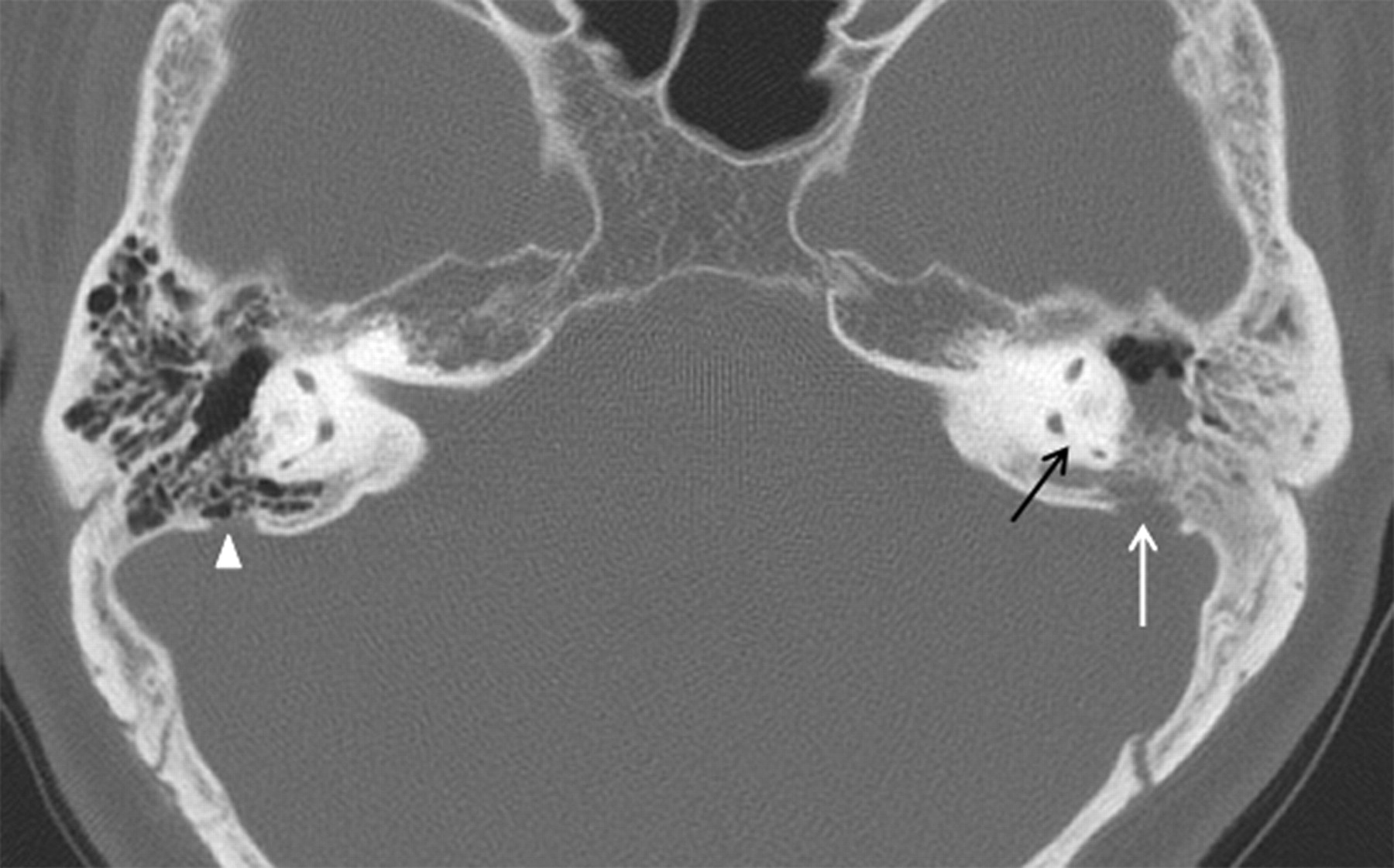
- Fig 1.
Subject 24. Axial CT scan at the level of the common crus (black arrow) shows a small bone defect caused by presumed AG (white arrow), located at the lateral third of the posterior wall of left temporal bone. Although a focal loss of the posterior wall of the mastoid air cells is also present, there is no evidence of tympanomastoid opacification. This 66-year-old woman presented with hemifacial spasm, and there was no clinical feature suggesting CSF leakage.
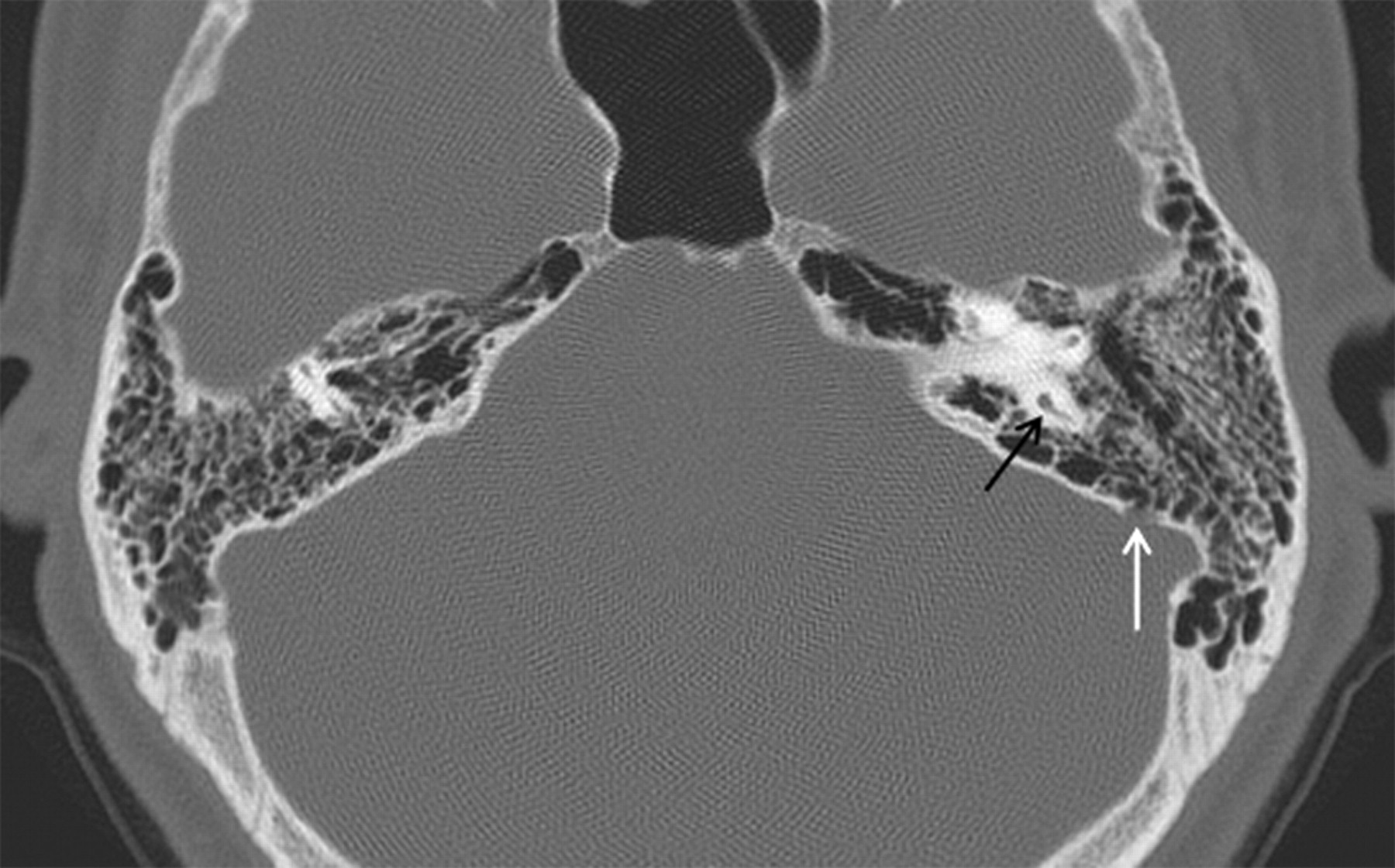
- Fig 2.
Subject 4. Axial CT scan at the level of the common crus (black arrow) shows a large bone defect caused by presumed AG (white arrow), located at the lateral third of the posterior wall of the left temporal bone. There also is a loss of the posterior wall of the mastoid air cells, which are opacified with fluid. This 41-year-old man presented with CSF otorrhea and recurrent septic meningitis. He underwent dural repair via intact canal wall mastoidectomy. Also noted is a small bone defect caused by presumed AG (arrowhead), located at the lateral third of the posterior wall of right temporal bone.


Tables
Prevalence of presumed AGs according to age of subjects
Age Total No. of Subjects (n = 1255) No. of Subjects Showing Presumed AGs on CT Scans (n = 30), n (%) <10 y 138 0 (0) <20 y 82 0 (0) <30 y 104 1 (0.1) <40 y 167 2 (1.2) <50 y 283 6 (2.1) <60 y 271 13 (4.8) <70 y 159 4 (2.5) <80 y 45 3 (6.7) <90 y 6 1 (16.7) Note:—AG indicates arachnoid granulation.
In this issue


Jump to section
Related Articles
Cited By...
- No citing articles found.