
Article Figures & Data
Figures
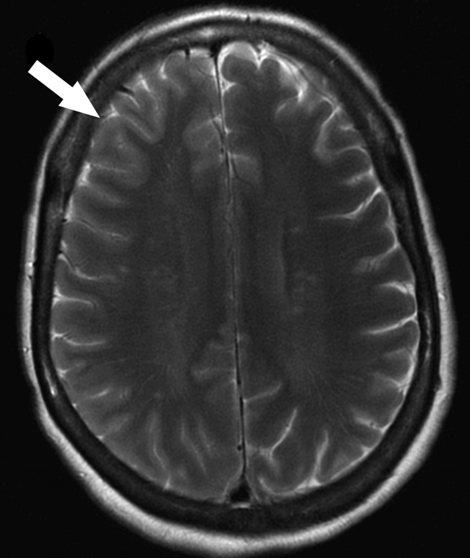
- Fig 1.
Patient 1. Subtle sulcal effacement and mild cortical edema are seen in the right frontal lobe on the axial T2-weighted image (arrow). Diffusion-weighted sequences (not shown) were normal.
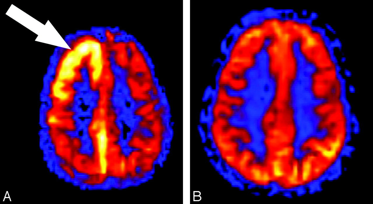
- Fig 2.
Patient 1. A, Quantitative CBF map generated from the ASL sequence shows significant hyperperfusion (arrow) within the cortex of the right cerebral hemisphere corresponding to the signal intensity abnormalities on the conventional MR imaging sequences. Region of interest placed over the frontal gray matter shows a mean CBF in the right hemisphere of 128.9 compared with the left frontal hemisphere mean CBF of 56.6 mL/100 g tissue/min. B, Quantitative CBF map obtained 6 days after the initial examination shows resolution of the hyperperfusion within the cortex of the right frontal and parietal lobes. Mean gray matter CBF in the right and left frontal lobe decreased to 57.7 and 61.6 mL/100 g tissue/min, respectively.
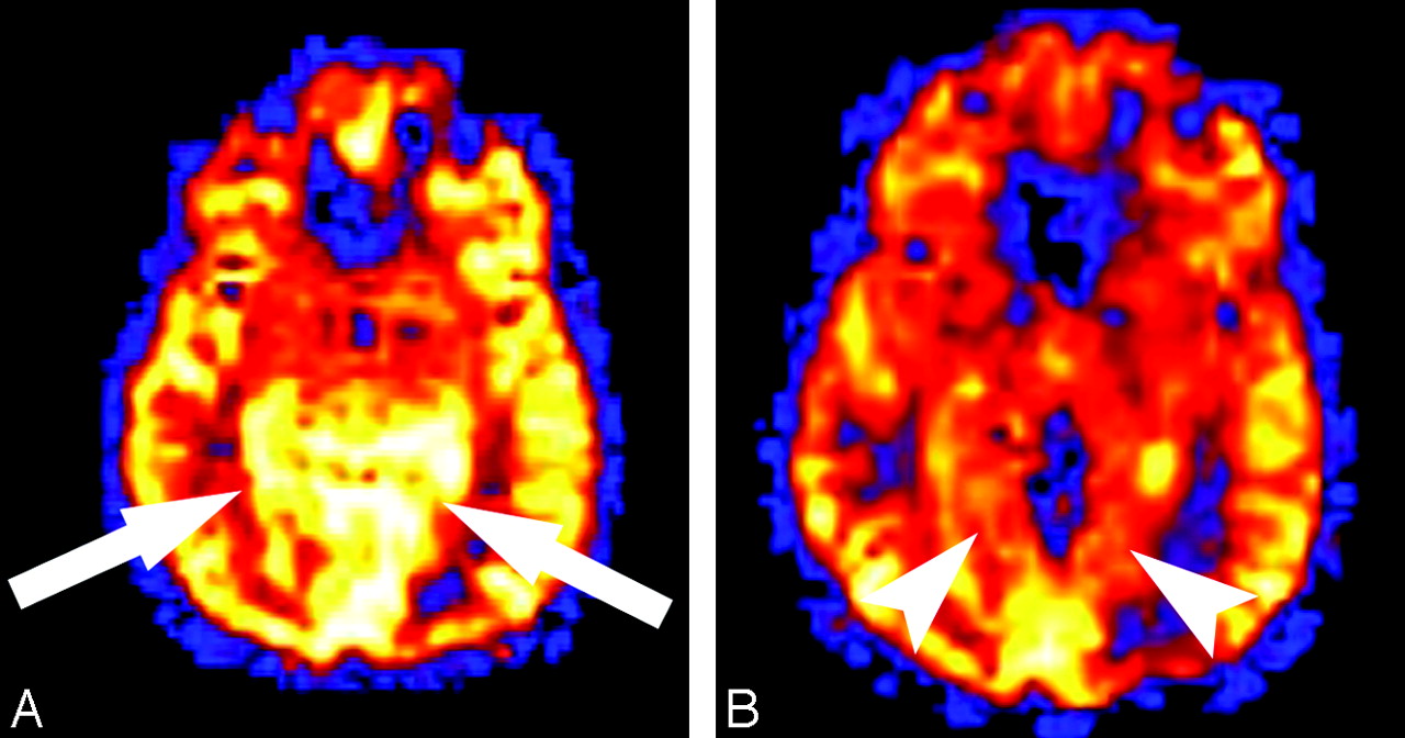
- Fig 3.
Patient 2. A, Quantitative ASL CBF map shows regional hyperperfusion (arrows) in the medial occipital lobes on a background of global hyperperfusion in this patient who underwent imaging examination during a migraine episode. Mean gray matter CBF measured 162.2 and 138.7 mL/100 g tissue/min, respectively, in the medial left and right occipital cortex. B, Quantitative CBF map obtained 6 months after the migraine episode shows resolution of the regional hyperperfusion in the medial occipital lobes (arrowheads). The underlying global hyperperfusion also resolved. Mean gray matter CBF decreased from 162.2 to 107.5 mL/100 g tissue/min in the medial left occipital cortex and from 138.7 to 112.9 mL/100 g tissue/min in the medial right occipital cortex.
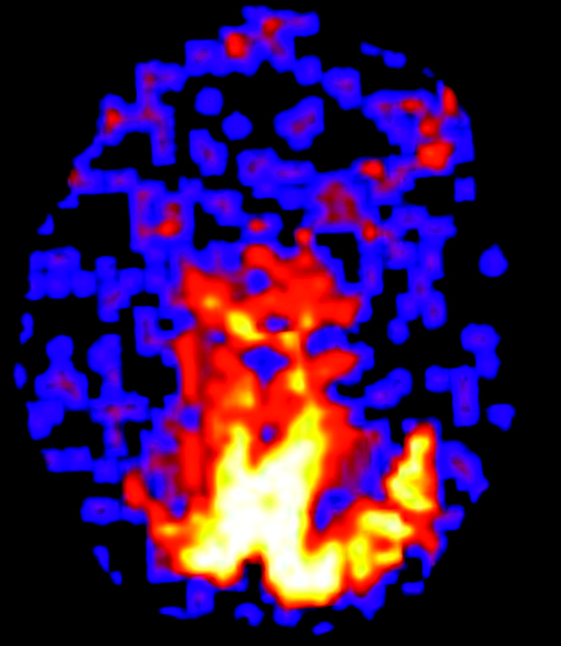
- Fig 4.
Patient 3. Quantitative CBF map shows marked regional hyperperfusion in the occipital lobes bilaterally. This 87-year-old patient had marked global cerebral atrophy accounting for the relative hypoperfusion of the remaining cortex.


In this issue


Jump to section
Related Articles
Cited By...
- The index vein pointing to the origin of the migraine aura symptom: A case series
- Time Course of Cerebral Perfusion Changes in Children with Migraine with Aura Mimicking Stroke
- Neurologic attack and dynamic perfusion abnormality in neuronal intranuclear inclusion disease
- Acute-Onset Migrainous Aura Mimicking Acute Stroke: MR Perfusion Imaging Features
- Blood Pressure and Vascular Dysfunction Underlie Elevated Cerebral Blood Flow in Systemic Lupus Erythematosus
- Susceptibility-Weighted Imaging in Migraine with Aura
- Elevated Cerebral Blood Flow and Volume in Systemic Lupus Measured by Dynamic Susceptibility Contrast Magnetic Resonance Imaging