
Article Figures & Data
Figures
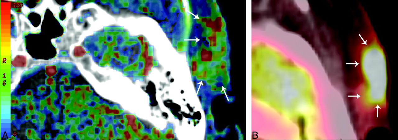
- Fig 1.
Tissue perfusion CT map (A) and PET/CT SUVmean map (B) of a 54-year-old woman with a SCCA in the skin on the left temporal side. Blood flow (A) map shows a hypervascularized area in the subcutaneous region with infiltration of the cutis (arrows). The increased perfusion of the neoplastic tissue is coupled with increased SUV uptake (B) in the FDG PET/CT imaging (arrows).
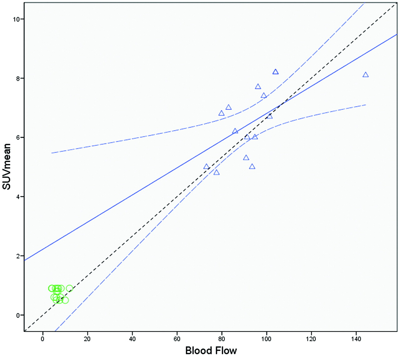
- Fig 2.
Regression analysis of the mean BF values in the tumor (triangles) and healthy tissue (circles) with the SUVmean. There is a positive significant correlation (r = 0.63; P = .011) in the tumor ROIs. Linear regression analysis showed SUVmean = 0.05 × BF + 2.22 (R2 = 0.45; P = .011) (regression line). The curved dashed lines show the 95% confidence intervals for the tumor ROIs means. The dashed line depicts the line of perfect agreement (equation line).
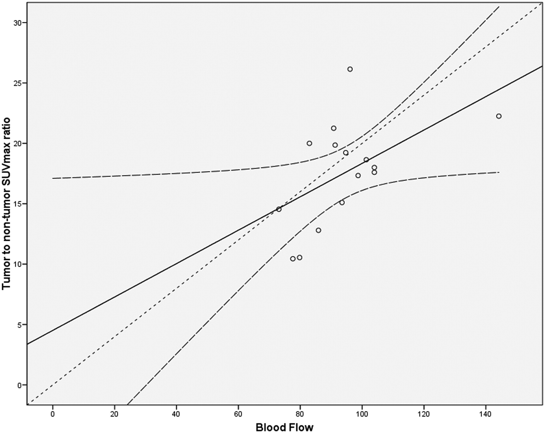
- Fig 3.
Regression analysis of the mean BF values in the tumor with the SUVmean ratios of tumor-to-nontumor tissue. There is a positive significant correlation (r = 0.64; P = .01) in the tumor ROIs. Linear regression analysis showed Ratio SUVmean = 0.14 × BF–3.48 (R2 = 0.42; P = .01) (regression line). The curved dashed lines show the 95% confidence intervals for means. The dashed line depicts the line of perfect agreement (equation line).


Tables
- Table 1:
Summary statistics mean (SD) and 95% confidence interval of the SUV values (SUVmax and SUVmean) and mean tissue perfusion parameters in healthy-appearing muscle tissue and neoplastic lesions
Tissue SUVmax SUVmean BF BV MTT PS Muscle 1.1 (± 0.43) 0.93 (± 0.47) 6.45 (± 2.09) 1.24 (± 0.77) 22.75 (± 3.31) 1.68 (± 1.03) (0.82, 1.37) (0.63, 1.23) (5.12, 7.78) (0.75, 1.73) (20.39, 25.11) (1.02, 2.34) Tumor 15.39 (± 2.54) 6.56 (± 1.2) 94.52 (± 16.7) 5.46 (± 1.24) 6.05 (± 1.38) 24.11 (± 8.39) (13.98, 16.8) (5.89, 7.22) (85.27, 103.77) (4.77, 6.14) (5.28, 6.81) (19.46, 28.76) Note:—SUVmax and SUVmean indicate maximal and mean standardized uptake values of glucose (g/mL); BF, blood flow (mL/min/100 g); BV, blood volume (mL/100 g); MTT, mean transit time (s); PS, permeability (mL/min/100 g).
- Table 2:
Pearson correlation coefficients between SUV values (SUVmax and SUVmean) and mean tissue perfusion parameters in healthy-appearing muscle tissue and neoplastic lesions
BF BV MTT PS SUVmax SUVmean BF 0.91* −0.94* 0.87* 0.97* 0.97* BV 0.91* −0.86* 0.82* 0.89* 0.88* MTT −0.94* −0.86* −0.84* −0.92* −0.92* PS 0.87* 0.82* −0.84* 0.92* 0.89* SUVmax 0.97* 0.89* −0.92* 0.92* 0.97* SUVmean 0.97* 0.88* −0.92* 0.89* 0.97* Note:—SUVmax and SUVmean indicate maximal and mean standardized uptake values of glucose (g/mL); BF, blood flow (mL/min/100 g); BV, blood volume (mL/100 g); MTT, mean transit time (s); PS, permeability (mL/min/100 g).
* Correlation is significant at the 0.01 level (2-tailed).
In this issue


Jump to section
Related Articles
Cited By...
- The Flow-Metabolic Phenotype of Primary Colorectal Cancer: Assessment by Integrated 18F-FDG PET/Perfusion CT with Histopathologic Correlation
- Prediction of Locoregional Control in Head and Neck Squamous Cell Carcinoma with Serial CT Perfusion during Radiotherapy
- CT Perfusion of the Neck: Internal Carotid Artery versus External Carotid Artery as the Reference Artery