
Article Figures & Data
Figures
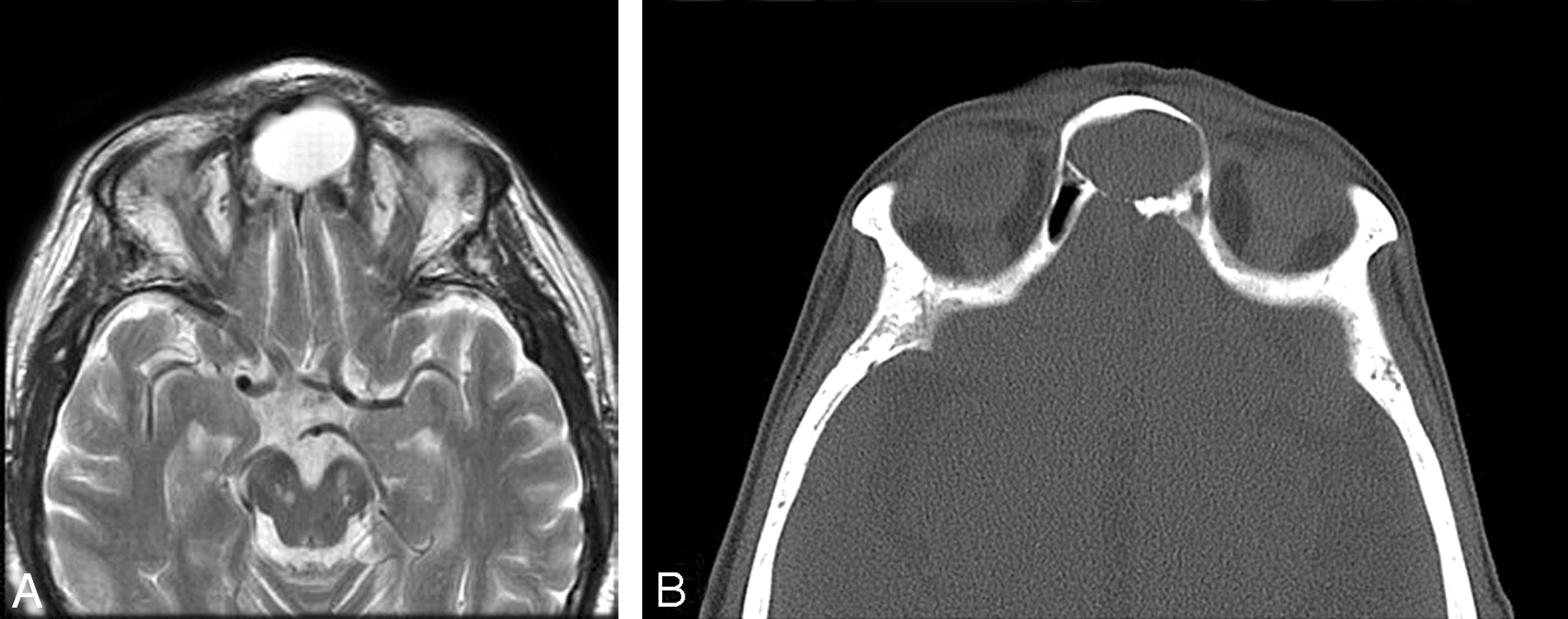
- Fig 1.
A 37-year-old woman presented with a feeling of pressure in her forehead. A, An axial T2-weighted MR image shows an expanded interfrontal sinus septal cell with high signal intensity. B, A CT scan shows the mucocele thinning the midline posterior frontal sinus table.
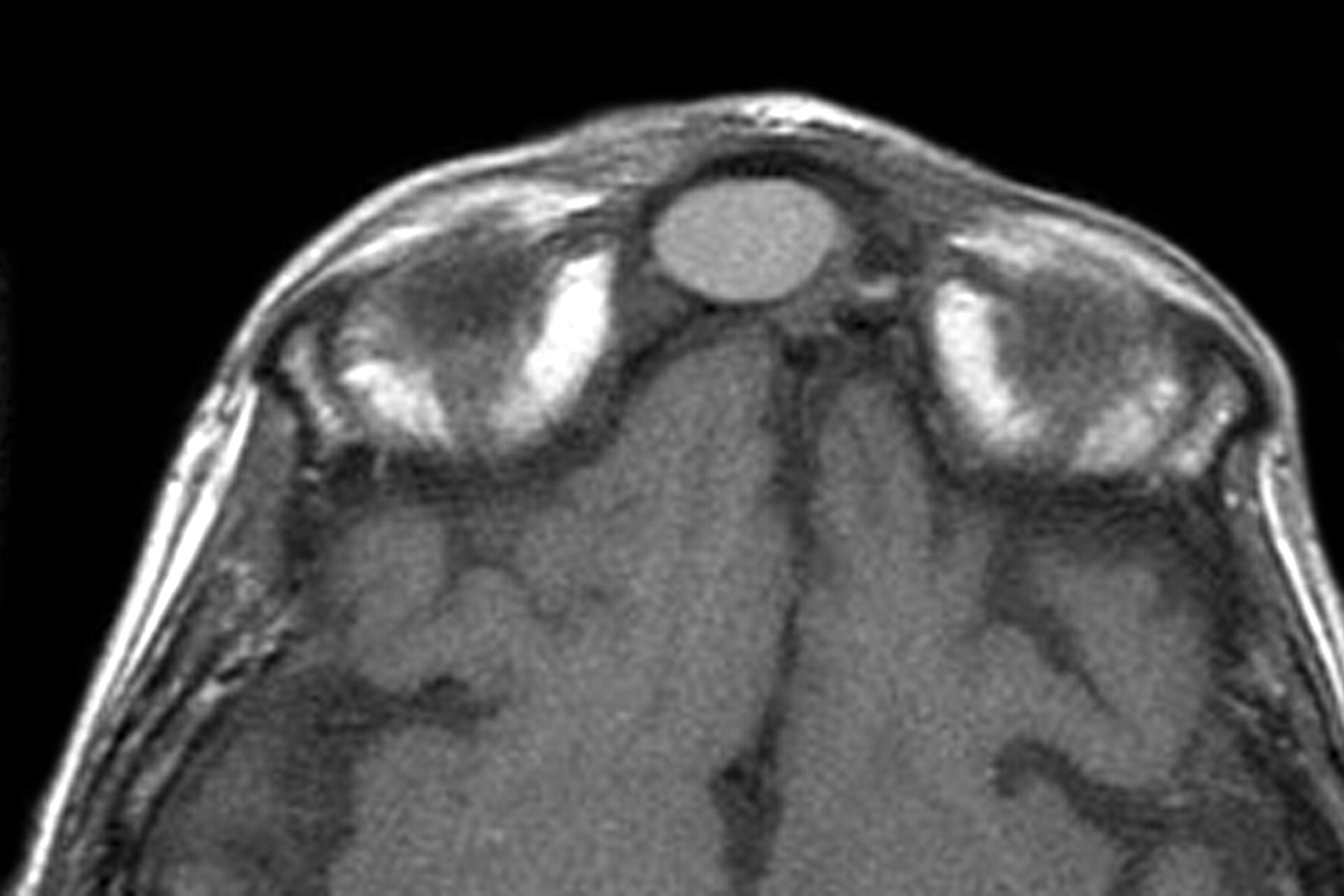
- Fig 2.
A 43-year-old man presented with a headache. An axial T1-weighted MR image shows an expanded interfrontal sinus septal cell with high (proteinaceous) signal intensity. There was thinning of the central posterior sinus table.
- Fig 3.
A 57-year-old woman presented with a slowly growing mass in her lower forehead. A coronal T2-weighted MR image shows an expanded interfrontal sinus septal cell with high signal intensity. This mucocele has obstructed the left frontal sinus proper. At surgery, the right frontal sinus drainage was patent and just behind the mucocele.
- Fig 4.
A 68-year-old woman presented with a painful forehead mass. An axial T2-weighted MR image shows a minimally expanded interfrontal sinus septal cell with high signal intensity. This mucocele extended through the midline anterior table and caused a Pott puffy tumor in the overlying scalp (arrow).
- Fig 5.
A 53-year-old man presented with a mass in his upper medial right orbit. A coronal T2-weighted MR image shows a right frontal sinus mucocele that has expanded the sinus and depressed the superomedial orbital rim. Inflammatory mucosal disease is present in the left frontal sinus, both ethmoid sinuses, and the right maxillary sinus. B, A 46-year-old woman presented with a headache. An axial T1-weighted MR image shows a mucocele expanding the right frontal sinus and thinning the posterior sinus table to the right side, off of the midline.
- Fig 6.
A 32-year-old man presented with a bulge in his medial right orbit and difficulty breathing through the right side of his nose. A coronal T2-weighted MR image shows a large right anterior ethmoid mucocele. It is clearly centered within the ethmoid complex, caudal to the frontal sinuses. Inflammatory mucosal disease is also present in the left ethmoid cells and the right antrum.


In this issue


Jump to section
Related Articles
Cited By...
- No citing articles found.