
Article Figures & Data
Figures
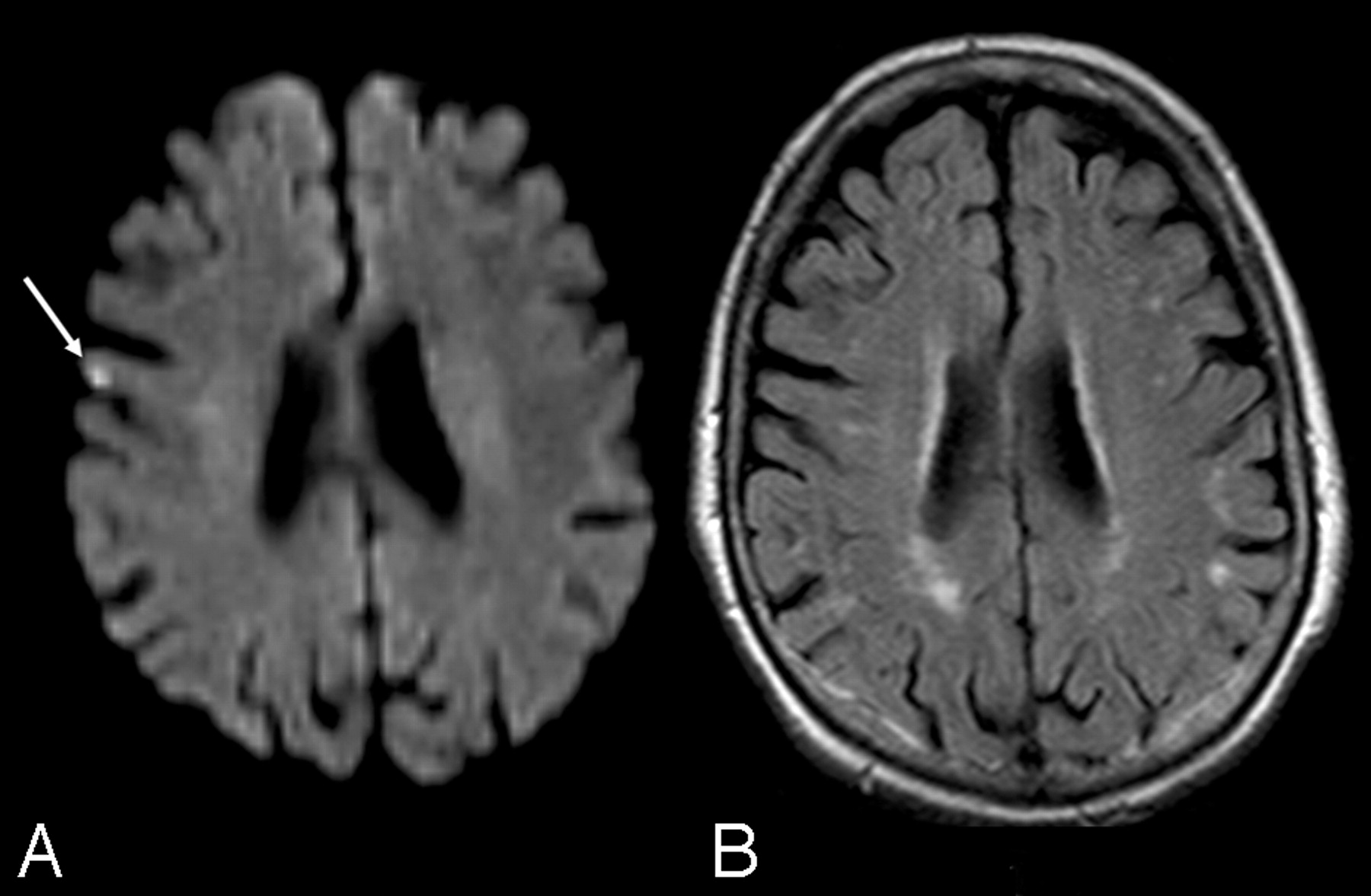
- Fig 1.
A, Ischemic lesion (arrow) seen on postoperative DWI. B, Delayed MR image in the same patient documents its disappearance.

Tables
Characteristics No. (%) Patients 110 Mean age (range) (years) 72.6 (56–86) Men 78 (71) Women 32 (29) Ipsilateral ICA stenosis 70%–90% 106 (96.3) >90% 4 (3.7) Plaque structure Type 5 12 (11) Types 3 and 4 72 (65) Types 1 and 2 26 (24) Contralateral ICA stenosis 0%–50% 68 (62) 50%–69% 24 (22) 70%–99% 12 (11) Occlusion 6 (5) Presenting symptoms TIA 24 (22) Stroke 6 (5) Asymptomatic 80 (73) Vascular risk factors Chronic renal failure 5 (4.5) Hypertension 92 (84) Cigarette smoking 49 (45) Diabetes 45 (41) Hyperlipidemia 60 (55) Chronic obstructive pulmonary disease 8 (7) Coronary artery disease 36 (33) Peripheral vascular disease 17 (15) Note:—TIA indicates transient ischemic attack; ICA, internal carotid artery.
- Table 2:
Location of postoperative DWI ischemic lesions and after a mean 6.2-month follow-up
Location Postoperative DWI MR Imaging Follow-Up P* Persisted Disappeared Lost Cortical 19 4 (25%) 12 (75%) 3 .02 Subcortical 10 7 (70%) 3 (30%) 0 Deep white matter 3 1 (33%) 2 (66%) 0 Basal ganglia 1 0 1 (100%) 0 Total 33 12 (40%) 18 (60%) 3 * P value by χ2 test.
Size Postoperative DWI Follow-Up MR Imaging P* Persisted Disappeared Lost 0–5 mm 19 3 (17%) 14 (83%) 2 .004 5–10 mm 14 9 (69%) 4 (31%) 1 Total 33 12 18 3 * P value by χ2 test.
Factors No. Patients with DWI Lesions No. Patients with Persistent MR Imaging Lesions P* Preoperative microinfarctual brain 6 3 (50%) .3 Normal brain 1 0 Age ≥80 years 3 3 (100%) .1 Age <80 years 10 5 (50%) Types 1 and 2 carotid plaque 1 0 .2 Types 3–5 carotid plaque 12 8 (67%) Asymptomatic 8 5 (62%) .9 Symptomatic 5 3 (60%) * P values by χ2 test.
In this issue


Jump to section
Related Articles
Cited By...
- Ischemic Brain Lesions After Carotid Artery Stenting Increase Future Cerebrovascular Risk
- Predictors of Acute and Persisting Ischemic Brain Lesions in Patients Randomized to Carotid Stenting or Endarterectomy
- Flow Reversal Versus Filter Protection: A Pilot Carotid Artery Stenting Randomized Trial
- Characteristics of Ischemic Brain Lesions After Stenting or Endarterectomy for Symptomatic Carotid Artery Stenosis: Results From the International Carotid Stenting Study-Magnetic Resonance Imaging Substudy
- Carotid Artery Stenting without Angioplasty and Cerebral Protection: A Single-Center Experience with up to 7 Years' Follow-Up
- Proximal Embolic Protection: A "Game Changer" for Carotid Stents