
Article Figures & Data
Figures
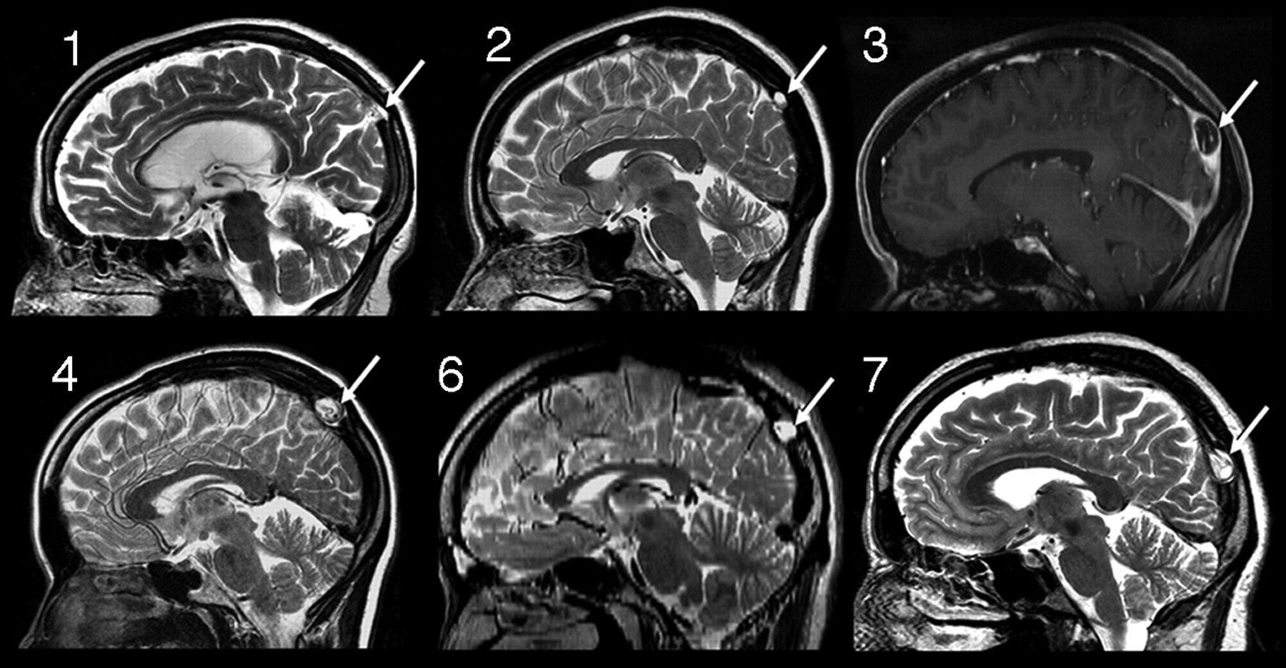
- Fig 1.
Sagittal images from cases 1, 2, 3, 4, 6, and 7 demonstrating the typical position of the dorsal SSS AG identified in this study (arrows). Case 3, Sagittal reconstruction of FSPGR postcontrast T1-weighted image (3T); case 6, sagittal reconstructed image from 2-mm axial FSE T2-WI (3T); all others, sagittal FSE T2-weighted images.
- Fig 2.
Case 4, 1.5T, axial T1WI (A), FLAIR (B), T2WI (C), DWI (D), and postcontrast T1WI (E and F). Typical appearance on multiple pulse sequences. Note the large AG with associated defect in the dura (arrows, C) along the rightward margin of the SSS. Note intrinsic vessels, which appear to be displaced cortical veins or channels (arrows, E and F). There is focal calvarial remodeling.
- Fig 3.
Case 3, 3T. Axial acquired TOF-MRV (A), CE-MRV (B), and segmented volume-rendered FSPGR sequence after contrast (C).Note multilumen SSS, intrinsic vessels (arrows, B and C), thin AG base along the SSS (arrowhead, C), and adjacent cortical vein (*C).


Tables
- Table 1:
Imaging and clinical findings in 12 patients with large arachnoid granulations within the dorsal SSS
Age Sex FS MRV Size, cm Distance Above Torcular, cm T1WI T2WI FLAIR DWI Location Relative to λ Stenosis, % Septation Clinical 64 F 3T 1.2 × 0.7 × 1.1 5.1 Mixed Hyper Iso Hypo λ 20 Yes MS follow-up, right leg weakness, initially called meningioma 42 F 3T 0.6 × 0.7 × 0.8 5.1 Mixed Hyper Iso Hypo λ 60 Yes Right temporal headaches 42 F 3T-MRV T, C 1.1 × 1.1 × 1.8 3.9 Mixed Hyper Iso Hypo −2 mm 86 Yes Headache, initially called thrombosis, 0.7T-MR imaging 35 F 1.5T 1.1 × 1.8 × 1.9 5.8 Hypo Hyper Iso Hypo λ 60 Yes Left temporal headache 42 M 1.5T 0.2 × 0.3 × 0.4 5.9 Hypo Hyper Iso Hypo λ 50 No Headache 28 M 3T T, C 0.8 × 0.9 × 1.1 5.2 Mixed Hyper Iso Hypo λ 50 No Seizures right frontal glioma 52 M 3T 0.7 × 1.1 × 1.7 3.0 Hypo Hyper Iso Hypo −2 mm 86 Yes Headaches 57 M 1.5T T 0.4 × 0.6 × 0.6 6.2 Hypo Hyper Hypo Hypo +12 mm 50 No Ischemic stroke 45 F 1.5T 0.4 × 0.6 × 0.6 3.5 Hypo Hyper Hypo Hypo λ 40 No Headache, Chiari I, initially called thrombosis 18 F 3 T 1.1 × 1.6 × 1.1 2.6 Hypo Hyper NA NA −3 mm 74 No Headache, blurry vision 2 M 1.5T 0.8 × 0.9 × 1.0 5.8 Hypo Hyper NA Hypo λ 58 No Developmental delay, initially called dermoid 17 F 1.5T T 1.3 × 1.0 × 1.0 2.8 Hypo Hyper Hypo Hypo λ 52 Yes Occipital headaches Note:—Hypo indicates hypointense relative to brain; Iso, isointense relative to brain; Hyper, hyperintense relative to brain; Mixed, areas of hypointensity and isointensity on T1WI; FS, magnetic field strength; Distance Above Torcular, linear distance above torcular; T, time-of-flight; C, elliptic centric encoded, contrast-enhanced MRV; SSS, superior sagittal sinus; M, male; F, female; NA, not applicable; MS, multiple sclerosis. Patient 10 did not have fluid-attenuated inversion recovery or diffusion-weighted MR imaging, and patient 11 did not have fluid-attenuated inversion recovery.
- Table 2:
Distribution of arachnoid granulations documented by imaging and anatomic studies
Reference Modality Patients Case Type No. NOS Vent. Sup. Dors. Inf. TS SS TH StS VoG Notes Leach et al1 CT with contrast 573 Unselected 168 NA 0 1 154 3 8 2 0 3- to 5-mm posterior fossa, 10-mm supratentorial section thickness Leach et al1 MR with contrast 100 Unselected 14 NA 0 1 13 0 0 0 0 5- to 6-mm section thickness Leach et al1 Anatomic 29 Unselected 91 NA NA 0 86 0 5 NA NA Did not assess anterior superior SSS, included posterior inferior Roche et al3 MR, CT, angiography 32 Selected 42 NA 0 0 36 4 2 0 0 Selected cases Liang et al4 MR with contrast 100 Unselected 433 NA 227 6 122 0 0 76 2 Assessed all dural sinuses, 3D MPRAGE, 1.3-mm section thickness Gailloud et al13 Angiography 57 Unselected 15 NA 0 2 12 0 1 0 0 All 12 TS AGs at vein of Labbe entrance site, angiographic study Browder et al6 Anatomic 295 Unselected 25 NA 0 2 23 0 0 0 0 Only counted large protuberances Ikushima et al19 MR 1118 Unselected 134 5 NA NA 115 3 8 3 0 Noncontrast MR Koshikawa et al20 MR 151 Unselected 162 NA NA NA 162 NA NA NA NA TS only Note:—TS indicates transverse sinus; SS, sigmoid sinus; TH, torcular herophili; StS, straight sinus; VoG, vein of Galen confluence with StS; Vent. Sup., ventral superior SSS; Dors. Inf., dorsal inferior SSS; NA, not applicable; AG, arachnoid granulation; MPRAGE, magnetization-prepared rapid acquisition of gradient echo; SSS, superior sagittal sinus; NOS, location not otherwise specified.


