Advertisement

Main menu

  • Home
  • Content
    • Current Issue
    • Accepted Manuscripts
    • Article Preview
    • Past Issue Archive
    • Video Articles
    • AJNR Case Collection
    • Case of the Week Archive
    • Case of the Month Archive
    • Classic Case Archive
  • Special Collections
    • AJNR Awards
    • Low-Field MRI
    • Alzheimer Disease
    • ASNR Foundation Special Collection
    • Photon-Counting CT
    • View All
  • Multimedia
    • AJNR Podcasts
    • AJNR SCANtastic
    • Trainee Corner
    • MRI Safety Corner
    • Imaging Protocols
  • For Authors
    • Submit a Manuscript
    • Submit a Video Article
    • Submit an eLetter to the Editor/Response
    • Manuscript Submission Guidelines
    • Statistical Tips
    • Fast Publishing of Accepted Manuscripts
    • Graphical Abstract Preparation
    • Imaging Protocol Submission
    • Author Policies
  • About Us
    • About AJNR
    • Editorial Board
    • Editorial Board Alumni
  • More
    • Become a Reviewer/Academy of Reviewers
    • Subscribers
    • Permissions
    • Alerts
    • Feedback
    • Advertisers
    • ASNR Home

User menu

  • Alerts
  • Log in

Search

  • Advanced search
American Journal of Neuroradiology

American Journal of Neuroradiology

ASHNR American Society of Functional Neuroradiology ASHNR American Society of Pediatric Neuroradiology ASSR
  • Alerts
  • Log in

Advanced Search

  • Home
  • Content
    • Current Issue
    • Accepted Manuscripts
    • Article Preview
    • Past Issue Archive
    • Video Articles
    • AJNR Case Collection
    • Case of the Week Archive
    • Case of the Month Archive
    • Classic Case Archive
  • Special Collections
    • AJNR Awards
    • Low-Field MRI
    • Alzheimer Disease
    • ASNR Foundation Special Collection
    • Photon-Counting CT
    • View All
  • Multimedia
    • AJNR Podcasts
    • AJNR SCANtastic
    • Trainee Corner
    • MRI Safety Corner
    • Imaging Protocols
  • For Authors
    • Submit a Manuscript
    • Submit a Video Article
    • Submit an eLetter to the Editor/Response
    • Manuscript Submission Guidelines
    • Statistical Tips
    • Fast Publishing of Accepted Manuscripts
    • Graphical Abstract Preparation
    • Imaging Protocol Submission
    • Author Policies
  • About Us
    • About AJNR
    • Editorial Board
    • Editorial Board Alumni
  • More
    • Become a Reviewer/Academy of Reviewers
    • Subscribers
    • Permissions
    • Alerts
    • Feedback
    • Advertisers
    • ASNR Home
  • Follow AJNR on Twitter
  • Visit AJNR on Facebook
  • Follow AJNR on Instagram
  • Join AJNR on LinkedIn
  • RSS Feeds

AJNR Awards, New Junior Editors, and more. Read the latest AJNR updates

Research ArticleBRAIN

Anoxic Injury-Associated Cerebral Hyperperfusion Identified with Arterial Spin-Labeled MR Imaging

J.M. Pollock, C.T. Whitlow, A.R. Deibler, H. Tan, J.H. Burdette, R.A. Kraft and J.A. Maldjian
American Journal of Neuroradiology August 2008, 29 (7) 1302-1307; DOI: https://doi.org/10.3174/ajnr.A1095
J.M. Pollock
  • Find this author on Google Scholar
  • Find this author on PubMed
  • Search for this author on this site
C.T. Whitlow
  • Find this author on Google Scholar
  • Find this author on PubMed
  • Search for this author on this site
A.R. Deibler
  • Find this author on Google Scholar
  • Find this author on PubMed
  • Search for this author on this site
H. Tan
  • Find this author on Google Scholar
  • Find this author on PubMed
  • Search for this author on this site
J.H. Burdette
  • Find this author on Google Scholar
  • Find this author on PubMed
  • Search for this author on this site
R.A. Kraft
  • Find this author on Google Scholar
  • Find this author on PubMed
  • Search for this author on this site
J.A. Maldjian
  • Find this author on Google Scholar
  • Find this author on PubMed
  • Search for this author on this site
  • Article
  • Figures & Data
  • Info & Metrics
  • Responses
  • References
  • PDF
Loading

Article Figures & Data

Figures

  • Tables
  • Fig 1.
    • Download figure
    • Open in new tab
    • Download powerpoint
    Fig 1.

    Segmentation map obtained by using the T1-weighted image shows CSF (blue), white matter (red), and GM (green). The green areas are used for the mean GM CBF measurements.

  • Fig 2.
    • Download figure
    • Open in new tab
    • Download powerpoint
    Fig 2.

    Axial diffusion-weighted, apparent diffusion coefficient (ADC), and quantitative CBF map from patient 2 shows the typical sequelae from anoxic injury, including diffuse bilateral symmetric restricted diffusion in the cerebral cortex (arrow) and the basal ganglia and thalami (arrowhead). The ADC image reflects the subacute nature of the ischemic change, because the imaging was done 9 days after the anoxic event. CBF map shows global hyperperfusion and the second highest average GM blood flow recorded in this study (190.6 mL/100 g of tissue per minute).
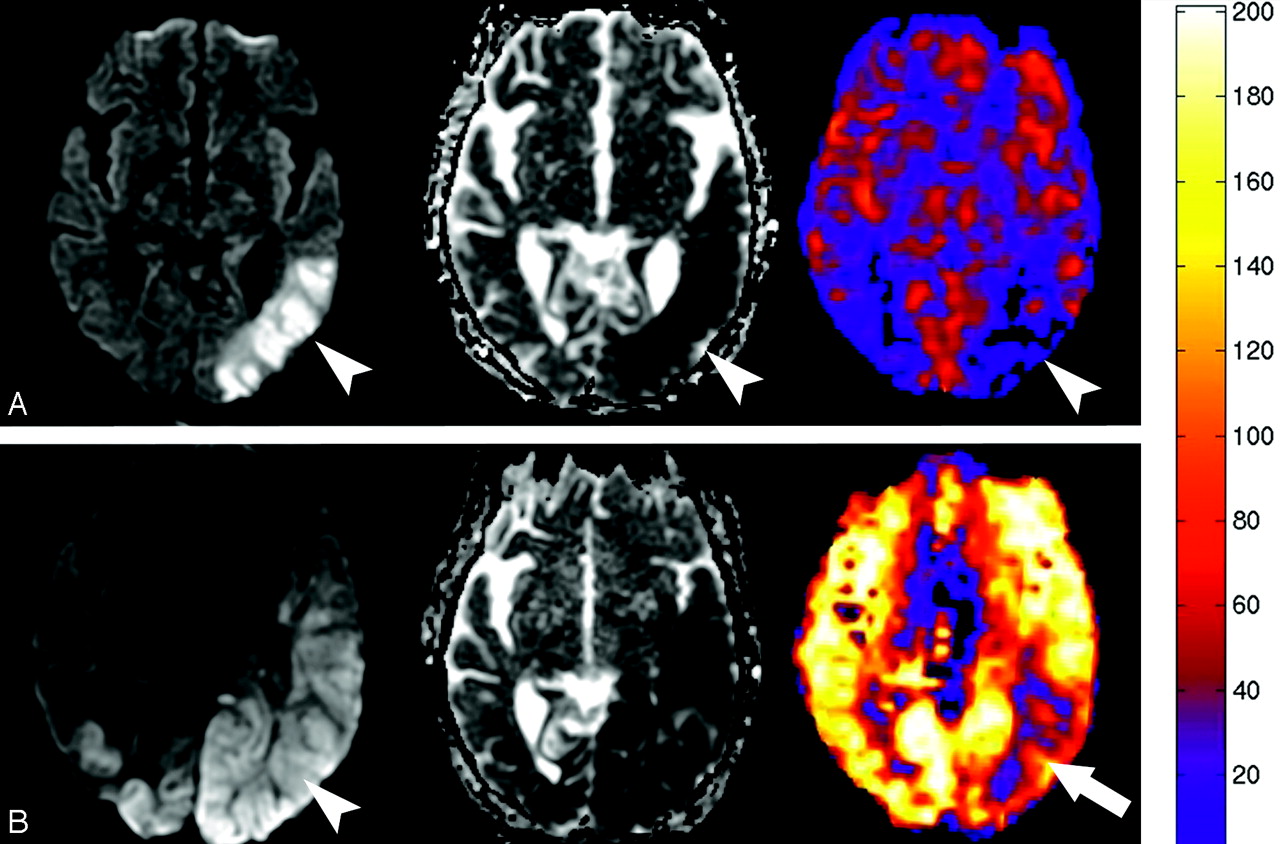
  • Fig 3.
    • Download figure
    • Open in new tab
    • Download powerpoint
    Fig 3.

    A, Diffusion, ADC, and quantitative CBF map generated from the PASL sequence obtained in patient 8 one day before the global anoxic event shows restricted diffusion and hypoperfusion in the left posterior watershed territory corresponding with areas of subacute infarction (arrowheads). B, Diffusion, ADC, and quantitative CBF map obtained 3 days after the episode of pulseless electrical activity in patient 8 shows interval worsening of the diffusion abnormality (arrowhead) with new marked global hyperperfusion. There is relative sparing of the previous subacute infarct in the left posterior MCA territory (arrow). Quantitative analysis performed by placing regions of interest over the entire section showed an average CBF increase from 31.4 to 188.6 mL/100 g of tissue per minute.
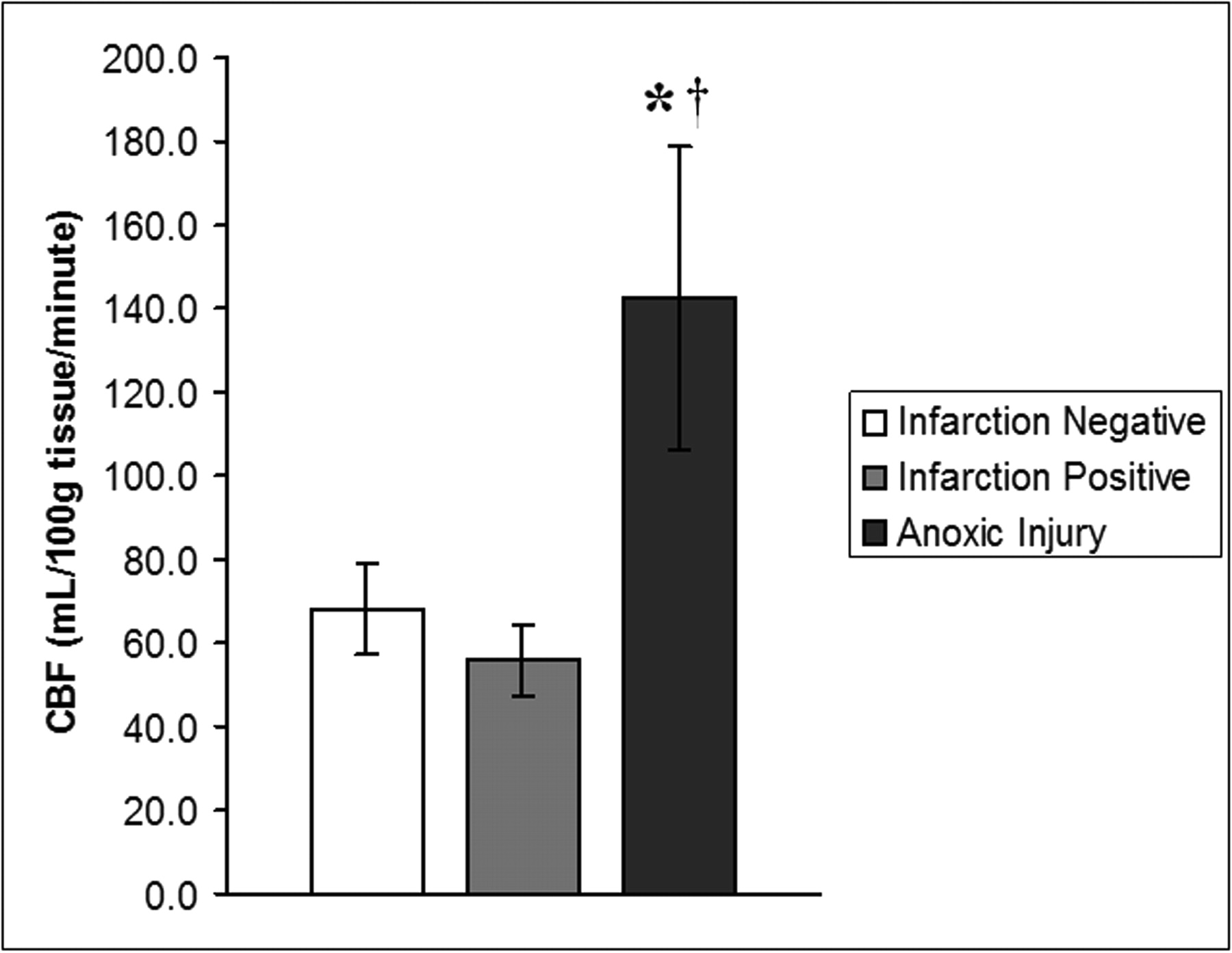
  • Fig 4.
    • Download figure
    • Open in new tab
    • Download powerpoint
    Fig 4.

    Graph demonstrates rates of global GM CBF (in milliliters per 100 grams per minute), expressed as means ± SDs for 3 groups (infarction negative, infarction positive, and anoxic injury). Rates of global CBF in the anoxic injury group are 109% and 154% higher than rates of CBF in the infarction-negative and infarction-positive control groups, respectively. Data show a statistically significant difference between the anoxic injury and infarction-negative (*) groups and infarction-positive (†) groups. There was no statistically significant difference in global rates of CBF between the infarction-positive and infarction-negative groups.

Tables

  • Figures
  • Clinical and imaging findings in 16 patients with anoxic injury

    Patient/SexAge, yEtiology of Anoxic EventEvent Location and DurationInterval between Event and Imaging, daysPco2, mm HgDiffusion Restriction LocationAverage GM CBF, mL/100 g/minHyperperfusion PatternOutcome
    1/M56HypoglycemiaOut, unknown240.8BG, diffuse cortex93.5UnilateralDied
    2/M11Septic ShockIn, unknown932.9BG, diffuse cortex190.6GlobalDied
    3/F56Cardiac arrestIn, 3 minutes735.4BG, posterior watershed79.9Global, sparing infarctDied
    4/M31ElectrocutionOut, unknown239.7BG, posterior watershed140.3GlobalPersistent Vegetative
    5/F69PEAOut, unknown242.9BG, diffuse watershed144.9GlobalDied
    6/F46Cardiac arrestOut, unknown654BG101.7GlobalSurvived
    7/M75Cardiac arrestIn, unknown633.6BG, periventricular142.7GlobalDied
    8/F37PEAIn, 30 seconds326.8Left MCA, bilateral PCA125.8Global, sparing infarctDied
    9/F71Cardiac arrestOut, unknown237.1Bilateral PCA107.8Global, sparing infarctDied
    10/M44Septic shockOut, unknown1336.9Mild diffuse cortical146.1GlobalDied
    11/F51Cardiac arrestIn, 30 minutes643.2BG, diffuse cortex126.2GlobalDied
    12/F64Cardiac arrestOut, unknown145.4Mild diffuse cortical188.1GlobalDied
    13/F1.5SeizureOut, 7 minutes337.9BG, white matter, cortical204.4GlobalDied
    14/M59Cardiac arrestIn, unknown537.3BG, posterior watershed177.5GlobalDied
    15/F78Cardiac arrestIn, 10 minutes134.9None161.7GlobalEncephalopathy
    16/F56Cardiac arrestOut, unknown639.0None150.7GlobalShort-term memory loss
    Average50.34.6 days38.6142.6
    • Note:—PEA indicates pulseless electrical activity; In, in hospital; Out, outside hospital; BG, basal ganglia; MCA, middle cerebral artery; PCA, posterior cerebral artery; M, male; F, female; GM, gray matter; CBF, cerebral blood flow; PCO2, partial pressure of carbon dioxide.

PreviousNext
Back to top

In this issue

American Journal of Neuroradiology: 29 (7)
American Journal of Neuroradiology
Vol. 29, Issue 7
August 2008
  • Table of Contents
  • Index by author
Advertisement
Print
Download PDF
Email Article

Thank you for your interest in spreading the word on American Journal of Neuroradiology.

NOTE: We only request your email address so that the person you are recommending the page to knows that you wanted them to see it, and that it is not junk mail. We do not capture any email address.

Enter multiple addresses on separate lines or separate them with commas.
Anoxic Injury-Associated Cerebral Hyperperfusion Identified with Arterial Spin-Labeled MR Imaging
(Your Name) has sent you a message from American Journal of Neuroradiology
(Your Name) thought you would like to see the American Journal of Neuroradiology web site.
CAPTCHA
This question is for testing whether or not you are a human visitor and to prevent automated spam submissions.
Cite this article
J.M. Pollock, C.T. Whitlow, A.R. Deibler, H. Tan, J.H. Burdette, R.A. Kraft, J.A. Maldjian
Anoxic Injury-Associated Cerebral Hyperperfusion Identified with Arterial Spin-Labeled MR Imaging
American Journal of Neuroradiology Aug 2008, 29 (7) 1302-1307; DOI: 10.3174/ajnr.A1095

Citation Manager Formats

  • BibTeX
  • Bookends
  • EasyBib
  • EndNote (tagged)
  • EndNote 8 (xml)
  • Medlars
  • Mendeley
  • Papers
  • RefWorks Tagged
  • Ref Manager
  • RIS
  • Zotero
0 Responses
Respond to this article
Share
Bookmark this article
Anoxic Injury-Associated Cerebral Hyperperfusion Identified with Arterial Spin-Labeled MR Imaging
J.M. Pollock, C.T. Whitlow, A.R. Deibler, H. Tan, J.H. Burdette, R.A. Kraft, J.A. Maldjian
American Journal of Neuroradiology Aug 2008, 29 (7) 1302-1307; DOI: 10.3174/ajnr.A1095
del.icio.us logo Twitter logo Facebook logo Mendeley logo
  • Tweet Widget
  • Facebook Like
  • Google Plus One
Purchase

Jump to section

  • Article
    • Abstract
    • Materials and Methods
    • Results
    • Discussion
    • Conclusion
    • Acknowledgments
    • Footnotes
    • References
  • Figures & Data
  • Info & Metrics
  • Responses
  • References
  • PDF

Related Articles

  • No related articles found.
  • PubMed
  • Google Scholar

Cited By...

  • Anoxic Brain Injury Detection with the Normalized Diffusion to ASL Perfusion Ratio: Implications for Blood-Brain Barrier Injury and Permeability
  • Arterial Spin Labeling Perfusion Magnetic Resonance Imaging Performed in Acute Perinatal Stroke Reveals Hyperperfusion Associated With Ischemic Injury
  • Medial Occipital Lobe Hyperperfusion Identified by Arterial Spin-Labeling: A Poor Prognostic Sign in Patients with Hypoxic-Ischemic Encephalopathy
  • Brain Perfusion in Encephalopathic Newborns after Therapeutic Hypothermia
  • Thalamocortical Dysfunction and Thalamic Injury after Asphyxial Cardiac Arrest in Developing Rats
  • Novel MRI Approaches for Assessing Cerebral Hemodynamics in Ischemic Cerebrovascular Disease
  • Crossref (46)
  • Google Scholar

This article has been cited by the following articles in journals that are participating in Crossref Cited-by Linking.

  • A neuroradiologist’s guide to arterial spin labeling MRI in clinical practice
    M. Grade, J. A. Hernandez Tamames, F. B. Pizzini, E. Achten, X. Golay, M. Smits
    Neuroradiology 2015 57 12
  • Arterial spin labeling MRI: Clinical applications in the brain
    Nicholas A. Telischak, John A. Detre, Greg Zaharchuk
    Journal of Magnetic Resonance Imaging 2015 41 5
  • Arterial Spin-Labeled MR Perfusion Imaging: Clinical Applications
    Jeffrey M. Pollock, Huan Tan, Robert A. Kraft, Christopher T. Whitlow, Jonathan H. Burdette, Joseph A. Maldjian
    Magnetic Resonance Imaging Clinics of North America 2009 17 2
  • CT and MR in non-neonatal hypoxic–ischemic encephalopathy: radiological findings with pathophysiological correlations
    Leonardo Guilhermino Gutierrez, Àlex Rovira, Luiz Antonio Pezzi Portela, Claudia da Costa Leite, Leandro Tavares Lucato
    Neuroradiology 2010 52 11
  • Arterial Spin-Label Imaging in Patients with Normal Bolus Perfusion-weighted MR Imaging Findings: Pilot Identification of the Borderzone Sign
    Greg Zaharchuk, Roland Bammer, Matus Straka, Ajit Shankaranarayan, David C. Alsop, Nancy J. Fischbein, Scott W. Atlas, Michael E. Moseley
    Radiology 2009 252 3
  • Alterations in Cerebral Blood Flow after Resuscitation from Cardiac Arrest
    Bistra Iordanova, Lingjue Li, Robert S. B. Clark, Mioara D. Manole
    Frontiers in Pediatrics 2017 5
  • Brain Perfusion in Encephalopathic Newborns after Therapeutic Hypothermia
    A. N. Massaro, M. Bouyssi-Kobar, T. Chang, L. G. Vezina, A. J. du Plessis, C. Limperopoulos
    American Journal of Neuroradiology 2013 34 8
  • Novel MRI Approaches for Assessing Cerebral Hemodynamics in Ischemic Cerebrovascular Disease
    Manus J. Donahue, Megan K. Strother, Jeroen Hendrikse
    Stroke 2012 43 3
  • Brain perfusion imaging: How does it work and what should I use?
    Blake E. McGehee, Jeffrey M. Pollock, Joseph A. Maldjian
    Journal of Magnetic Resonance Imaging 2012 36 6
  • Novel Contrast‐Enhanced Ultrasound Evaluation in Neonatal Hypoxic Ischemic Injury: Clinical Application and Future Directions
    Misun Hwang, Robert M. De Jong, Stephan Herman, Renee Boss, Becky Riggs, Aylin Tekes‐Brady, Melissa Spevak, Andrea Poretti, Bruno P. Soares, Christopher R. Bailey, Emily Dunn, Samuel S. Shin, Shai Shrot, Thierry A.G.M. Huisman
    Journal of Ultrasound in Medicine 2017 36 11

More in this TOC Section

  • Enhanced Axonal Metabolism during Early Natalizumab Treatment in Relapsing-Remitting Multiple Sclerosis
  • Progression of Microstructural Damage in Spinocerebellar Ataxia Type 2: A Longitudinal DTI Study
  • SWI or T2*: Which MRI Sequence to Use in the Detection of Cerebral Microbleeds? The Karolinska Imaging Dementia Study
Show more BRAIN

Similar Articles

Advertisement

Indexed Content

  • Current Issue
  • Accepted Manuscripts
  • Article Preview
  • Past Issues
  • Editorials
  • Editor's Choice
  • Fellows' Journal Club
  • Letters to the Editor
  • Video Articles

Cases

  • Case Collection
  • Archive - Case of the Week
  • Archive - Case of the Month
  • Archive - Classic Case

More from AJNR

  • Trainee Corner
  • Imaging Protocols
  • MRI Safety Corner
  • Book Reviews

Multimedia

  • AJNR Podcasts
  • AJNR Scantastics

Resources

  • Turnaround Time
  • Submit a Manuscript
  • Submit a Video Article
  • Submit an eLetter to the Editor/Response
  • Manuscript Submission Guidelines
  • Statistical Tips
  • Fast Publishing of Accepted Manuscripts
  • Graphical Abstract Preparation
  • Imaging Protocol Submission
  • Evidence-Based Medicine Level Guide
  • Publishing Checklists
  • Author Policies
  • Become a Reviewer/Academy of Reviewers
  • News and Updates

About Us

  • About AJNR
  • Editorial Board
  • Editorial Board Alumni
  • Alerts
  • Permissions
  • Not an AJNR Subscriber? Join Now
  • Advertise with Us
  • Librarian Resources
  • Feedback
  • Terms and Conditions
  • AJNR Editorial Board Alumni

American Society of Neuroradiology

  • Not an ASNR Member? Join Now

© 2025 by the American Society of Neuroradiology All rights, including for text and data mining, AI training, and similar technologies, are reserved.
Print ISSN: 0195-6108 Online ISSN: 1936-959X

Powered by HighWire