Advertisement
  • Main menu
  • User menu
  • Search
  • English ▼
    • English
    • Afrikaans
    • Albanian
    • Amharic
    • Arabic
    • Armenian
    • Azerbaijani
    • Basque
    • Belarusian
    • Bengali
    • Bosnian
    • Bulgarian
    • Catalan
    • Cebuano
    • Chichewa
    • Chinese (Simplified)
    • Chinese (Traditional)
    • Corsican
    • Croatian
    • Czech
    • Danish
    • Dutch
    • Esperanto
    • Estonian
    • Filipino
    • Finnish
    • French
    • Frisian
    • Galician
    • Georgian
    • German
    • Greek
    • Gujarati
    • Haitian Creole
    • Hausa
    • Hawaiian
    • Hebrew
    • Hindi
    • Hmong
    • Hungarian
    • Icelandic
    • Igbo
    • Indonesian
    • Irish
    • Italian
    • Japanese
    • Javanese
    • Kannada
    • Kazakh
    • Khmer
    • Korean
    • Kurdish (Kurmanji)
    • Kyrgyz
    • Lao
    • Latin
    • Latvian
    • Lithuanian
    • Luxembourgish
    • Macedonian
    • Malagasy
    • Malay
    • Malayalam
    • Maltese
    • Maori
    • Marathi
    • Mongolian
    • Myanmar (Burmese)
    • Nepali
    • Norwegian
    • Pashto
    • Persian
    • Polish
    • Portuguese
    • Punjabi
    • Romanian
    • Russian
    • Samoan
    • Scottish Gaelic
    • Serbian
    • Sesotho
    • Shona
    • Sindhi
    • Sinhala
    • Slovak
    • Slovenian
    • Somali
    • Spanish
    • Sudanese
    • Swahili
    • Swedish
    • Tajik
    • Tamil
    • Telugu
    • Thai
    • Turkish
    • Ukrainian
    • Urdu
    • Uzbek
    • Vietnamese
    • Welsh
    • Xhosa
    • Yiddish
    • Yoruba
    • Zulu

Main menu

  • Home
  • Content
    • Current Issue
    • Accepted Manuscripts
    • Article Preview
    • Past Issue Archive
    • Video Articles
    • AJNR Case Collection
    • Case of the Week Archive
    • Case of the Month Archive
    • Classic Case Archive
  • Special Collections
    • Advancing NeuroMRI with High-Relaxivity Contrast Agents
    • AJNR Awards
    • Low-Field MRI
    • Alzheimer Disease
    • ASNR Foundation Special Collection
    • Photon-Counting CT
    • View All
  • Multimedia
    • AJNR Podcasts
    • AJNR SCANtastic
    • Trainee Corner
    • MRI Safety Corner
    • Imaging Protocols
  • For Authors
    • Submit a Manuscript
    • Submit a Video Article
    • Submit an eLetter to the Editor/Response
    • Manuscript Submission Guidelines
    • Statistical Tips
    • Fast Publishing of Accepted Manuscripts
    • Graphical Abstract Preparation
    • Imaging Protocol Submission
    • Author Policies
  • About Us
    • About AJNR
    • Editorial Board
    • Editorial Board Alumni
  • More
    • Become a Reviewer/Academy of Reviewers
    • Subscribers
    • Permissions
    • Alerts
    • Feedback
    • Advertisers
    • ASNR Home

User menu

  • Alerts
  • Log in

Search

  • Advanced search
American Journal of Neuroradiology

American Journal of Neuroradiology

ASHNR American Society of Functional Neuroradiology ASHNR American Society of Pediatric Neuroradiology ASSR
  • Alerts
  • Log in

Advanced Search

English ▼
  • English
  • Afrikaans
  • Albanian
  • Amharic
  • Arabic
  • Armenian
  • Azerbaijani
  • Basque
  • Belarusian
  • Bengali
  • Bosnian
  • Bulgarian
  • Catalan
  • Cebuano
  • Chichewa
  • Chinese (Simplified)
  • Chinese (Traditional)
  • Corsican
  • Croatian
  • Czech
  • Danish
  • Dutch
  • Esperanto
  • Estonian
  • Filipino
  • Finnish
  • French
  • Frisian
  • Galician
  • Georgian
  • German
  • Greek
  • Gujarati
  • Haitian Creole
  • Hausa
  • Hawaiian
  • Hebrew
  • Hindi
  • Hmong
  • Hungarian
  • Icelandic
  • Igbo
  • Indonesian
  • Irish
  • Italian
  • Japanese
  • Javanese
  • Kannada
  • Kazakh
  • Khmer
  • Korean
  • Kurdish (Kurmanji)
  • Kyrgyz
  • Lao
  • Latin
  • Latvian
  • Lithuanian
  • Luxembourgish
  • Macedonian
  • Malagasy
  • Malay
  • Malayalam
  • Maltese
  • Maori
  • Marathi
  • Mongolian
  • Myanmar (Burmese)
  • Nepali
  • Norwegian
  • Pashto
  • Persian
  • Polish
  • Portuguese
  • Punjabi
  • Romanian
  • Russian
  • Samoan
  • Scottish Gaelic
  • Serbian
  • Sesotho
  • Shona
  • Sindhi
  • Sinhala
  • Slovak
  • Slovenian
  • Somali
  • Spanish
  • Sudanese
  • Swahili
  • Swedish
  • Tajik
  • Tamil
  • Telugu
  • Thai
  • Turkish
  • Ukrainian
  • Urdu
  • Uzbek
  • Vietnamese
  • Welsh
  • Xhosa
  • Yiddish
  • Yoruba
  • Zulu
  • Home
  • Content
    • Current Issue
    • Accepted Manuscripts
    • Article Preview
    • Past Issue Archive
    • Video Articles
    • AJNR Case Collection
    • Case of the Week Archive
    • Case of the Month Archive
    • Classic Case Archive
  • Special Collections
    • Advancing NeuroMRI with High-Relaxivity Contrast Agents
    • AJNR Awards
    • Low-Field MRI
    • Alzheimer Disease
    • ASNR Foundation Special Collection
    • Photon-Counting CT
    • View All
  • Multimedia
    • AJNR Podcasts
    • AJNR SCANtastic
    • Trainee Corner
    • MRI Safety Corner
    • Imaging Protocols
  • For Authors
    • Submit a Manuscript
    • Submit a Video Article
    • Submit an eLetter to the Editor/Response
    • Manuscript Submission Guidelines
    • Statistical Tips
    • Fast Publishing of Accepted Manuscripts
    • Graphical Abstract Preparation
    • Imaging Protocol Submission
    • Author Policies
  • About Us
    • About AJNR
    • Editorial Board
    • Editorial Board Alumni
  • More
    • Become a Reviewer/Academy of Reviewers
    • Subscribers
    • Permissions
    • Alerts
    • Feedback
    • Advertisers
    • ASNR Home
  • Follow AJNR on Twitter
  • Visit AJNR on Facebook
  • Follow AJNR on Instagram
  • Join AJNR on LinkedIn
  • RSS Feeds

AJNR Awards, New Junior Editors, and more. Read the latest AJNR updates


Improved Turnaround Times | Median time to first decision: 12 days

Research ArticleBRAIN

Cerebral Blood Flow by Using Pulsed Arterial Spin-Labeling in Elderly Subjects with White Matter Hyperintensities

A.J. Bastos-Leite, J.P.A. Kuijer, S.A.R.B. Rombouts, E. Sanz-Arigita, E.C. van Straaten, A.A. Gouw, W.M. van der Flier, P. Scheltens and F. Barkhof
American Journal of Neuroradiology August 2008, 29 (7) 1296-1301; DOI: https://doi.org/10.3174/ajnr.A1091
A.J. Bastos-Leite
  • Find this author on Google Scholar
  • Find this author on PubMed
  • Search for this author on this site
J.P.A. Kuijer
  • Find this author on Google Scholar
  • Find this author on PubMed
  • Search for this author on this site
S.A.R.B. Rombouts
  • Find this author on Google Scholar
  • Find this author on PubMed
  • Search for this author on this site
E. Sanz-Arigita
  • Find this author on Google Scholar
  • Find this author on PubMed
  • Search for this author on this site
E.C. van Straaten
  • Find this author on Google Scholar
  • Find this author on PubMed
  • Search for this author on this site
A.A. Gouw
  • Find this author on Google Scholar
  • Find this author on PubMed
  • Search for this author on this site
W.M. van der Flier
  • Find this author on Google Scholar
  • Find this author on PubMed
  • Search for this author on this site
P. Scheltens
  • Find this author on Google Scholar
  • Find this author on PubMed
  • Search for this author on this site
F. Barkhof
  • Find this author on Google Scholar
  • Find this author on PubMed
  • Search for this author on this site
  • Article
  • Figures & Data
  • Info & Metrics
  • Responses
  • References
  • PDF
Loading

Article Figures & Data

Figures

  • Tables
  • Fig 1.
    • Download figure
    • Open in new tab
    • Download powerpoint
    Fig 1.

    CBF images representing the 6 analyzed sections from subject 4.
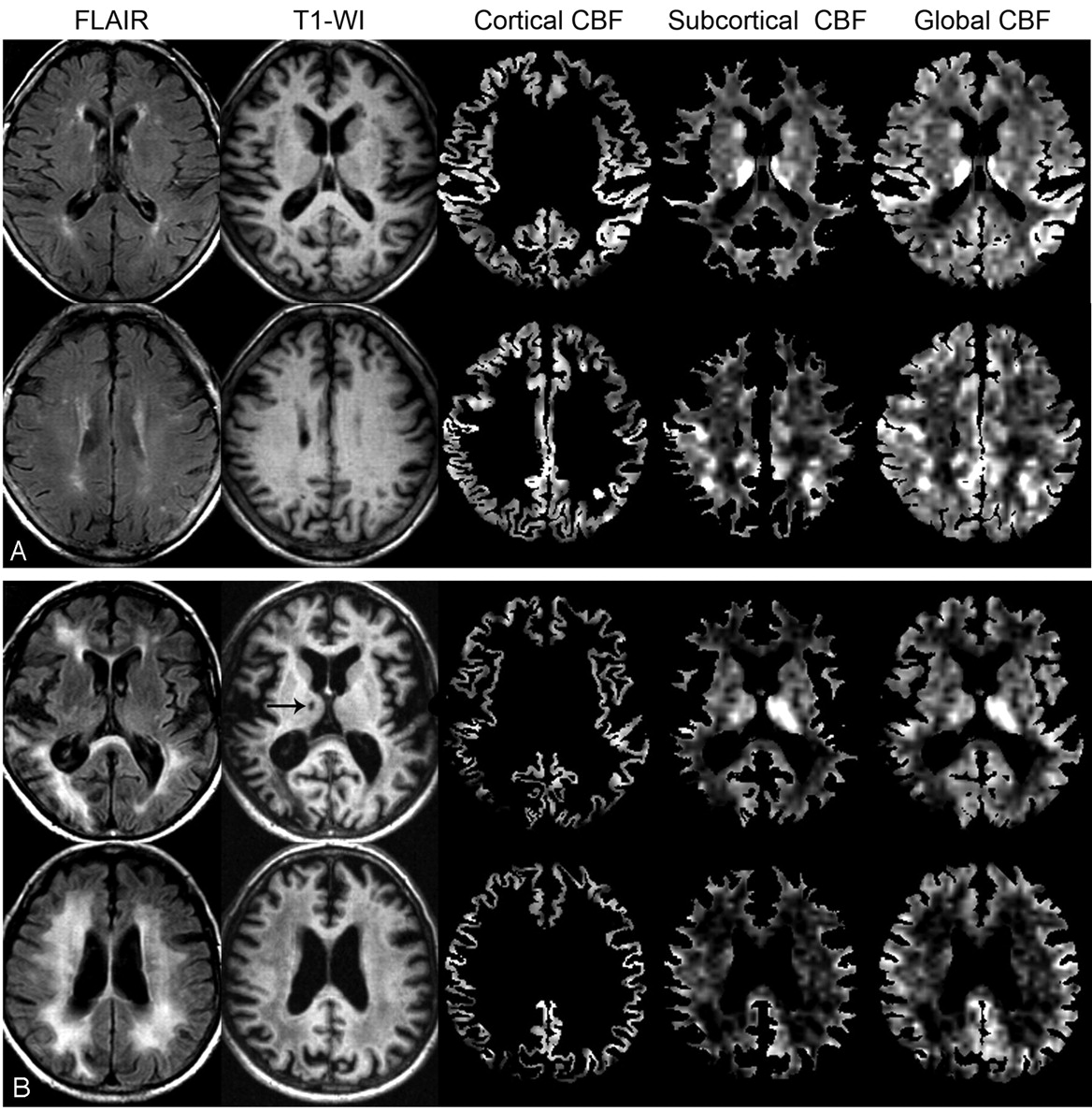
  • Fig 2.
    • Download figure
    • Open in new tab
    • Download powerpoint
    Fig 2.

    A, Axial FLAIR images, T1-weighted images, cortical gray matter CBF images, subcortical CBF images, and global CBF images combining all gray and white matter at the level of the basal ganglia and thalamus (above) and at the level of cerebral white matter (below) from subject 4. B, Corresponding images from subject 18. On the CBF images, note that the cerebral cortex, the cortical-subcortical transition, and the thalamus are highly perfused structures. Also note the perfusion differences between subject 18 (with diffuse confluent WMH on FLAIR images) and subject 4 (with punctiform WMH on FLAIR images). Finally, on the subcortical and global CBF images from subject 18, note the relative hypoperfusion of the right thalamus, where a lacunar infarct occurs (arrow).

Tables

  • Figures
    • View popup
    Table 1:

    Characteristics of subjects (n = 21), CBF measurements in mL/100 mL/min, and WMH score

    SubjectsSexAgeDiagnosisMedication and DrugsCortical CBFSubcortical CBFGlobal CBFWMH†
    1F73–Diuretic64.137.150.51
    2M70–Diuretic65.738.452.11
    3F78––69.640.454.81
    4F79–Diuretic68.548.658.01
    5F76ADAA, nicotine57.634.244.42
    6F82ADβ-Bl61.634.446.22
    7F80––62.137.147.92
    8F78––55.541.447.92
    9M75–Diuretic, CA66.839.451.42
    10F66–AA70.751.560.02
    11M73––72.547.860.42
    12F70–α2-agonist81.248.162.92
    13F70–β-Bl, nitrate77.153.364.42
    14F72–CA*, nitrate106.160.278.92
    15M80–β-Bl, CA51.326.736.23
    16M70–β-Bl, CA48.726.937.83
    17F84–Diuretic, β-Bl, AA, nitrate47.635.040.63
    18M76VaDDiuretic, ACE inhibitor62.331.740.93
    19M84–ACE inhibitor60.635.246.33
    20M71–Diuretic, β-Bl65.337.049.43
    21F79––69.142.953.13
    Mean (SD)–75.5 (5.1)––65.9 (12.6)40.3 (8.6)51.6 (10.1)2.1 (0.7)
    • Note:—AD indicates Alzheimer disease; VaD, vascular dementia; AA, angiotensin antagonist; β-Bl, β-blocker; CA*, calcium antagonist (*diltiazem); ACE inhibitor, angiotensin-converting enzyme inhibitor; WMH, white matter hyperintensities; CBF, cerebral blood flow.

    • † Higher values indicate greater severity.

    • View popup
    Table 2:

    Comparisons of CBF in mL/100 mL/min between groups of subjects with different grades of WMH

    Mean CBF (SD)Relative Difference (%)P value
    WMH Score 1 or 2 (n = 14)WMH Score 3 (n = 7)
    Global55.7 (9.2)43.5 (6.3)21.9<.01
    Subcortical43.7 (7.9)33.6 (5.8)23.1<.01
    Cortical69.9 (12.6)57.9 (8.6)17.2<.05
PreviousNext
Back to top

In this issue

American Journal of Neuroradiology: 29 (7)
American Journal of Neuroradiology
Vol. 29, Issue 7
August 2008
  • Table of Contents
  • Index by author
Advertisement
Print
Download PDF
Email Article
Cite this article
0 Responses
Respond to this article
Share
Bookmark this article
Cerebral Blood Flow by Using Pulsed Arterial Spin-Labeling in Elderly Subjects with White Matter Hyperintensities
A.J. Bastos-Leite, J.P.A. Kuijer, S.A.R.B. Rombouts, E. Sanz-Arigita, E.C. van Straaten, A.A. Gouw, W.M. van der Flier, P. Scheltens, F. Barkhof
American Journal of Neuroradiology Aug 2008, 29 (7) 1296-1301; DOI: 10.3174/ajnr.A1091
del.icio.us logo Twitter logo Facebook logo Mendeley logo
  • Tweet Widget
Purchase

Jump to section

  • Article
    • Abstract
    • Materials and Methods
    • Results
    • Discussion
    • Conclusion
    • Footnotes
    • References
  • Figures & Data
  • Info & Metrics
  • Responses
  • References
  • PDF

Related Articles

  • No related articles found.
  • PubMed
  • Google Scholar

Cited By...

  • Increased resting cerebral blood flow in adult Fabry disease: MRI arterial spin labeling study
  • In search of a putative imaging biomarker for Fabry disease: Go with the flow?
  • Arterial Spin-Labeling Perfusion MR Imaging Demonstrates Regional CBF Decrease in Idiopathic Normal Pressure Hydrocephalus
  • Intravoxel Incoherent Motion Imaging in Small Vessel Disease: Microstructural Integrity and Microvascular Perfusion Related to Cognition
  • White Matter Hyperintensity Volume and Cerebral Perfusion in Older Individuals with Hypertension Using Arterial Spin-Labeling
  • Longitudinal Relationship Between Cerebral Small-Vessel Disease and Cerebral Blood Flow: The Second Manifestations of Arterial Disease-Magnetic Resonance Study
  • Clinical Application of Arterial Spin-Labeling MR Imaging in Patients with Carotid Stenosis: Quantitative Comparative Study with Single-Photon Emission CT
  • Crossref
  • Google Scholar

This article has not yet been cited by articles in journals that are participating in Crossref Cited-by Linking.

More in this TOC Section

  • Evaluating the Effects of White Matter Multiple Sclerosis Lesions on the Volume Estimation of 6 Brain Tissue Segmentation Methods
  • Quiet PROPELLER MRI Techniques Match the Quality of Conventional PROPELLER Brain Imaging Techniques
  • Predictors of Reperfusion in Patients with Acute Ischemic Stroke
Show more Brain

Similar Articles

  • Intracranial Pulsatility in Pseudoxanthoma
  • Pulsatility Attenuation along the Carotid Siphon in Pseudoxanthoma Elasticum
  • Collateral Development in PEDS Stroke
  • Cerebral Aneurysms in a Patient with Osteogenesis Imperfecta and Exon 28 Polymorphism of COL1A2
  • CT and MR Imaging of Progressive Dural Involvement by Nephrogenic Systemic Fibrosis
See more
Advertisement

Indexed Content

  • Current Issue
  • Accepted Manuscripts
  • Article Preview
  • Past Issues
  • Editorials
  • Editor's Choice
  • Fellows' Journal Club
  • Letters to the Editor
  • Video Articles

Cases

  • Case Collection
  • Archive - Case of the Week
  • Archive - Case of the Month
  • Archive - Classic Case

More from AJNR

  • Trainee Corner
  • Imaging Protocols
  • MRI Safety Corner
  • Book Reviews

Multimedia

  • AJNR Podcasts
  • AJNR Scantastics

Resources

  • Turnaround Time
  • Submit a Manuscript
  • Submit a Video Article
  • Submit an eLetter to the Editor/Response
  • Manuscript Submission Guidelines
  • Statistical Tips
  • Fast Publishing of Accepted Manuscripts
  • Graphical Abstract Preparation
  • Imaging Protocol Submission
  • Evidence-Based Medicine Level Guide
  • Publishing Checklists
  • Author Policies
  • Become a Reviewer/Academy of Reviewers
  • News and Updates

About Us

  • About AJNR
  • Editorial Board
  • Editorial Board Alumni
  • Alerts
  • Permissions
  • Not an AJNR Subscriber? Join Now
  • Advertise with Us
  • Librarian Resources
  • Feedback
  • Terms and Conditions
  • AJNR Editorial Board Alumni

American Society of Neuroradiology

  • Not an ASNR Member? Join Now

© 2025 by the American Society of Neuroradiology All rights, including for text and data mining, AI training, and similar technologies, are reserved.
Print ISSN: 0195-6108 Online ISSN: 1936-959X

Powered by HighWire
Email this Article

Thank you for your interest in spreading the word on American Journal of Neuroradiology.

NOTE: We only request your email address so that the person you are recommending the page to knows that you wanted them to see it, and that it is not junk mail. We do not capture any email address.

Enter multiple addresses on separate lines or separate them with commas.
Cerebral Blood Flow by Using Pulsed Arterial Spin-Labeling in Elderly Subjects with White Matter Hyperintensities
(Your Name) has sent you a message from American Journal of Neuroradiology
(Your Name) thought you would like to see the American Journal of Neuroradiology web site.
CAPTCHA
This question is for testing whether or not you are a human visitor and to prevent automated spam submissions.
Cite this article
A.J. Bastos-Leite, J.P.A. Kuijer, S.A.R.B. Rombouts, E. Sanz-Arigita, E.C. van Straaten, A.A. Gouw, W.M. van der Flier, P. Scheltens, F. Barkhof
Cerebral Blood Flow by Using Pulsed Arterial Spin-Labeling in Elderly Subjects with White Matter Hyperintensities
American Journal of Neuroradiology Aug 2008, 29 (7) 1296-1301; DOI: 10.3174/ajnr.A1091

Citation Manager Formats

  • BibTeX
  • Bookends
  • EasyBib
  • EndNote (tagged)
  • EndNote 8 (xml)
  • Medlars
  • Mendeley
  • Papers
  • RefWorks Tagged
  • Ref Manager
  • RIS
  • Zotero

We use cookies on this site to enhance your user experience. By clicking any link on this page you are giving your consent for us to set cookies.